

Volume 12, Issue 3 (2026)
Pharm Biomed Res 2026, 12(3): 207-218 |
Back to browse issues page



Download citation:
BibTeX | RIS | EndNote | Medlars | ProCite | Reference Manager | RefWorks
Send citation to:


BibTeX | RIS | EndNote | Medlars | ProCite | Reference Manager | RefWorks
Send citation to:
Beheshti A, Tavakoli M, Karbasi V, Payeghadr M. A Simple, Accurate, and Cost-effective HPLC-UV for Determination of Nanoliposome Components as Drug Carriers. Pharm Biomed Res 2026; 12 (3) :207-218
URL: http://pbr.mazums.ac.ir/article-1-752-en.html
URL: http://pbr.mazums.ac.ir/article-1-752-en.html


1- Department of Chemistry, Payame Noor University, Tehran, Iran.
2- Quality Control Lab, Tofigh Daru Research and Engineering Co, Tehran, Iran.
3- Department of Chemistry, Payame Noor University, Tehran, Iran. & Quality Control Lab, Tofigh Daru Research and Engineering Co, Tehran, Iran.
2- Quality Control Lab, Tofigh Daru Research and Engineering Co, Tehran, Iran.
3- Department of Chemistry, Payame Noor University, Tehran, Iran. & Quality Control Lab, Tofigh Daru Research and Engineering Co, Tehran, Iran.
Keywords: Nanoliposomes, Drug carrier, Lipid analysis, High-performance liquid chromatography (HPLC), Method development
Full-Text [PDF 864 kb]
(65 Downloads)
| Abstract (HTML) (290 Views)
Preparation of standard and test solutions
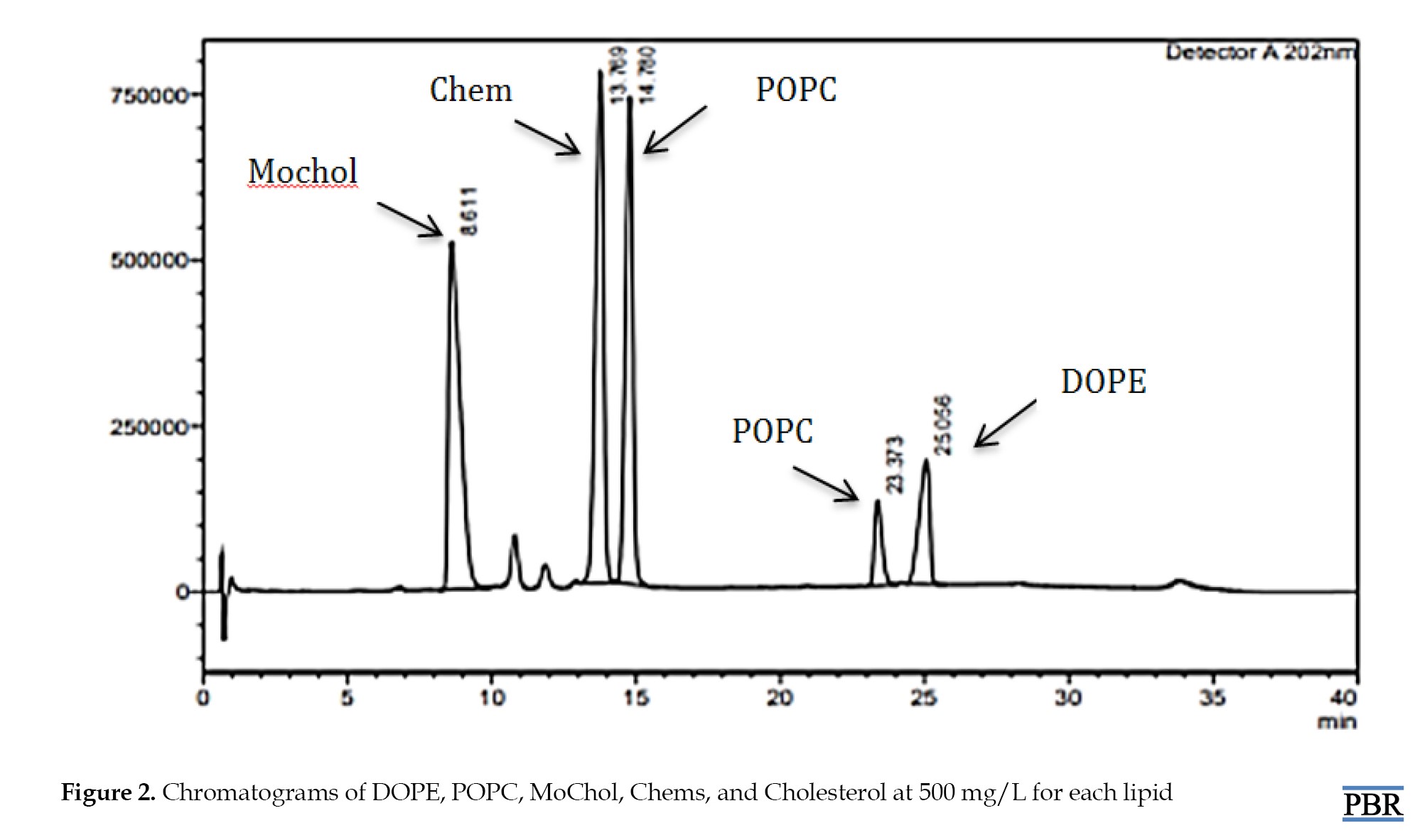
The stock standard solution (2000 mg/L) of DOPE, POPC, MoChol, Chems, and cholesterol was prepared by weighing approximately 7.5 mg of each standard and transferring it into a 5 mL volumetric amber flask. The diluent was added, and the mixture was sonicated to dissolve. All diluted solutions were prepared by diluting a concentrated stock solution. The results from different concentration levels (1-2000 ppm) were used to create a calibration target curve for each component. The sample chromatogram showed high-resolution separations of these lipids (Figure 2).
Full-Text: (33 Views)
Introduction
Due to unique characteristics and versatile applications, nanoliposomes known as lipid nanoparticles, have garnered significant attention in drug delivery [1]. With a lipid bilayer shell surrounding an aqueous core, these nanosized vesicles provide an ideal platform for nanoencapsulation and deliver various therapeutic (biologically active) compounds [2-4]. Composed primarily of naturally occurring phospholipids, nanoliposomes are biocompatible, biodegradable and mimic the lipid composition of cell membranes. This reduces the risk of immune responses or adverse reactions upon administration [3]. Furthermore, the nanoliposome components are metabolized and eliminated from the body, minimizing potential toxicity and long-term accumulation. The nanoliposomes’ lipid bilayer structure provides a barrier against enzymatic degradation and environmental factors, preserving the integrity of encapsulated drugs, particularly sensitive or labile compounds, during storage and transportation [2, 4]. The hydrophobic drugs’ solubility and bioavailability can be improved by facilitating their incorporation into nano carriers, thereby enhancing their therapeutic efficacy and reducing the required dosage [4]. In addition, nanocarriers can facilitate targeted drug delivery to specific cells or tissues through surface functionalization. Therefore, they can actively deliver to diseased sites or overexpressed receptors. This new strategy not only enhances drug accumulation and efficacy but also minimizes off-target effects. This targeted approach improves therapeutic outcomes while reducing systemic toxicity and adverse reactions [5-8]. Nanoliposomes can be engineered to exhibit sustained-release kinetics, prolonging the duration of drug action and minimizing the need for frequent dosing [4]. By modulating the size, surface properties, and lipid composition of nanoliposomes, it is possible to control the rate and extent of drug release, achieving desired pharmacokinetic profiles and therapeutic effects [9-12]. The composition and structure of spherical or multilayered spherical vesicles can be tailored with protective coatings or structural changes to optimize drug encapsulation, stability, and release kinetics. This modification makes them highly adaptable for a wide range of pharmaceutical formulations. Accurate determination of nanoliposomes components is crucial for ensuring product quality, efficacy, maintaining batch-to-batch consistency and meeting regulatory requirements [13, 14]. The analytical challenges associated with lipid determination become more pronounced. Labour-intensive and time-consuming traditional analytical methods, prone to variability result, in an unsuitable high-throughput process. Additionally, multiple lipid components with similar chemical properties pose challenges for selective quantification, further complicating nanocarrier analysis. Various analytical methods such as liquid chromatography with ultraviolet detection (HPLC-UV-Vis) [14-16] or gas chromatography [11, 17] as well as nuclear magnetic resonance spectroscopy [18, 19] and mass spectrometry [20] have been utilized before in nanoliposome component determination. While these methods offer high sensitivity and selectivity, they often require complex sample preparation and laborious instrumentation, limiting their suitability for routine high-throughput analysis. Despite progress in determining lipid content in nanoliposomes, there is a lack of simultaneous separation and analysis of multiple lipid components in nanoliposomes [21-23]. This study aimed to develop a robust, simple, accurate and cost-effective analytical method for the determination of nanoliposome components as a drug carrier. Leveraging the unique absorbance properties of lipid components, we optimized chromatographic conditions to achieve simultaneous separation and determination of α-(3-O-cholesteryloxy)-δ-(N-ethylmorpholine)-succineamide (MoChol, cationic), cholesteryl-hemisuccinate (Chems, anionic), cholesterol, dioleoyl-phosphatidylethanolamine (DOPE) and palmitoyl-oleoylphosphatidylcholine (POPC). The method development process focused on appropriate chromatographic conditions to maximize sensitivity, selectivity, and reproducibility. Validation studies were conducted in accordance with the International Council for Harmonization (ICH) Guidelines to demonstrate the reliability and robustness of the proposed method.
Experimental
Instrumentation and reagents
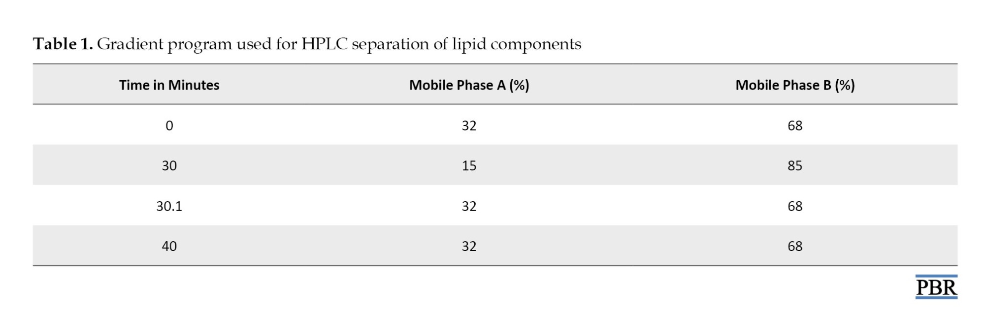
HPLC-based separation and analysis were performed on a Shimadzu Prominence system (Shimadzu Corporation, Kyoto, Japan) equipped with an LC-20AD pump, an online DGU-20A degassing system, a CTO-20A column oven, and an SPD-20A UV–visible detector. The separation was achieved using a Hector C18-M column (250×4.6 mm, 5 μm particle size). The system was controlled using Lab Solutions software, version 5.51 (Shimadzu), which was also applied for data processing. Column temperature was kept constant at 50 °C, and the flow rate was set at 1.0 mL/min with a mobile phase gradient (Table 1), where mobile phase A was 0.1% trifluoroacetic acid (TFA) in Water and mobile phase B was 0.1% TFA in ethanol.
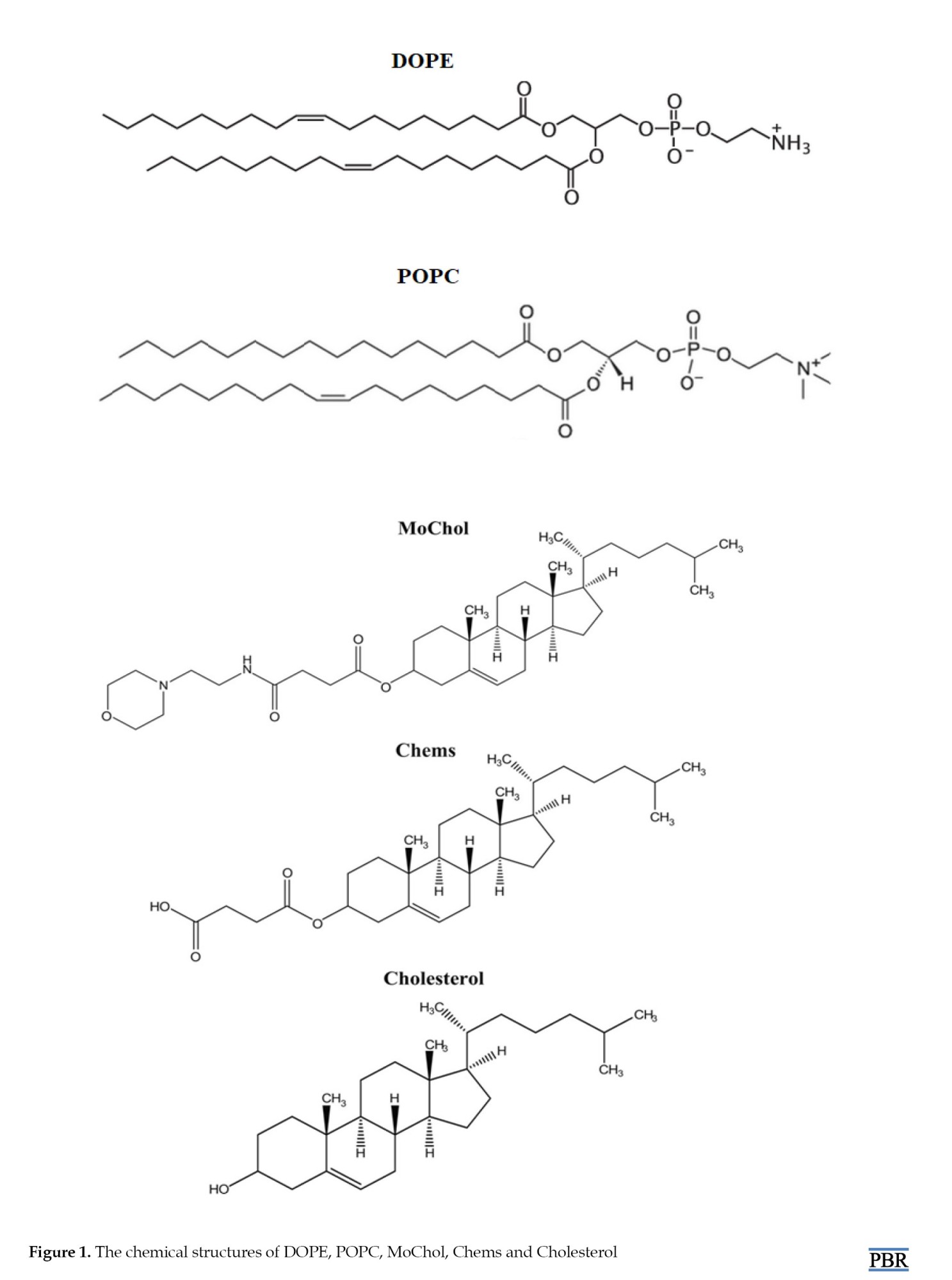
The injection was performed using a 20 μL injection loop and the detector wavelength was adjusted to 202 nm. Ethanol was used as diluent. These chromatographic conditions were selected based on an optimization process, considering factors such as peak shape, retention time, and resolution between lipid components. MoChol, TFA, and ethanol (HPLC grade) were purchased from Merck (Germany). Cholesterol, DOPE, POPC and Chems were purchased from Sigma-Aldrich, Switzerland. In-house purified Water provided by a Milli-Q purification system (Millipore) was used. The anti-BCL-2 DNA oligonucleotide loaded nanoliposome was purchased from Polymon Company. Figure 1 shows the chemical structures of MoChol, Chems, DOPE, POPC, and cholesterol.
Due to unique characteristics and versatile applications, nanoliposomes known as lipid nanoparticles, have garnered significant attention in drug delivery [1]. With a lipid bilayer shell surrounding an aqueous core, these nanosized vesicles provide an ideal platform for nanoencapsulation and deliver various therapeutic (biologically active) compounds [2-4]. Composed primarily of naturally occurring phospholipids, nanoliposomes are biocompatible, biodegradable and mimic the lipid composition of cell membranes. This reduces the risk of immune responses or adverse reactions upon administration [3]. Furthermore, the nanoliposome components are metabolized and eliminated from the body, minimizing potential toxicity and long-term accumulation. The nanoliposomes’ lipid bilayer structure provides a barrier against enzymatic degradation and environmental factors, preserving the integrity of encapsulated drugs, particularly sensitive or labile compounds, during storage and transportation [2, 4]. The hydrophobic drugs’ solubility and bioavailability can be improved by facilitating their incorporation into nano carriers, thereby enhancing their therapeutic efficacy and reducing the required dosage [4]. In addition, nanocarriers can facilitate targeted drug delivery to specific cells or tissues through surface functionalization. Therefore, they can actively deliver to diseased sites or overexpressed receptors. This new strategy not only enhances drug accumulation and efficacy but also minimizes off-target effects. This targeted approach improves therapeutic outcomes while reducing systemic toxicity and adverse reactions [5-8]. Nanoliposomes can be engineered to exhibit sustained-release kinetics, prolonging the duration of drug action and minimizing the need for frequent dosing [4]. By modulating the size, surface properties, and lipid composition of nanoliposomes, it is possible to control the rate and extent of drug release, achieving desired pharmacokinetic profiles and therapeutic effects [9-12]. The composition and structure of spherical or multilayered spherical vesicles can be tailored with protective coatings or structural changes to optimize drug encapsulation, stability, and release kinetics. This modification makes them highly adaptable for a wide range of pharmaceutical formulations. Accurate determination of nanoliposomes components is crucial for ensuring product quality, efficacy, maintaining batch-to-batch consistency and meeting regulatory requirements [13, 14]. The analytical challenges associated with lipid determination become more pronounced. Labour-intensive and time-consuming traditional analytical methods, prone to variability result, in an unsuitable high-throughput process. Additionally, multiple lipid components with similar chemical properties pose challenges for selective quantification, further complicating nanocarrier analysis. Various analytical methods such as liquid chromatography with ultraviolet detection (HPLC-UV-Vis) [14-16] or gas chromatography [11, 17] as well as nuclear magnetic resonance spectroscopy [18, 19] and mass spectrometry [20] have been utilized before in nanoliposome component determination. While these methods offer high sensitivity and selectivity, they often require complex sample preparation and laborious instrumentation, limiting their suitability for routine high-throughput analysis. Despite progress in determining lipid content in nanoliposomes, there is a lack of simultaneous separation and analysis of multiple lipid components in nanoliposomes [21-23]. This study aimed to develop a robust, simple, accurate and cost-effective analytical method for the determination of nanoliposome components as a drug carrier. Leveraging the unique absorbance properties of lipid components, we optimized chromatographic conditions to achieve simultaneous separation and determination of α-(3-O-cholesteryloxy)-δ-(N-ethylmorpholine)-succineamide (MoChol, cationic), cholesteryl-hemisuccinate (Chems, anionic), cholesterol, dioleoyl-phosphatidylethanolamine (DOPE) and palmitoyl-oleoylphosphatidylcholine (POPC). The method development process focused on appropriate chromatographic conditions to maximize sensitivity, selectivity, and reproducibility. Validation studies were conducted in accordance with the International Council for Harmonization (ICH) Guidelines to demonstrate the reliability and robustness of the proposed method.
Experimental
Instrumentation and reagents
HPLC-based separation and analysis were performed on a Shimadzu Prominence system (Shimadzu Corporation, Kyoto, Japan) equipped with an LC-20AD pump, an online DGU-20A degassing system, a CTO-20A column oven, and an SPD-20A UV–visible detector. The separation was achieved using a Hector C18-M column (250×4.6 mm, 5 μm particle size). The system was controlled using Lab Solutions software, version 5.51 (Shimadzu), which was also applied for data processing. Column temperature was kept constant at 50 °C, and the flow rate was set at 1.0 mL/min with a mobile phase gradient (Table 1), where mobile phase A was 0.1% trifluoroacetic acid (TFA) in Water and mobile phase B was 0.1% TFA in ethanol.
The injection was performed using a 20 μL injection loop and the detector wavelength was adjusted to 202 nm. Ethanol was used as diluent. These chromatographic conditions were selected based on an optimization process, considering factors such as peak shape, retention time, and resolution between lipid components. MoChol, TFA, and ethanol (HPLC grade) were purchased from Merck (Germany). Cholesterol, DOPE, POPC and Chems were purchased from Sigma-Aldrich, Switzerland. In-house purified Water provided by a Milli-Q purification system (Millipore) was used. The anti-BCL-2 DNA oligonucleotide loaded nanoliposome was purchased from Polymon Company. Figure 1 shows the chemical structures of MoChol, Chems, DOPE, POPC, and cholesterol.
Preparation of standard and test solutions
The stock standard solution (2000 mg/L) of DOPE, POPC, MoChol, Chems, and cholesterol was prepared by weighing approximately 7.5 mg of each standard and transferring it into a 5 mL volumetric amber flask. The diluent was added, and the mixture was sonicated to dissolve. All diluted solutions were prepared by diluting a concentrated stock solution. The results from different concentration levels (1-2000 ppm) were used to create a calibration target curve for each component. The sample chromatogram showed high-resolution separations of these lipids (Figure 2).
Additionally, 25 μL of synthesized nanoliposome was added to 1000 μL of Otyl β-D-glucopyanoside water solution (2% w/w) to prepare the nanoliposome test solution, which was vortexed for 30 s before injection.
Materials and Methods
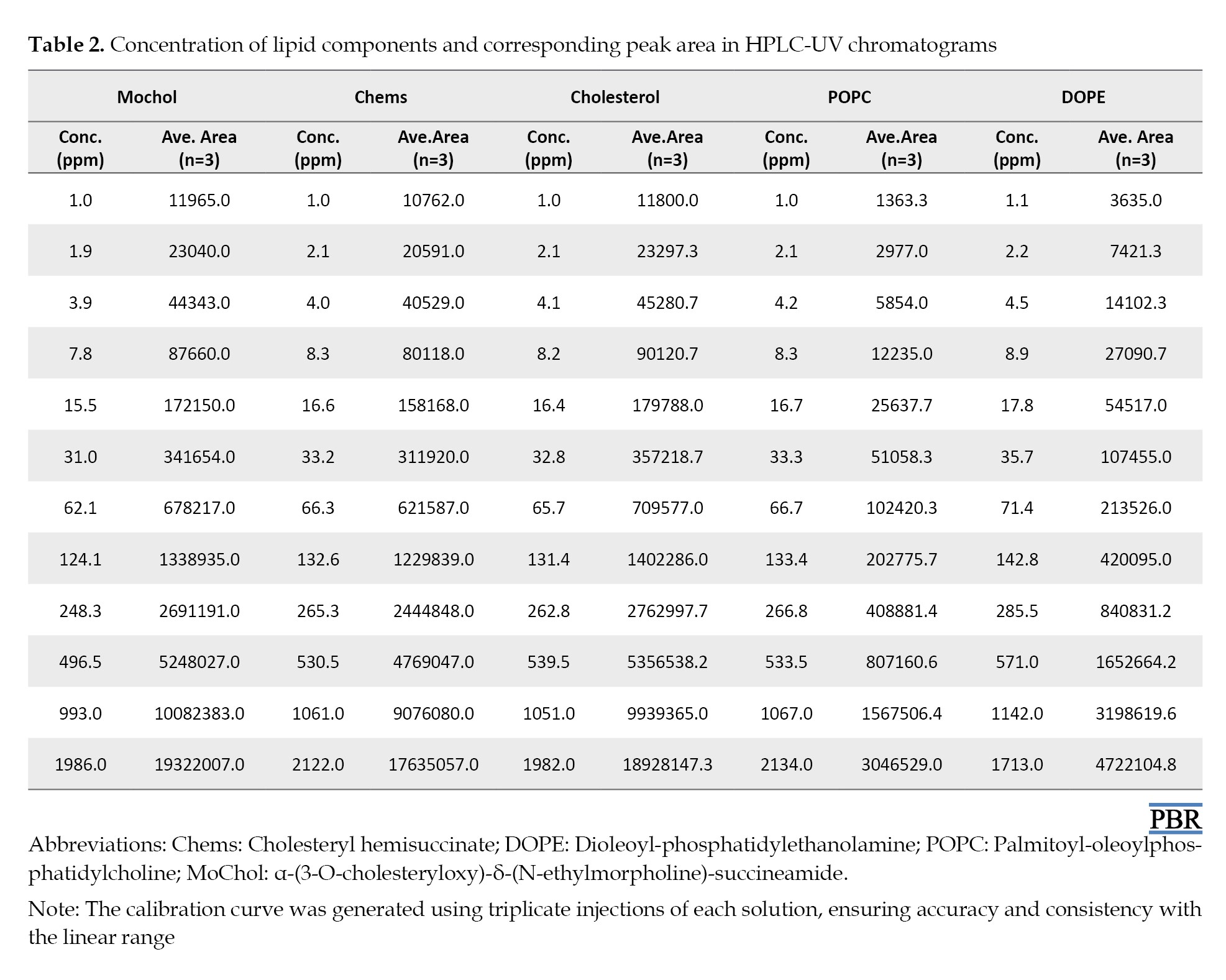
HPLC with refractive index (RI) or evaporative light scattering (ELSD) detector is commonly utilized for lipid composition analysis. Either RI or ELSD detectors have drawbacks, including the laborious parameter optimization, sample decomposition, detector response fluctuations and the need to add internal standards. Furthermore, these costly techniques are limitedly accessible, which hinders their widespread application in lipid analysis. In addition, despite their versatility, RI detectors have limitations: Their response is dependent on temperature, pressure fluctuation, flow rate instability, and mobile phase composition, which make them inappropriate for gradient elution [24] because the lipids lack chromophores, their detection in the UV range is limited to approximately 200–210 nm [25] which restricts the solvents, buffers or additives choice in sampling procedure and HPLC analysis [26] as a mobile phase modifier, TFA forms ion pairs with basic analytes, suppress their interaction with column stationary phase and reduce peak tailing [27]. In the current study, TFA addition overcame the limitation in selecting an appropriate additive. Consequently, a gradient HPLC method was developed for cholesterol, MoChol, Chems, POPC and DOPE analysis in nanoliposomes. The standard peak response (average of three replicates) was plotted versus the lipid concentration in the range of 1 to 2000 mg L−1 (Table 2).
The quantitative data obtained using the developed HPLC-UV method demonstrate the method’s linearity, reliability, and accuracy. Parameters including the slope, intercept, calibration equation and linear range were established through linear regression analysis and are presented in Table 3.
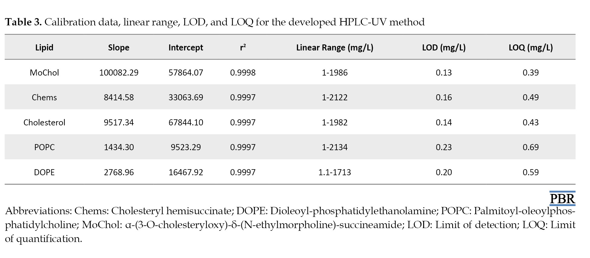
These data highlight the method’s robustness, providing a reliable tool for lipid component quantification in nanoliposome formulations. The developed method ensures consistent product quality and is essential for large-scale manufacturing.
Results
Method validation
To ensure the results are trustworthy and precise, key parameters in validation according to ICH guidelines were assessed. This typically involves evaluating accuracy, precision, limit of detection (LOD), limit of quantification (LOQ), specificity, and robustness [28].
Accuracy
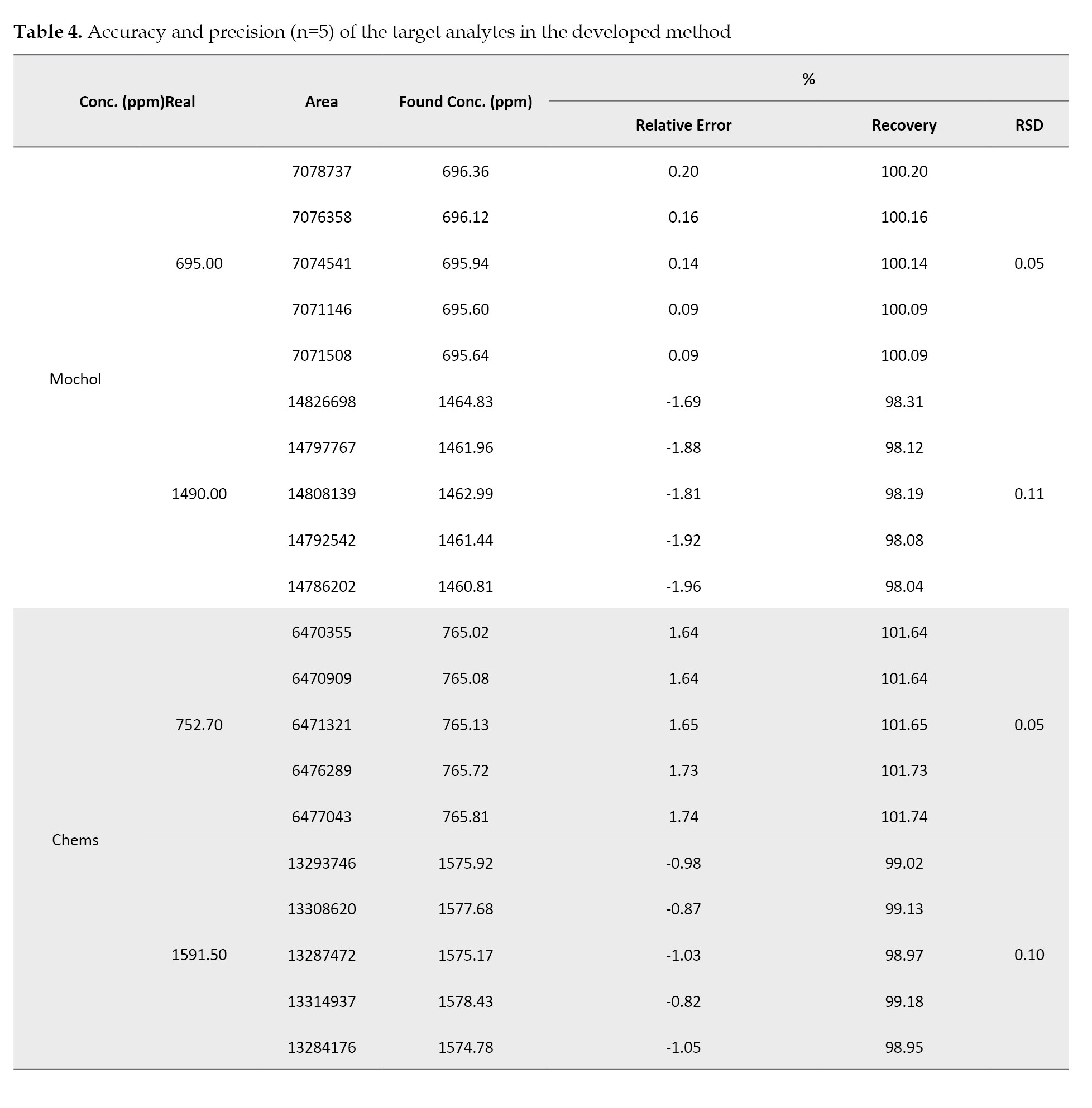
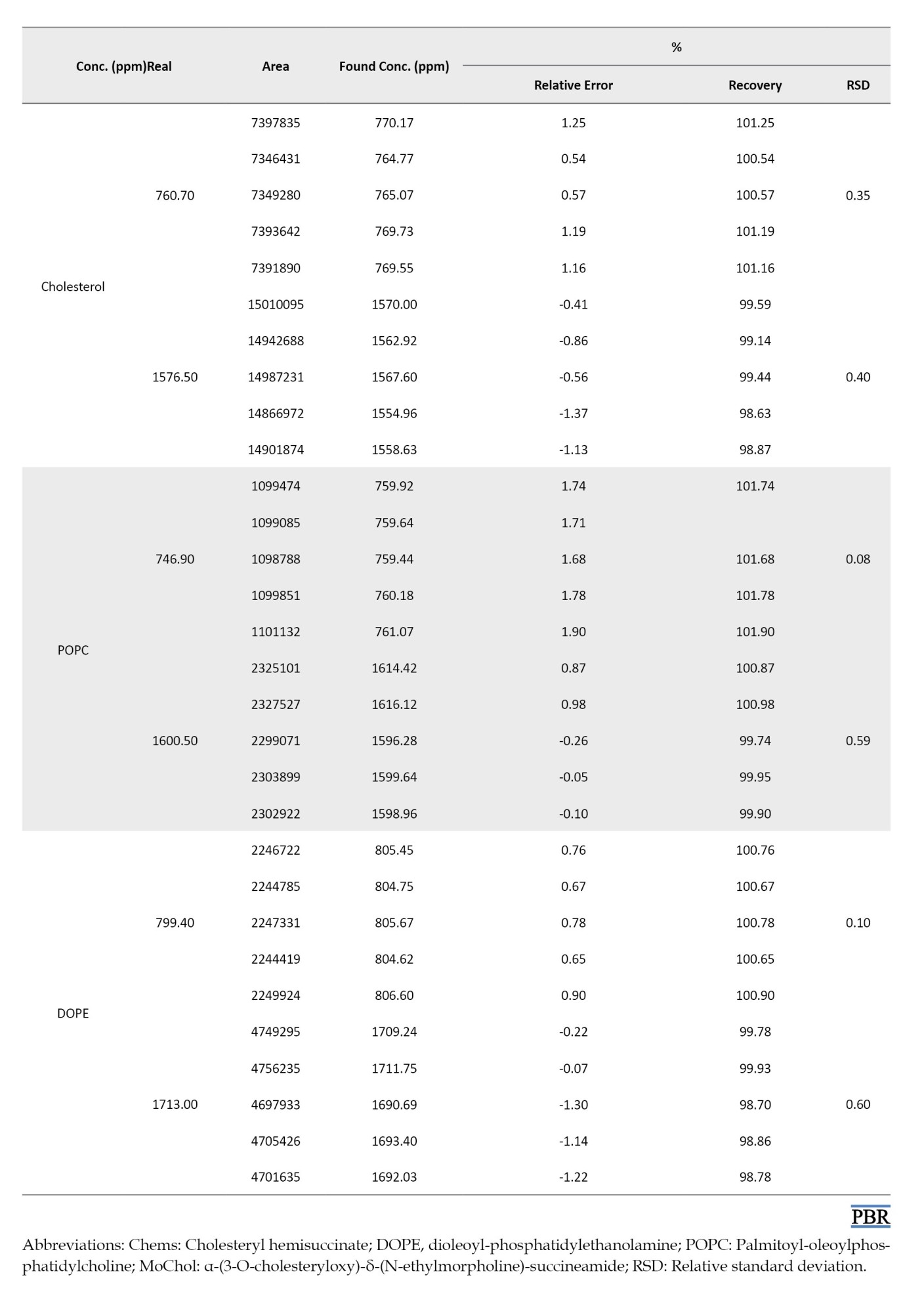
Accuracy refers to how closely the results obtained from an analytical method match the true or accepted value. It measures the method’s exactness and is assessed by comparing measured values to known reference values. In this study, accuracy was assessed by analyzing five replicate standard solutions at approximately 700 ppm and 1600 ppm. The recovery percentages shown in Table 4 were close to the range of 98–102%.
Precision
Precision refers to the consistency or reproducibility of the results and is monitored by the relative standard deviation percentage (RSD%) obtained from multiple standard or sample injections. As shown in Table 4, the proposed method precision was assessed by measuring the repeatability of peak areas for samples used in the accuracy test, meeting the criterion (RSD≤1%), indicating reproducible results.
Limit of detection and limit of quantification
LOD is the lowest concentration of an analyte that can be reliably detected but not necessarily quantified, while the LOQ is the lowest concentration that can be quantified accurately and precisely. These parameters were calculated using the y-intercept standard deviation value and the linearity plot slope. The calibration curve was created using samples with lower analyte content near the LOD. Table 3 presents the obtained LOD and LOQ values with the proposed method.
Specificity
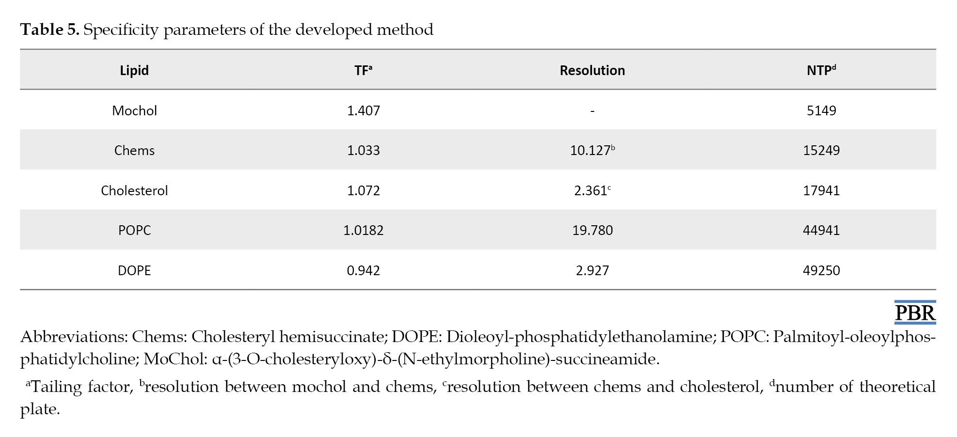
As defined by the ICH guideline, specificity refers to the method’s ability to measure the analyte in the presence of impurities, by-products, and matrix components accurately and selectively. Specificity of validation ensures that each peak response corresponds to a single component without overlap. Parameters commonly studied to evaluate specificity include resolution, tailing factor (TF) and number of theoretical plates (NTP). In the present study, it is crucial to achieve a suitable resolution between MoChol, Chems, and Cholesterol, as these are structurally cholesterol-related derivatives. The structural similarity between these lipids makes them difficult to distinguish during chromatographic separation. In addition, because their degradation can affect nanoliposome stability and efficacy, their specific separation is mandatory. Our method provides reliable and precise quantification by achieving a resolution >2.0. Table 5 presents the specificity parameters at 250 mg/L, demonstrating that the resolution between the five lipids exceeds 2.0, ensuring the integrity and performance of the proposed method.
Robustness
Method robustness refers to the ability to withstand minor but deliberate deviations in flow rate, mobile phase composition, and column temperature without significantly affecting results. It was evaluated by examining system suitability parameters, as shown in Table 6.
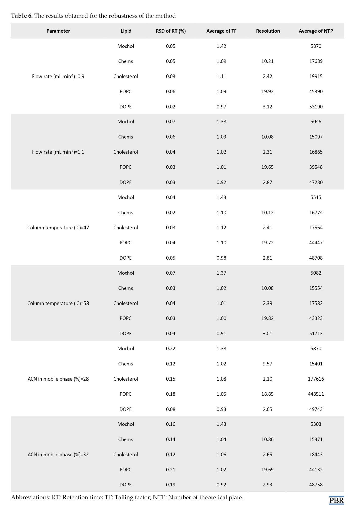
The results indicate that the method remains stable, with NTP values consistently above 5,000, TF values below 1.5 for each lipid, and resolution exceeding 2. This indicates that the method remains unaffected by parameter variations and is reliable under normal usage.
Discussion
The purchased nanoliposome real sample contained the following amounts (mg/L) of API: 1.53, MoChol: 23.34, Chems: 9.45, POPC: 4.03, DOPE: 15.31, and Cholesterol: 0.9. Also, it exhibits these characteristics: an average particle size of 124.4 nm, a polydispersity index of 0.13, a zeta potential of −32.4 mV, a pH of 7.56, and an endotoxin content of 3.44 EEU mL−1. MoChol and Chems may undergo degradation through oxidation or hydrolysis under storage-related conditions. Oxidation produces peroxides that degrade MoChol and Chems into Cholesterol and other products. In addition, hydrolysis or enzymatic catalysis breaks ester bonds in the formulation or environment. MoChol and Chems’ integrity in the nanoliposome is compromized by this degradation process, affecting its stability and efficacy. The nanoliposome compositions were determined after storage for 8 months at −20 °C to compare the stability of different batches. Preparation of the real sample involved adding nanoliposomes (25 μL) to Otyl β-D-glucopyanoside solution in water (1000 μL, 2% w/w) to achieve concentrations within the linear calibration ranges. The solution was vortexed for 30 s and injected into the HPLC. Table 7 presents the quantitative results.
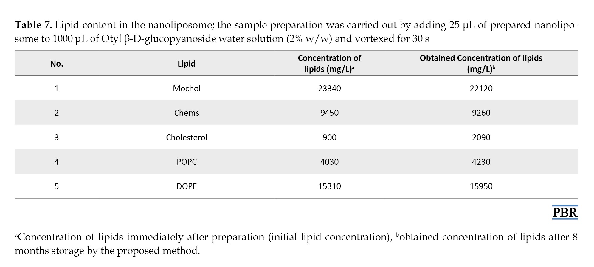
Slight degradation of MoChol and Chems over 8 months was revealed, while Cholesterol concentration significantly increased. The observed compositional changes underline the importance of this analytical approach for monitoring formulation stability.
Conclusion
In the current study, an HPLC-UV method for the simultaneous determination of lipid components in nanoliposome formulations was developed and validated. The method addressed significant technical challenges in lipid analysis. By optimizing chromatographic conditions, specifically the mobile phase composition, the method achieved high sensitivity, selectivity, and reproducibility. Key parameters, including linearity, precision, accuracy, and specificity, were thoroughly evaluated according to ICH guidelines. Linear regression analysis demonstrated strong correlation coefficients, and the method’s precision met the predefined acceptance criterion of ≤1%. The accuracy, validated through recovery studies, was within the acceptable range of 98–102%. In addition to technical robustness, the method effectively detected minor degradation products, demonstrating its capability to monitor nanoliposome stability. The observed degradation product underlines the importance of this analytical approach in ensuring drug delivery systems’ stability and efficacy. In addition, the method’s ability to separate and quantify cholesterol derivatives and two phospholipids with sufficient resolution is crucial, due to their similarities. Specificity parameters, such as NTP, resolution, and TF, confirmed the method’s adequacy in distinguishing these compounds without peak overlap. The developed HPLC-UV method offers a reliable, cost-effective way for routine analysis, facilitating quality control and regulatory compliance. Its robustness and sensitivity ensure consistent product quality, minimizing the risk of batch failure or deviations. This study’s findings significantly contribute to the field of nanomedicine by providing a standardized analytical framework for future research and development in lipid-based drug delivery systems.
Ethical Considerations
Compliance with ethical guidelines
There were no ethical considerations to be considered in this research.
Funding
This research was financially supported by Tofigh Daru Research and Engineering Company, Tehran, Iran.
Authors' contributions
Supervision and conceptualization: Abolghasem Beheshti and Mahmood Payeghadr; Review and editing: Abolghasem Beheshti and Mehdi Tavakoli; Methodology: Abolghasem Beheshti, Mahmood Payeghadr and Vahid Karbasi; Resources, project administration and funding acquisition: Abolghasem Beheshti; Investigation, software, data curation, formal analysis, visualization: Vahid Karbasi; Writing the original draft, validation, formal analysis: Mehdi Tavakoli.
Conflict of interest
The authors declared no conflict of interest.
Acknowledgments
Authors thank Tofigh Daru Research and Engineering Company for the financial and technical support of this project.
Materials and Methods
HPLC with refractive index (RI) or evaporative light scattering (ELSD) detector is commonly utilized for lipid composition analysis. Either RI or ELSD detectors have drawbacks, including the laborious parameter optimization, sample decomposition, detector response fluctuations and the need to add internal standards. Furthermore, these costly techniques are limitedly accessible, which hinders their widespread application in lipid analysis. In addition, despite their versatility, RI detectors have limitations: Their response is dependent on temperature, pressure fluctuation, flow rate instability, and mobile phase composition, which make them inappropriate for gradient elution [24] because the lipids lack chromophores, their detection in the UV range is limited to approximately 200–210 nm [25] which restricts the solvents, buffers or additives choice in sampling procedure and HPLC analysis [26] as a mobile phase modifier, TFA forms ion pairs with basic analytes, suppress their interaction with column stationary phase and reduce peak tailing [27]. In the current study, TFA addition overcame the limitation in selecting an appropriate additive. Consequently, a gradient HPLC method was developed for cholesterol, MoChol, Chems, POPC and DOPE analysis in nanoliposomes. The standard peak response (average of three replicates) was plotted versus the lipid concentration in the range of 1 to 2000 mg L−1 (Table 2).
The quantitative data obtained using the developed HPLC-UV method demonstrate the method’s linearity, reliability, and accuracy. Parameters including the slope, intercept, calibration equation and linear range were established through linear regression analysis and are presented in Table 3.
These data highlight the method’s robustness, providing a reliable tool for lipid component quantification in nanoliposome formulations. The developed method ensures consistent product quality and is essential for large-scale manufacturing.
Results
Method validation
To ensure the results are trustworthy and precise, key parameters in validation according to ICH guidelines were assessed. This typically involves evaluating accuracy, precision, limit of detection (LOD), limit of quantification (LOQ), specificity, and robustness [28].
Accuracy
Accuracy refers to how closely the results obtained from an analytical method match the true or accepted value. It measures the method’s exactness and is assessed by comparing measured values to known reference values. In this study, accuracy was assessed by analyzing five replicate standard solutions at approximately 700 ppm and 1600 ppm. The recovery percentages shown in Table 4 were close to the range of 98–102%.
Precision
Precision refers to the consistency or reproducibility of the results and is monitored by the relative standard deviation percentage (RSD%) obtained from multiple standard or sample injections. As shown in Table 4, the proposed method precision was assessed by measuring the repeatability of peak areas for samples used in the accuracy test, meeting the criterion (RSD≤1%), indicating reproducible results.
Limit of detection and limit of quantification
LOD is the lowest concentration of an analyte that can be reliably detected but not necessarily quantified, while the LOQ is the lowest concentration that can be quantified accurately and precisely. These parameters were calculated using the y-intercept standard deviation value and the linearity plot slope. The calibration curve was created using samples with lower analyte content near the LOD. Table 3 presents the obtained LOD and LOQ values with the proposed method.
Specificity
As defined by the ICH guideline, specificity refers to the method’s ability to measure the analyte in the presence of impurities, by-products, and matrix components accurately and selectively. Specificity of validation ensures that each peak response corresponds to a single component without overlap. Parameters commonly studied to evaluate specificity include resolution, tailing factor (TF) and number of theoretical plates (NTP). In the present study, it is crucial to achieve a suitable resolution between MoChol, Chems, and Cholesterol, as these are structurally cholesterol-related derivatives. The structural similarity between these lipids makes them difficult to distinguish during chromatographic separation. In addition, because their degradation can affect nanoliposome stability and efficacy, their specific separation is mandatory. Our method provides reliable and precise quantification by achieving a resolution >2.0. Table 5 presents the specificity parameters at 250 mg/L, demonstrating that the resolution between the five lipids exceeds 2.0, ensuring the integrity and performance of the proposed method.
Robustness
Method robustness refers to the ability to withstand minor but deliberate deviations in flow rate, mobile phase composition, and column temperature without significantly affecting results. It was evaluated by examining system suitability parameters, as shown in Table 6.
The results indicate that the method remains stable, with NTP values consistently above 5,000, TF values below 1.5 for each lipid, and resolution exceeding 2. This indicates that the method remains unaffected by parameter variations and is reliable under normal usage.
Discussion
The purchased nanoliposome real sample contained the following amounts (mg/L) of API: 1.53, MoChol: 23.34, Chems: 9.45, POPC: 4.03, DOPE: 15.31, and Cholesterol: 0.9. Also, it exhibits these characteristics: an average particle size of 124.4 nm, a polydispersity index of 0.13, a zeta potential of −32.4 mV, a pH of 7.56, and an endotoxin content of 3.44 EEU mL−1. MoChol and Chems may undergo degradation through oxidation or hydrolysis under storage-related conditions. Oxidation produces peroxides that degrade MoChol and Chems into Cholesterol and other products. In addition, hydrolysis or enzymatic catalysis breaks ester bonds in the formulation or environment. MoChol and Chems’ integrity in the nanoliposome is compromized by this degradation process, affecting its stability and efficacy. The nanoliposome compositions were determined after storage for 8 months at −20 °C to compare the stability of different batches. Preparation of the real sample involved adding nanoliposomes (25 μL) to Otyl β-D-glucopyanoside solution in water (1000 μL, 2% w/w) to achieve concentrations within the linear calibration ranges. The solution was vortexed for 30 s and injected into the HPLC. Table 7 presents the quantitative results.
Slight degradation of MoChol and Chems over 8 months was revealed, while Cholesterol concentration significantly increased. The observed compositional changes underline the importance of this analytical approach for monitoring formulation stability.
Conclusion
In the current study, an HPLC-UV method for the simultaneous determination of lipid components in nanoliposome formulations was developed and validated. The method addressed significant technical challenges in lipid analysis. By optimizing chromatographic conditions, specifically the mobile phase composition, the method achieved high sensitivity, selectivity, and reproducibility. Key parameters, including linearity, precision, accuracy, and specificity, were thoroughly evaluated according to ICH guidelines. Linear regression analysis demonstrated strong correlation coefficients, and the method’s precision met the predefined acceptance criterion of ≤1%. The accuracy, validated through recovery studies, was within the acceptable range of 98–102%. In addition to technical robustness, the method effectively detected minor degradation products, demonstrating its capability to monitor nanoliposome stability. The observed degradation product underlines the importance of this analytical approach in ensuring drug delivery systems’ stability and efficacy. In addition, the method’s ability to separate and quantify cholesterol derivatives and two phospholipids with sufficient resolution is crucial, due to their similarities. Specificity parameters, such as NTP, resolution, and TF, confirmed the method’s adequacy in distinguishing these compounds without peak overlap. The developed HPLC-UV method offers a reliable, cost-effective way for routine analysis, facilitating quality control and regulatory compliance. Its robustness and sensitivity ensure consistent product quality, minimizing the risk of batch failure or deviations. This study’s findings significantly contribute to the field of nanomedicine by providing a standardized analytical framework for future research and development in lipid-based drug delivery systems.
Ethical Considerations
Compliance with ethical guidelines
There were no ethical considerations to be considered in this research.
Funding
This research was financially supported by Tofigh Daru Research and Engineering Company, Tehran, Iran.
Authors' contributions
Supervision and conceptualization: Abolghasem Beheshti and Mahmood Payeghadr; Review and editing: Abolghasem Beheshti and Mehdi Tavakoli; Methodology: Abolghasem Beheshti, Mahmood Payeghadr and Vahid Karbasi; Resources, project administration and funding acquisition: Abolghasem Beheshti; Investigation, software, data curation, formal analysis, visualization: Vahid Karbasi; Writing the original draft, validation, formal analysis: Mehdi Tavakoli.
Conflict of interest
The authors declared no conflict of interest.
Acknowledgments
Authors thank Tofigh Daru Research and Engineering Company for the financial and technical support of this project.
References
- Puri A, Loomis K, Smith B, Lee JH, Yavlovich A, Heldman E, et al. Lipid-based nanoparticles as pharmaceutical drug carriers: from concepts to clinic. Crit Rev Ther Drug Carrier Syst. 2009; 26(6):523-80. [DOI:10.1615/CritRevTherDrugCarrierSyst.v26.i6.10] [PMID] [PMCID]
- Eskandari V, Sadeghi M, Hadi A. Physical and chemical properties of nano-liposome, application in nano medicine. J Comput Appl Mech. 2021; 52(4):751-67. [DOI:10.22059/jcamech.2021.336004.677]
- Beltrán-Gracia E, López-Camacho A, Higuera-Ciapara I, Velázquez-Fernández JB, Vallejo-Cardona, AA. Nanomedicine review: Clinical developments in liposomal applications. Cancer Nanotechnol. 2019; 10(1):11. [DOI:10.1186/s12645-019-0055-y] [PMCID]
- Akbarzadeh A, Rezaei-Sadabady R, Davaran S, Joo SW, Zarghami N, Hanifehpour Y, et al. Liposome: Classification, preparation, and applications. Nanoscale Res Lett. 2013; 8(1):102. [DOI:10.1186/1556-276X-8-102] [PMID] [PMCID]
- Khan AA, Allemailem KS, Almatroodi SA, Almatroudi A, Rahmani AH. Recent strategies towards the surface modification of liposomes: An innovative approach for different clinical applications. 3 Biotech. 2020; 10(4):163. [DOI:10.1007/s13205-020-2144-3] [PMID] [PMCID]
- Eloy JO, Petrilli R, Trevizan LNF, Chorilli M. Immunoliposomes: A review on functionalization strategies and targets for drug delivery. Colloids Surf B Biointerfaces. 2017; 159:454-67. [DOI:10.1016/j.colsurfb.2017.07.085] [PMID]
- Mufamadi MS, Choonara YE, Kumar, P., Modi G, Naidoo D, Vuuren SV, et al. Ligand-functionalized nanoliposomes for targeted delivery of galantamine. Int J Pharm. 2013; 448(1):267-81. [DOI:10.1016/j.ijpharm.2013.03.037] [PMID]
- Riaz MK, Riaz MA, Zhang X, Lin C, Wong KH, Chen X, et al. Surface functionalization and targeting strategies of liposomes in solid tumor therapy: A review. Int J Mol Sci. 2018; 19(1):195. [DOI:10.3390/ijms19010195] [PMID] [PMCID]
- Kothalawala N, Mudalige TK, Sisco P, Linder SW. Novel analytical methods to assess the chemical and physical properties of liposomes. J Chromatogr B Analyt Technol Biomed Life Sci. 2018; 1091:14-20. [DOI:10.1016/j.jchromb.2018.05.028] [PMID]
- Shi Y, Li X. High-performance liquid chromatography coupled with tandem mass spectrometry method for the identification and quantification of lipids in liposomes. Methods Mol Biol. 2023; 2622:227-239. [DOI:10.1007/978-1-0716-2954-3_20] [PMID]
- Edwards KA, Baeumner AJ. Analysis of liposomes. Talanta. 2006; 68(5):1432-41. [DOI:10.1016/j.talanta.2005.08.031] [PMID]
- Holzer M, Burghardt A, Schubert R. Quantitative high-performance thin-layer chromatography determination of common liposome components and critical parameters influencing the analysis results. J Liposome Res. 2010; 20(2):124-33. [DOI:10.3109/08982100903218884] [PMID]
- Jiang Y, Li W, Wang Z, Lu J. Lipid-based nanotechnology: liposome. Pharmaceutics. 2023; 16(1):34. [DOI:10.3390/pharmaceutics16010034] [PMID] [PMCID]
- Beheshti A, Ghaffari S, Farahani H. Determination of cholesterol and its derivatives in nanoliposomes as drug delivery conveyances by HPLC-UV: A simple, accurate and cost-effective method development and validation approach. J Chromatogr Sci. 2019; 57(5):469-75. [DOI:10.1093/chromsci/bmz021] [PMID]
- Wang C, Siriwardane DA, Jiang W, Mudalige T. Quantitative analysis of cholesterol oxidation products and desmosterol in parenteral liposomal pharmaceutical formulations. Int J Pharm. 2019; 569:118576. [DOI:10.1016/j.ijpharm.2019.118576] [PMID]
- Abucayon EG, Sweeney S, Matyas GR. A reliable quantification of cholesterol and 25-hydroxycholesterol in liposomal adjuvant formulation by liquid chromatography high-resolution tandem mass spectrometry. ACS Omega. 2024; 9(17):19637-644. [DOI:10.1021/acsomega.4c01524] [PMID] [PMCID]
- Kuznetcova DV, Linder M, Jeandel C, Paris C, Desor F, Baranenko DA, et al. Nanoliposomes and nanoemulsions based on chia seed lipids: Preparation and characterization. Int J Mol Sci. 2020; 21(23):9079. [DOI:10.3390/ijms21239079] [PMID] [PMCID]
- Franz AH, Samoshina NM, Samoshin, VV. A convenient method for the relative and absolute quantification of lipid components in liposomes by (1)H- and (31)P NMR-spectroscopy. Chem Phys Lipids. 2024; 261:105395. [DOI:10.1016/j.chemphyslip.2024.105395] [PMID]
- Gouilleux B, Christensen NV, Malmos KG, Vosegaard T. Analytical evaluation of low-field 31P NMR spectroscopy for lipid analysis. Anal Chem. 2019; 91(4):3035-42. [DOI:10.1021/acs.analchem.8b05416] [PMID]
- Su C, Liu Y, He Y, Gu J. Analytical methods for investigating in vivo fate of nanoliposomes: A review. J Pharm Anal. 2018; 8(4):219-25. [DOI:10.1016/j.jpha.2018.07.002] [PMID] [PMCID]
- Mousli Y, Brachet M, Chain JL, Ferey L. A rapid and quantitative reversed-phase HPLC-DAD/ELSD method for lipids involved in nanoparticle formulations. J Pharm Biomed Anal. 2022; 220:115011. [DOI:10.1016/j.jpba.2022.115011] [PMID] [PMCID]
- Yu X, Yu C, Wu X, Cui Y, Liu X, Jin Y, et al. Validation of an HPLC-CAD method for determination of lipid content in LNP-Encapsulated COVID-19 mRNA Vaccines. Vaccines. 2023; 11(5):937. [DOI:10.3390/vaccines11050937] [PMID] [PMCID]
- Fan Y, Marioli M, Zhang K. Analytical characterization of liposomes and other lipid nanoparticles for drug delivery. J Pharm Biomed Anal. 2021; 192:113642. [DOI:10.1016/j.jpba.2020.113642] [PMID]
- Becker JH, Wu SC. Fever--an update. J Am Podiatr Med Assoc. 2010; 100(4):281-90. [DOI:10.1533/9781845699611.4.281] [PMID]
- Lee KW, Porter CJ, Boyd BJ. A simple quantitative approach for the determination of long and medium chain lipids in bio-relevant matrices by high performance liquid chromatography with refractive index detection. AAPS PharmSciTech. 2013; 14(3):927-34. [DOI:10.1208/s12249-013-9976-7] [PMID] [PMCID]
- Naguib IA, Abdallah FF. Ultraviolet cutoff area and predictive ability of partial least squares regression method: A pharmaceutical case study. Spectrochim Acta A Mol Biomol Spectrosc. 2020; 231:118116. [DOI:10.1016/j.saa.2020.118116] [PMID]
- Zhong Z, Ji Q, Zhang JA. Analysis of cationic liposomes by reversed-phase HPLC with evaporative light-scattering detection. J Pharm Biomed Anal. 2010; 51(4):947-51. [DOI:10.1016/j.jpba.2009.10.001] [PMID]
- European Medicines Agency. ICH Q2(R2) Guideline on validation of analytical procedures-Scientific guideline [Internet]. 2023 [Updated 2026 September 30]. Available from: [Link]
Type of Study: Original Research |
Subject:
Drug Analysis
| Rights and permissions | |
 |
This work is licensed under a Creative Commons Attribution-NonCommercial 4.0 International License. |


This is an open access article distributed under the terms of the Creative Commons Attribution License (CC-By-NC), which permits use, distribution, and reproduction in any medium, provided the original work is properly cited and is not used for commercial purposes.
Contact Information