
Volume 12, Issue 3 (2026)
Pharm Biomed Res 2026, 12(3): 179-186 |
Back to browse issues page



Download citation:
BibTeX | RIS | EndNote | Medlars | ProCite | Reference Manager | RefWorks
Send citation to:


BibTeX | RIS | EndNote | Medlars | ProCite | Reference Manager | RefWorks
Send citation to:
Tahir A, Muhammad A S, Makeri Ayuba G, Abubakar Saeed A, Samaila Lassi A, Abdullahi Kobi K, et al . Phytochemical Composition and Broad-spectrum Antimicrobial Activity of Guiera senegalensis Methanolic Leaf Extract Against Clinical Isolates. Pharm Biomed Res 2026; 12 (3) :179-186
URL: http://pbr.mazums.ac.ir/article-1-706-en.html
URL: http://pbr.mazums.ac.ir/article-1-706-en.html
Albashir Tahir *1 

 , Aishatu Sani Muhammad1 , Gideon Makeri Ayuba1 , Al-ameen Abubakar Saeed1 , Aminu Samaila Lassi2 , Khadija Abdullahi Kobi3 , Nazif Yakubu4 , Saddam Ahmad1
1- Department of Pharmacology, Faculty of Basic Medical Sciences, Sa’adu Zungur University, Bauchi, Nigeria.
2- Department of Public Health, Faculty of Allied Health Sciences, Abubakar Tafawa Balewa University, Bauchi, Nigeria.
3- Department of Nursing, Faculty of Allied Health Sciences, Abubakar Tafawa Balewa University, Bauchi, Nigeria.
4- Department of Microbiology, Faculty of Sciences, Sa’adu Zungur University, Bauchi, Nigeria.
Keywords: Guiera senegalensis, antimicrobial activity, antifungal, antibacterial, bacterial isolates
Full-Text [PDF 625 kb]
(59 Downloads)
| Abstract (HTML) (314 Views)
Full-Text: (26 Views)
Introduction
The global rise in antimicrobial resistance, related to both bacterial and fungal pathogens, has become an urgent public health concern, threatening the efficacy of existing therapies, leading to more severe and difficult-to-treat infections, increased mortality rates, and higher healthcare costs [1]. As conventional antibiotics and antifungal agents continue to lose effectiveness due to resistance, there is a growing interest in alternative therapeutic methods, particularly herbal medicines. Historically, plants have served as critical reservoirs of bioactive compounds with potent pharmacological properties, many of which have been successfully translated into modern drugs [2]. One of these plants is Guiera senegalensis, abundant in the savanna and semi-arid zones of West and Central Africa [3]. Known locally as “Sabara” among the Hausa and “Geloki” among the Fulani populations of Nigeria, G. senegalensis has long been used in African traditional medicine to treat a wide variety of ailments, including bacterial infections, fungal diseases, malaria, and respiratory disorders [3, 4]. Various parts of this plant, particularly the leaves, roots, bark, and galls, are used in traditional remedies, either alone or in combination with other herbs, to treat conditions ranging from gastrointestinal infections and skin inflammation to systemic febrile illnesses [4].
Phytochemical study of G. senegalensis has revealed a diverse array of secondary metabolites, including alkaloids, flavonoids, tannins, terpenoids, saponins, and phenolic compounds, many of which have been shown to have antimicrobial properties [4, 5]. These bioactive constituents contribute not only to the plant’s defense mechanisms but also to its therapeutic potential in human and veterinary medicine. In particular, its leaf extract has shown inhibitory activity against a variety of microbial species, suggesting that G. senegalensis can be a valuable source of natural antimicrobial agents [5].
Despite its extensive traditional use and reported bioactivity, there is a paucity of well-designed scientific studies that simultaneously evaluate the antibacterial and antifungal efficacy of G. senegalensis against clinically relevant pathogens. This study aims to evaluate the phytochemical composition as well as the in vitro antibacterial and antifungal activities of the methanol leaf extract of G. senegalensis against clinical isolates.
Materials and Methods
Materials
The chemicals and reagents used in this study included: Ammonium chloride, ammonium hydroxide, chloroform, perchloric acid, sodium chloride, sodium hydroxide, DPPH, distilled water, dilute hydrochloric acid (HCl), benzene, nutrient agar, Mueller-Hinton agar, Simmon’s citrate agar, nutrient broth, Kovac’s reagent, and hydrogen peroxide.
Bacterial and fungal isolates were obtained from the Federal University of Health Sciences Teaching Hospital, Azare, Nigeria. The isolates were transported to the Microbiology Laboratory, Sa’adu Zungur University, Bauchi State, for re-identification and further analysis.
Fresh, mature, and disease-free leaves of G. senegalensis were collected from Gadau Campus of Sa’adu Zungur University, Bauchi State, on February 22, 2025, using a sterilized cutlass. The harvested leaves were kept in sterile plastic containers and transported to the laboratory. The plant was authenticated at the university’s herbarium with voucher specimen number 002. Leaves were washed with tap water, shade-dried at room temperature for seven days, ground into fine powder using a clean mortar and pestle, and stored in an airtight container until use.
Extract preparation and phytochemical screening
A total of 150 g of the powdered G. senegalensis leaves was macerated in 1 L of 70% methanol for 72 hours with stirring every 12 hours. The mixture was filtered using Whatman No. 1 filter paper. The filtrate was concentrated in an oven at 45–50 °C until complete evaporation. The dried extract was stored in a labelled container for subsequent use. Phytochemical screening of the methanol leaf extract was carried out using standard methods described by Trease and Evans (2002) [6] and Harborne (1998) [7] to detect the presence of alkaloids, flavonoids, saponins, tannins, steroids, and glycosides.
Antibacterial study
All media were prepared following the manufacturer’s instructions. A total of 2.8 g of nutrient agar was dissolved in 100 mL of distilled water and autoclaved at 121°C for 15 minutes. The sterilized medium was poured into sterile Petri dishes and allowed to solidify.
A smear of each isolate was prepared on a clean slide, heat-fixed, and stained sequentially with crystal violet, iodine, alcohol (decolorizer), and safranin. Slides were examined under oil immersion at ×100 magnification.
Biochemical identification
Peptone water inoculated with isolates was incubated at 37 °C for 24 hours. A few drops of Kovac’s reagent were added. A red or pink ring indicates a positive indole test result. A thick suspension of the isolate was mixed with undiluted human plasma on a slide. Clot formation within 5–10 seconds indicates a positive coagulase test result. A drop of hydrogen peroxide was added to a slide containing a loopful of the isolate. Bubble formation indicates a positive catalase test reaction. Simmon’s citrate agar slants were inoculated and incubated at 37 °C for 24 hours. A color change from green to blue indicates a positive citrate utilization test result.
Antimicrobial susceptibility testing
The in vitro antibacterial activity was evaluated using the agar disk diffusion method. Bacterial cultures grown in nutrient broth were inoculated onto Mueller-Hinton agar plates. Sterile 6-mm disks were impregnated with different concentrations (500, 250, 125, 62.5, 31.25, and 15.62 µg/mL) of the extract and placed on inoculated plates. Plates were incubated at 37 °C for 24 hours, and the zone of inhibition (ZOI) was measured in millimeters. Disks with methanol were used as negative controls.
Determination of minimum inhibitory concentration
The minimum inhibitory concentration (MIC) was determined using the method described by the clinical and laboratory standards institute (CLSI). Serial dilutions of the extract were prepared and inoculated with test organisms in nutrient broth. Tubes were incubated at 37 °C for 24 hours. The lowest concentration showing no visible growth was recorded as the MIC [8].
Determination of minimum bactericidal concentration
Tubes showing no growth in the MIC assay were sub-cultured onto nutrient agar plates. Plates were incubated at 37 °C for 24 hours. The lowest concentration showing no bacterial growth was recorded as the minimum bactericidal concentration (MBC).
Antifungal study
Preparation of potato dextrose agar
Potato dextrose agar (PDA) was prepared by dissolving 19.5 g of PDA powder in 250 mL of distilled water, then autoclaving at 121°C for 20 minutes. Under aseptic conditions, 20 mL of the molten medium was poured into sterile Petri dishes and allowed to solidify. Fungal isolates were inoculated using the spread plate method [9].
Antifungal sensitivity testing
The agar well diffusion method was used for antifungal sensitivity testing. PDA plates were inoculated uniformly with fungal isolates and allowed to dry. A sterile cork borer (6 mm) was used to make four wells and one central control well. Each well was filled with 0.1 mL of the extract at various concentrations. Ethanol served as the control. Plates were incubated at 37 °C for 72 hours, and ZOI was measured in millimeters.
Preparation of potato dextrose broth
To determine the MIC for antifungal activity, 6.25 g of potato dextrose broth (PDB) was dissolved in 125 mL of distilled water and autoclaved at 121 °C for 20 minutes. The medium was allowed to cool and used for tube dilution assays.
MIC determination
The MIC was determined using the tube dilution method [9]. Serial two-fold dilutions of the extract (from 250 µg/mL down to 1.95 µg/mL) were prepared in PDB. Each tube was inoculated with 0.1 mL of the fungal isolate and incubated at 37 °C for 72 hours. The MIC was recorded as the lowest concentration showing no visible fungal growth (i.e. a clear tube).
Results
Qualitative phytochemical analysis of the methanolic leaf extract
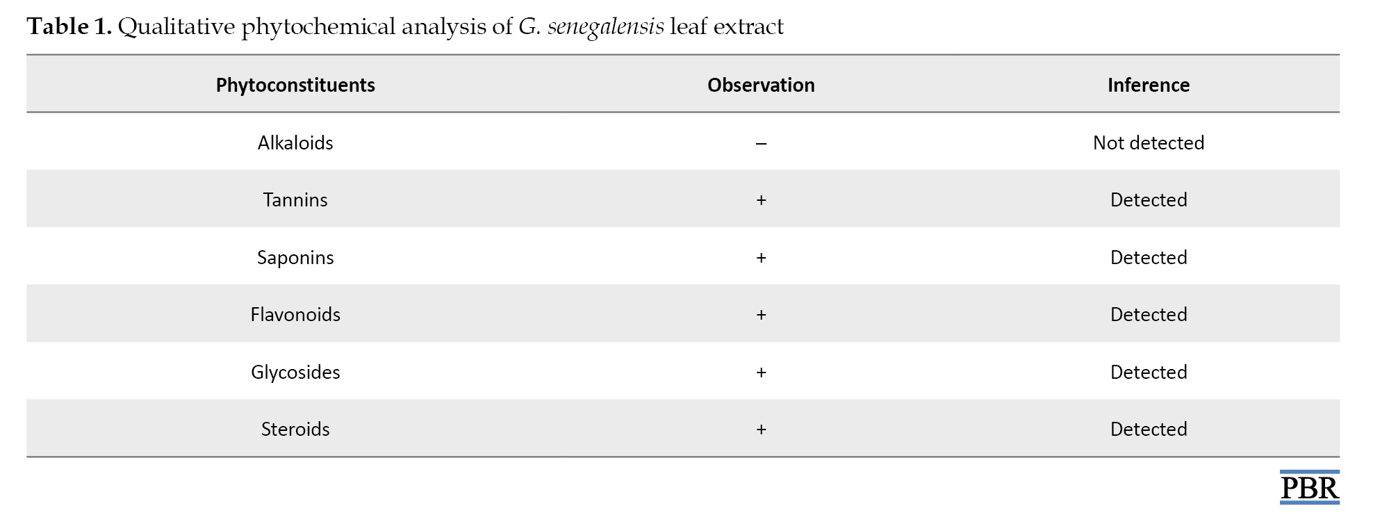
Phytochemical screening of the methanolic leaf extract of G. senegalensis revealed the presence of tannins, saponins, flavonoids, glycosides, and steroids, while alkaloids were absent (Table 1).
Antibacterial study
Morphological and biochemical identification of bacterial isolates
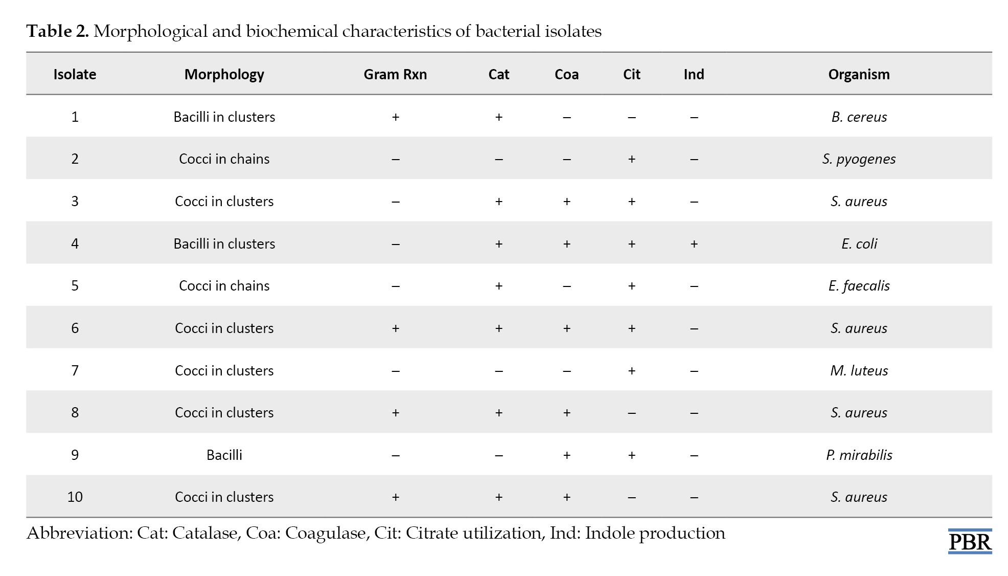
Ten bacterial isolates were identified through Gram staining and biochemical tests. The results revealed both Gram-positive and Gram-negative organisms with varied morphology and enzymatic profiles. Staphylococcus aureus was the most frequently identified species (Table 2).
Antibacterial activity of the extract
The antibacterial activity of the leaf extract was concentration-dependent, with maximum inhibition observed at 500 µg/mL. Escherichia coli exhibited the largest ZOI (29 mm), followed by S. aureus and S. pyogenes (Table 3).
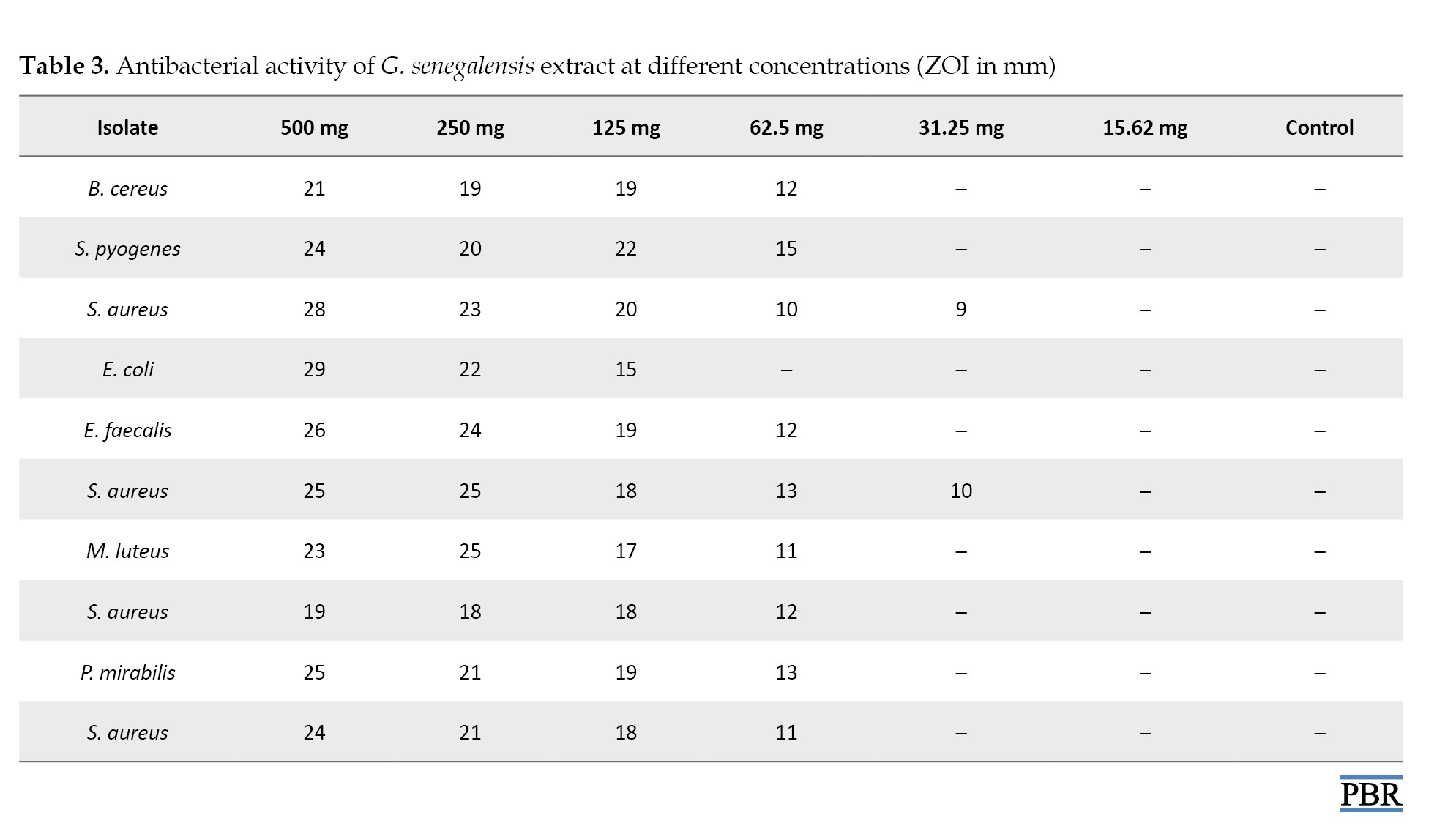
No inhibition was recorded at concentrations ≤31.25 µg/mL.
MIC and MBC of the extract
MIC values ranged from 31.25 to 125 µg/mL, while MBC values ranged from 62.5 to 250 µg/mL (Table 4).
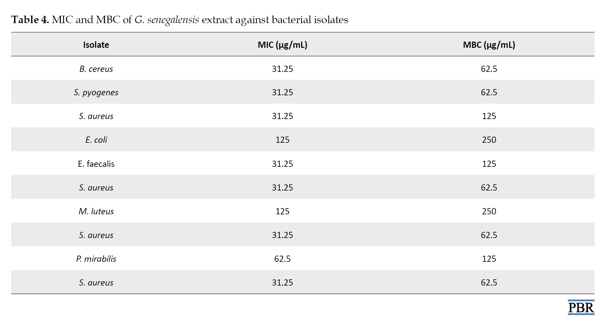
Gram-positive bacteria were generally more sensitive to the extract than Gram-negative ones. E. coli and Micrococcus luteus had the highest MBC values (250 µg/mL).
Antifungal study
Antifungal activity of the extract
The methanolic leaf extract of G. senegalensis demonstrated dose-dependent antifungal activity against Aspergillus niger and Fusarium oxysporum (Table 5).
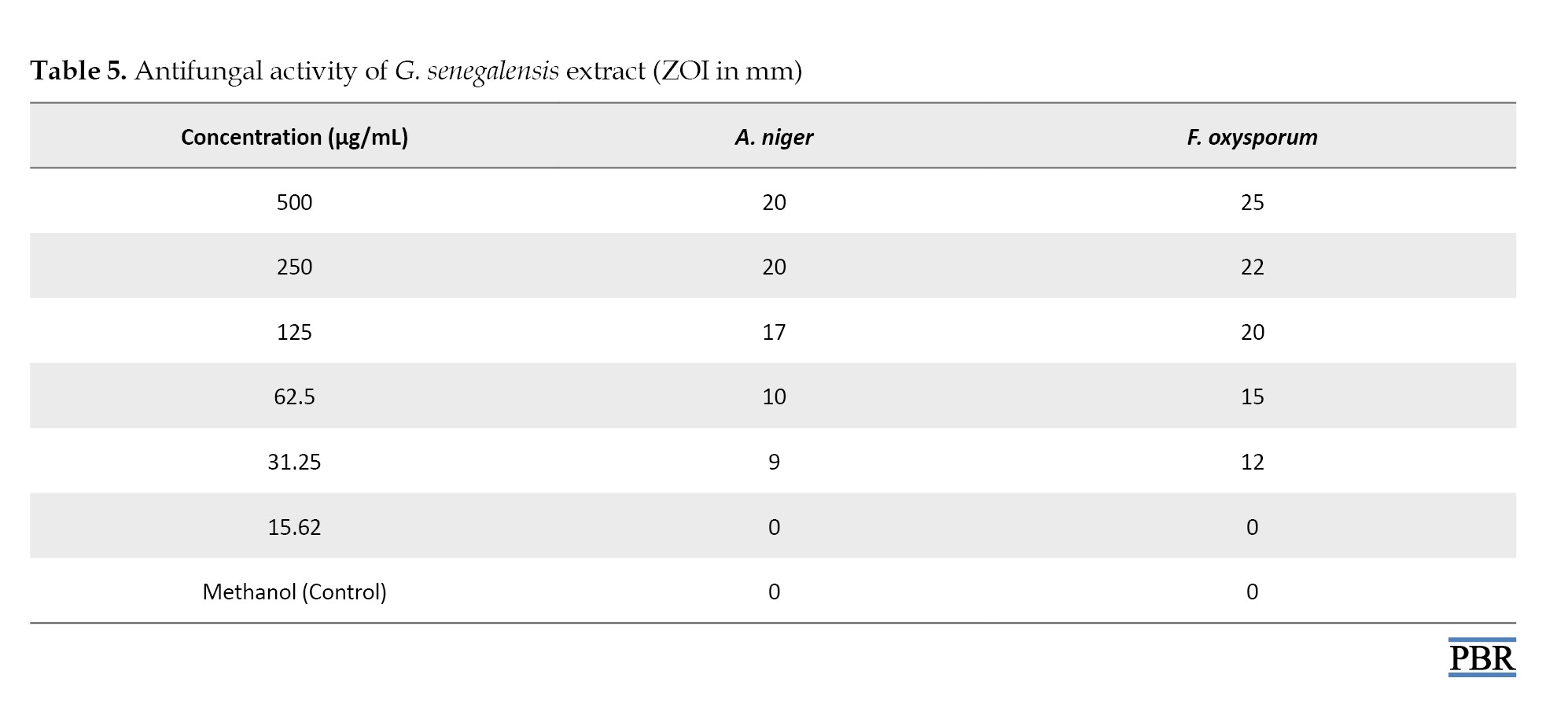
Maximum inhibition was observed at 500 µg/mL, with F. oxysporum exhibiting a larger ZOI than A. niger. No inhibition was observed at 15.62 µg/mL or in the control sample.
MIC against fungal isolates
The MIC was determined using broth dilution. For A. niger, the MIC was 62.5 µg/mL, while for F. oxysporum, the MIC was 31.25 µg/mL (Table 6).
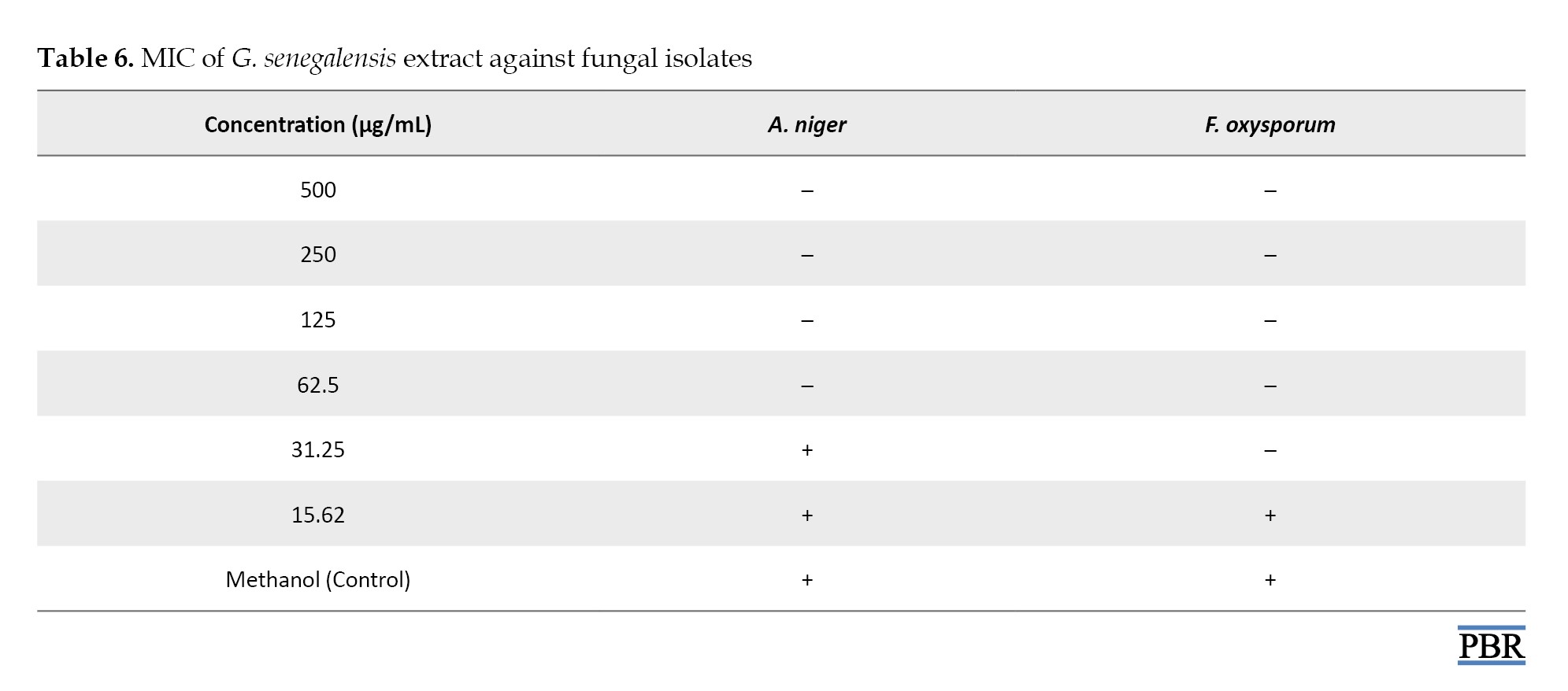
No inhibition was observed in the methanol control.
Discussion
This study explored the phytochemical profile and in vitro antimicrobial activity of the methanol-leaf extract of G. senegalensis, testing its effects on clinical bacterial and fungal isolates. Phytochemical screening identified flavonoids, alkaloids, saponins, tannins, glycosides, and steroids (Table 1), findings consistent with recent phytochemical profiling reports [3, 5, 10]. These compounds are widely associated with antimicrobial functions, including membrane disruption, enzyme inhibition, and interference with the structural integrity of pathogens [11].
Our findings demonstrated concentration-dependent antibacterial activity, with strong inhibition zones at 500 µg/mL (Table 3). E. coli was the most susceptible species (29 mm), followed by S. aureus and S. pyogenes. Antibacterial evaluations from other studies indicate MICs in the range of 40–200 µg/mL and MBCs of 20–200 µg/mL depending on extract type and bacterial strain. In this study, MICs ranged 31.25–125 µg/mL, and MBCs ranged 62.5–250 µg/mL (Table 4). Particularly, Gram-positive isolates, S. aureus, B. cereus, and S. pyogenes, had the lowest MICs (31.25 µg/mL), indicating heightened sensitivity, consistent with broader trends: Gram-positive bacteria are generally more susceptible to plant extracts due to cell wall structural differences [12]. Moreover, fractionated extracts evaluated by Shehu et al. (2025) revealed that the methanol fraction often produced larger inhibition zones (up to ~24 mm) against S. aureus and similar pathogens, confirming the strong activity of unrefined extracts [5]. These comparative data consolidate the antibacterial legitimacy of G. senegalensis.
The methanolic extract also exhibited dose-dependent antifungal activity against A. niger and F. oxysporum, with inhibition zones up to 25 mm and 20 mm at 500 µg/mL, respectively (Table 5). MIC values were 31.25 µg/mL for F. oxysporum and 62.5 µg/mL for A. niger (Table 6). These findings align with earlier reports by Hadiza et al. (2021), who recorded moderate to good activity against agricultural fungal pathogens using concentrations of 125–250 µg/mL, with zones up to ~14 mm for S. cruentum [13].
In a more detailed mechanistic study, an ethyl acetate fraction (EAF) from G. senegalensis leaves displayed strong antifungal efficacy against yeasts (C. neoformans, C. krusei, S. cerevisiae), achieving MICs as low as 8–16 µg/mL and MFCs up to 125–500 µg/mL, demonstrating potency on par with standard antifungals like penconazole and fluconazole [14]. These MIC values (µg range) are significantly lower than those in our crude extract (mg range), highlighting the enhanced efficacy achievable through fractionation. The primary active molecule in that work was guieranone A, possibly aided by chlorophyll-related compounds in the EAF, suggesting that minor phytochemicals may synergistically contribute to antifungal activity.
Conclusion
The methanolic leaf extract of G. senegalensis exhibits robust antibacterial and antifungal activity with broader effects on Gram-positive bacteria. These findings validate its ethnomedicinal potential and support its further development as a natural antimicrobial agent, especially in resource-limited settings. It is recommended that future studies focus on isolating and characterizing the active phytochemicals in G. senegalensis, particularly those responsible for its strong antibacterial and antifungal effects. In vivo and toxicological evaluations are essential to establish safety and therapeutic potential. Mechanistic studies should explore how the extract disrupts microbial cells.
Ethical Considerations
Compliance with ethical guidelines
The ethical protocol for the study was approved by the Faculty of Basic Medical Sciences’ Research and Ethics Committee (FBMSREC), Bauchi State University Gadau (Code: BASUG/FBMS/REC/VOL.08/01047).
Funding
This research did not receive any grant from funding agencies in the public, commercial, or non-profit sectors.
Authors' contributions
Conceptualization: Albashir Tahir, Saddam Ahmad, and Nazif Yakubu; Methodology: Albashir Tahir, and Khadija Abdullahi Kobi; Experimentation: Nazif Yakubu, Aishatu Sani Muhammad, Gideon Makeri Ayuba, and Al-ameen Abubakar Saeed; Writing the original draft: Albashir Tahir,Aminu Samaila Lassi, and Nazif Yakubu; review and editing: Albashir Tahir, Aminu Samaila Lassi, and Nazif Yakubu; Supervision: Albashir Tahir, and Saddam Ahmad; Final approval: All authors.
Conflict of interest
The authors declare no conflict of interest.
The global rise in antimicrobial resistance, related to both bacterial and fungal pathogens, has become an urgent public health concern, threatening the efficacy of existing therapies, leading to more severe and difficult-to-treat infections, increased mortality rates, and higher healthcare costs [1]. As conventional antibiotics and antifungal agents continue to lose effectiveness due to resistance, there is a growing interest in alternative therapeutic methods, particularly herbal medicines. Historically, plants have served as critical reservoirs of bioactive compounds with potent pharmacological properties, many of which have been successfully translated into modern drugs [2]. One of these plants is Guiera senegalensis, abundant in the savanna and semi-arid zones of West and Central Africa [3]. Known locally as “Sabara” among the Hausa and “Geloki” among the Fulani populations of Nigeria, G. senegalensis has long been used in African traditional medicine to treat a wide variety of ailments, including bacterial infections, fungal diseases, malaria, and respiratory disorders [3, 4]. Various parts of this plant, particularly the leaves, roots, bark, and galls, are used in traditional remedies, either alone or in combination with other herbs, to treat conditions ranging from gastrointestinal infections and skin inflammation to systemic febrile illnesses [4].
Phytochemical study of G. senegalensis has revealed a diverse array of secondary metabolites, including alkaloids, flavonoids, tannins, terpenoids, saponins, and phenolic compounds, many of which have been shown to have antimicrobial properties [4, 5]. These bioactive constituents contribute not only to the plant’s defense mechanisms but also to its therapeutic potential in human and veterinary medicine. In particular, its leaf extract has shown inhibitory activity against a variety of microbial species, suggesting that G. senegalensis can be a valuable source of natural antimicrobial agents [5].
Despite its extensive traditional use and reported bioactivity, there is a paucity of well-designed scientific studies that simultaneously evaluate the antibacterial and antifungal efficacy of G. senegalensis against clinically relevant pathogens. This study aims to evaluate the phytochemical composition as well as the in vitro antibacterial and antifungal activities of the methanol leaf extract of G. senegalensis against clinical isolates.
Materials and Methods
Materials
The chemicals and reagents used in this study included: Ammonium chloride, ammonium hydroxide, chloroform, perchloric acid, sodium chloride, sodium hydroxide, DPPH, distilled water, dilute hydrochloric acid (HCl), benzene, nutrient agar, Mueller-Hinton agar, Simmon’s citrate agar, nutrient broth, Kovac’s reagent, and hydrogen peroxide.
Bacterial and fungal isolates were obtained from the Federal University of Health Sciences Teaching Hospital, Azare, Nigeria. The isolates were transported to the Microbiology Laboratory, Sa’adu Zungur University, Bauchi State, for re-identification and further analysis.
Fresh, mature, and disease-free leaves of G. senegalensis were collected from Gadau Campus of Sa’adu Zungur University, Bauchi State, on February 22, 2025, using a sterilized cutlass. The harvested leaves were kept in sterile plastic containers and transported to the laboratory. The plant was authenticated at the university’s herbarium with voucher specimen number 002. Leaves were washed with tap water, shade-dried at room temperature for seven days, ground into fine powder using a clean mortar and pestle, and stored in an airtight container until use.
Extract preparation and phytochemical screening
A total of 150 g of the powdered G. senegalensis leaves was macerated in 1 L of 70% methanol for 72 hours with stirring every 12 hours. The mixture was filtered using Whatman No. 1 filter paper. The filtrate was concentrated in an oven at 45–50 °C until complete evaporation. The dried extract was stored in a labelled container for subsequent use. Phytochemical screening of the methanol leaf extract was carried out using standard methods described by Trease and Evans (2002) [6] and Harborne (1998) [7] to detect the presence of alkaloids, flavonoids, saponins, tannins, steroids, and glycosides.
Antibacterial study
All media were prepared following the manufacturer’s instructions. A total of 2.8 g of nutrient agar was dissolved in 100 mL of distilled water and autoclaved at 121°C for 15 minutes. The sterilized medium was poured into sterile Petri dishes and allowed to solidify.
A smear of each isolate was prepared on a clean slide, heat-fixed, and stained sequentially with crystal violet, iodine, alcohol (decolorizer), and safranin. Slides were examined under oil immersion at ×100 magnification.
Biochemical identification
Peptone water inoculated with isolates was incubated at 37 °C for 24 hours. A few drops of Kovac’s reagent were added. A red or pink ring indicates a positive indole test result. A thick suspension of the isolate was mixed with undiluted human plasma on a slide. Clot formation within 5–10 seconds indicates a positive coagulase test result. A drop of hydrogen peroxide was added to a slide containing a loopful of the isolate. Bubble formation indicates a positive catalase test reaction. Simmon’s citrate agar slants were inoculated and incubated at 37 °C for 24 hours. A color change from green to blue indicates a positive citrate utilization test result.
Antimicrobial susceptibility testing
The in vitro antibacterial activity was evaluated using the agar disk diffusion method. Bacterial cultures grown in nutrient broth were inoculated onto Mueller-Hinton agar plates. Sterile 6-mm disks were impregnated with different concentrations (500, 250, 125, 62.5, 31.25, and 15.62 µg/mL) of the extract and placed on inoculated plates. Plates were incubated at 37 °C for 24 hours, and the zone of inhibition (ZOI) was measured in millimeters. Disks with methanol were used as negative controls.
Determination of minimum inhibitory concentration
The minimum inhibitory concentration (MIC) was determined using the method described by the clinical and laboratory standards institute (CLSI). Serial dilutions of the extract were prepared and inoculated with test organisms in nutrient broth. Tubes were incubated at 37 °C for 24 hours. The lowest concentration showing no visible growth was recorded as the MIC [8].
Determination of minimum bactericidal concentration
Tubes showing no growth in the MIC assay were sub-cultured onto nutrient agar plates. Plates were incubated at 37 °C for 24 hours. The lowest concentration showing no bacterial growth was recorded as the minimum bactericidal concentration (MBC).
Antifungal study
Preparation of potato dextrose agar
Potato dextrose agar (PDA) was prepared by dissolving 19.5 g of PDA powder in 250 mL of distilled water, then autoclaving at 121°C for 20 minutes. Under aseptic conditions, 20 mL of the molten medium was poured into sterile Petri dishes and allowed to solidify. Fungal isolates were inoculated using the spread plate method [9].
Antifungal sensitivity testing
The agar well diffusion method was used for antifungal sensitivity testing. PDA plates were inoculated uniformly with fungal isolates and allowed to dry. A sterile cork borer (6 mm) was used to make four wells and one central control well. Each well was filled with 0.1 mL of the extract at various concentrations. Ethanol served as the control. Plates were incubated at 37 °C for 72 hours, and ZOI was measured in millimeters.
Preparation of potato dextrose broth
To determine the MIC for antifungal activity, 6.25 g of potato dextrose broth (PDB) was dissolved in 125 mL of distilled water and autoclaved at 121 °C for 20 minutes. The medium was allowed to cool and used for tube dilution assays.
MIC determination
The MIC was determined using the tube dilution method [9]. Serial two-fold dilutions of the extract (from 250 µg/mL down to 1.95 µg/mL) were prepared in PDB. Each tube was inoculated with 0.1 mL of the fungal isolate and incubated at 37 °C for 72 hours. The MIC was recorded as the lowest concentration showing no visible fungal growth (i.e. a clear tube).
Results
Qualitative phytochemical analysis of the methanolic leaf extract
Phytochemical screening of the methanolic leaf extract of G. senegalensis revealed the presence of tannins, saponins, flavonoids, glycosides, and steroids, while alkaloids were absent (Table 1).
Antibacterial study
Morphological and biochemical identification of bacterial isolates
Ten bacterial isolates were identified through Gram staining and biochemical tests. The results revealed both Gram-positive and Gram-negative organisms with varied morphology and enzymatic profiles. Staphylococcus aureus was the most frequently identified species (Table 2).
Antibacterial activity of the extract
The antibacterial activity of the leaf extract was concentration-dependent, with maximum inhibition observed at 500 µg/mL. Escherichia coli exhibited the largest ZOI (29 mm), followed by S. aureus and S. pyogenes (Table 3).
No inhibition was recorded at concentrations ≤31.25 µg/mL.
MIC and MBC of the extract
MIC values ranged from 31.25 to 125 µg/mL, while MBC values ranged from 62.5 to 250 µg/mL (Table 4).
Gram-positive bacteria were generally more sensitive to the extract than Gram-negative ones. E. coli and Micrococcus luteus had the highest MBC values (250 µg/mL).
Antifungal study
Antifungal activity of the extract
The methanolic leaf extract of G. senegalensis demonstrated dose-dependent antifungal activity against Aspergillus niger and Fusarium oxysporum (Table 5).
Maximum inhibition was observed at 500 µg/mL, with F. oxysporum exhibiting a larger ZOI than A. niger. No inhibition was observed at 15.62 µg/mL or in the control sample.
MIC against fungal isolates
The MIC was determined using broth dilution. For A. niger, the MIC was 62.5 µg/mL, while for F. oxysporum, the MIC was 31.25 µg/mL (Table 6).
No inhibition was observed in the methanol control.
Discussion
This study explored the phytochemical profile and in vitro antimicrobial activity of the methanol-leaf extract of G. senegalensis, testing its effects on clinical bacterial and fungal isolates. Phytochemical screening identified flavonoids, alkaloids, saponins, tannins, glycosides, and steroids (Table 1), findings consistent with recent phytochemical profiling reports [3, 5, 10]. These compounds are widely associated with antimicrobial functions, including membrane disruption, enzyme inhibition, and interference with the structural integrity of pathogens [11].
Our findings demonstrated concentration-dependent antibacterial activity, with strong inhibition zones at 500 µg/mL (Table 3). E. coli was the most susceptible species (29 mm), followed by S. aureus and S. pyogenes. Antibacterial evaluations from other studies indicate MICs in the range of 40–200 µg/mL and MBCs of 20–200 µg/mL depending on extract type and bacterial strain. In this study, MICs ranged 31.25–125 µg/mL, and MBCs ranged 62.5–250 µg/mL (Table 4). Particularly, Gram-positive isolates, S. aureus, B. cereus, and S. pyogenes, had the lowest MICs (31.25 µg/mL), indicating heightened sensitivity, consistent with broader trends: Gram-positive bacteria are generally more susceptible to plant extracts due to cell wall structural differences [12]. Moreover, fractionated extracts evaluated by Shehu et al. (2025) revealed that the methanol fraction often produced larger inhibition zones (up to ~24 mm) against S. aureus and similar pathogens, confirming the strong activity of unrefined extracts [5]. These comparative data consolidate the antibacterial legitimacy of G. senegalensis.
The methanolic extract also exhibited dose-dependent antifungal activity against A. niger and F. oxysporum, with inhibition zones up to 25 mm and 20 mm at 500 µg/mL, respectively (Table 5). MIC values were 31.25 µg/mL for F. oxysporum and 62.5 µg/mL for A. niger (Table 6). These findings align with earlier reports by Hadiza et al. (2021), who recorded moderate to good activity against agricultural fungal pathogens using concentrations of 125–250 µg/mL, with zones up to ~14 mm for S. cruentum [13].
In a more detailed mechanistic study, an ethyl acetate fraction (EAF) from G. senegalensis leaves displayed strong antifungal efficacy against yeasts (C. neoformans, C. krusei, S. cerevisiae), achieving MICs as low as 8–16 µg/mL and MFCs up to 125–500 µg/mL, demonstrating potency on par with standard antifungals like penconazole and fluconazole [14]. These MIC values (µg range) are significantly lower than those in our crude extract (mg range), highlighting the enhanced efficacy achievable through fractionation. The primary active molecule in that work was guieranone A, possibly aided by chlorophyll-related compounds in the EAF, suggesting that minor phytochemicals may synergistically contribute to antifungal activity.
Conclusion
The methanolic leaf extract of G. senegalensis exhibits robust antibacterial and antifungal activity with broader effects on Gram-positive bacteria. These findings validate its ethnomedicinal potential and support its further development as a natural antimicrobial agent, especially in resource-limited settings. It is recommended that future studies focus on isolating and characterizing the active phytochemicals in G. senegalensis, particularly those responsible for its strong antibacterial and antifungal effects. In vivo and toxicological evaluations are essential to establish safety and therapeutic potential. Mechanistic studies should explore how the extract disrupts microbial cells.
Ethical Considerations
Compliance with ethical guidelines
The ethical protocol for the study was approved by the Faculty of Basic Medical Sciences’ Research and Ethics Committee (FBMSREC), Bauchi State University Gadau (Code: BASUG/FBMS/REC/VOL.08/01047).
Funding
This research did not receive any grant from funding agencies in the public, commercial, or non-profit sectors.
Authors' contributions
Conceptualization: Albashir Tahir, Saddam Ahmad, and Nazif Yakubu; Methodology: Albashir Tahir, and Khadija Abdullahi Kobi; Experimentation: Nazif Yakubu, Aishatu Sani Muhammad, Gideon Makeri Ayuba, and Al-ameen Abubakar Saeed; Writing the original draft: Albashir Tahir,Aminu Samaila Lassi, and Nazif Yakubu; review and editing: Albashir Tahir, Aminu Samaila Lassi, and Nazif Yakubu; Supervision: Albashir Tahir, and Saddam Ahmad; Final approval: All authors.
Conflict of interest
The authors declare no conflict of interest.
References
- Belay WY, Getachew M, Tegegne BA, Teffera ZH, Dagne A, Zeleke TK, et al. Antimicrobial resistance with a focus on antibacterial, antifungal, antimalarial, and antiviral drugs resistance, its threat, global priority pathogens, prevention, and control strategies: A review. Ther Adv Infect Dis. 2025; 12:20499361251340144. [DOI:10.1177/20499361251340144] [PMID] [PMCID]
- Dar RA, Shahnawaz M, Ahanger MA, Majid IU. Exploring the diverse bioactive compounds from medicinal plants: A review. J Phytopharm. 2023; 12(3):189-95. [DOI:10.31254/phyto.2023.12307]
- Hassan UA, Tahir JI, Akanji CF, Adamu A, Tahir A. Analgesic and anti-inflammatory effects of ethanol leaf extract of Guiera senegalensis in murine models. Sci Phytochem. 2025; 4(1):49-54. [DOI:10.58920/sciphy0401300]
- Dirar AI, Devkota HP. Ethnopharmacological uses, phytochemistry and pharmacological activities of Guiera senegalensis J.F. Gmel. (Combretaceae). J Ethnopharmacol. 2021; 267:113433. [DOI:10.1016/j.jep.2020.113433] [PMID]
- Abubakar S, Umar G, Hannatu ML, Augie AL. Phytochemical composition and antimicrobial properties of guiera senegalensis leaves extract obtained from yabo community in Sokoto State, Nigeria. Int J Sci Glob Sustain. 2025; 11(1):25-32. [DOI:10.57233/ijsgs.v11i1.778]
- Trease GE, Evans WC. Pharmacognosy (9th ed.). Saunders Publishers; 2002. [Link]
- Harborne JB. Phytochemical methods: A guide to modern techniques of plant analysis (3rd ed.). London: Chapman and Hall; 1998. [Link]
- Franklin R. Cockerill III. Methods for dilution antimicrobial susceptibility tests for bacteria that grow aerobically: Approved standard. Malvern: Clinical and Laboratory Standards Institute; 2012. [Link]
- Prescott LM, Harley JP, Klein DA. Microbiology: Food and industrial microbiology. 5th edition, Boston: McGraw-Hill; 2002. [Link]
- Abubakar K, Mikailu AY, Sidi AH, Tukur M, Abubakar M, Yusuf MM. Phytochemical and antibacterial studies of methanolic extract and fractions of Guiera senegalensis leaves. Ajol. 2023; 5(2):177-83. [DOI:10.4314/cajost.v5i2.12]
- Jadimurthy R, Jagadish S, Nayak SC, Kumar S, Mohan CD, Rangappa KS. Phytochemicals as invaluable sources of potent antimicrobial agents to combat antibiotic resistance. Life. 2023; 13(4):948. [DOI:10.3390/life13040948] [PMID] [PMCID]
- Khameneh B, Eskin NM, Iranshahy M, Fazly Bazzaz BS. Phytochemicals: A promising weapon in the arsenal against antibiotic-resistant bacteria. Antibiotics. 2021; 10(9):1044. [DOI:10.3390/antibiotics10091044]
- Hadiza MM, Kutama AS, Umar S. Antifungal Activity of Gueira senegalensis L. and Mangifera indica L. on Sorghumloose smut pathogen (Sporisorium cruentum (KUHN) POTTER). J Microbiol Res. 2021; 6(1):189-94. [DOI:10.47430/ujmr.2161.025]
- Moreira R, Ferreres F, Gil-Izquierdo Á, Gomes NGM, Araújo L, Pinto E, et al. Antifungal activity of guiera senegalensis: From the chemical composition to the mitochondrial toxic effects and tyrosinase inhibition. Antibiotics. 2023; 12(5):869. [DOI:10.3390/antibiotics12050869] [PMID] [PMCID]
Type of Study: Original Research |
Subject:
Ehtnopharmacology
| Rights and permissions | |
 |
This work is licensed under a Creative Commons Attribution-NonCommercial 4.0 International License. |


This is an open access article distributed under the terms of the Creative Commons Attribution License (CC-By-NC), which permits use, distribution, and reproduction in any medium, provided the original work is properly cited and is not used for commercial purposes.
Contact Information