

Volume 12, Issue 1 (2026)
Pharm Biomed Res 2026, 12(1): 27-36 |
Back to browse issues page



Download citation:
BibTeX | RIS | EndNote | Medlars | ProCite | Reference Manager | RefWorks
Send citation to:


BibTeX | RIS | EndNote | Medlars | ProCite | Reference Manager | RefWorks
Send citation to:
Shokrzadeh M, Modanloo M, Fasihbeiki M, Zamani E, Alinia M, Shaki F. Atorvastatin Mitigates Cisplatin-induced Genotoxicity and Oxidative Stress in Human Lymphocytes: Insights From the Micronucleus Assay. Pharm Biomed Res 2026; 12 (1) :27-36
URL: http://pbr.mazums.ac.ir/article-1-677-en.html
URL: http://pbr.mazums.ac.ir/article-1-677-en.html
Mohammad Shokrzadeh1 

 , Mona Modanloo2 , Mina Fasihbeiki1 , Ehsan Zamani3 , Mona Alinia3 , Fatemeh Shaki *1
, Mona Modanloo2 , Mina Fasihbeiki1 , Ehsan Zamani3 , Mona Alinia3 , Fatemeh Shaki *1
1- Pharmaceutical Sciences Research Center, Hemoglobinopathy Institute, Mazandaran University of Medical Sciences, Sari, Iran.
2- Department of Toxicology and Pharmacology, Faculty of Pharmacy, Mazandaran University of Medical Sciences, Sari, Iran.
3- Department of Pharmacology and Toxicology, School of Pharmacy, Guilan University of Medical Sciences, Rasht, Iran.
2- Department of Toxicology and Pharmacology, Faculty of Pharmacy, Mazandaran University of Medical Sciences, Sari, Iran.
3- Department of Pharmacology and Toxicology, School of Pharmacy, Guilan University of Medical Sciences, Rasht, Iran.
Keywords: Cisplatin (Cis), Genotoxicity, Lymphocytes, Atorvastatin (Atv), Micronucleus (MN) test, Oxidative stress
Full-Text [PDF 1131 kb]
(246 Downloads)
| Abstract (HTML) (739 Views)
Micronucleus (MN) assay
MN assay was performed to assess genotoxicity in human lymphocytes, as previously described [24].
Twenty hours after exposure, cytochalasin B (5 mg/mL) was added to the cells to arrest the cell cycle in cytokinesis. The cell culture medium was harvested and centrifuged after 28 hours. Next, the supernatant was gently removed, and the cells were treated with a mild hypotonic solution (0.075 M potassium chloride [KCL] for 5 minutes). After fixing the samples with glacial acetic acid, they were refrigerated for 24 hours. The slides were transferred to a freezer and kept until frozen. Next, three drops of the cell suspension were placed on each microscopic slide from a distance of 10 cm, then the drops were smeared. The microscopic slides were air-dried at room temperature. In the next step, the slides were stained with Giemsa stain. First, a 2% Giemsa solution was prepared using phosphate buffer. The slides were then incubated in this solution for 10 min. Next, the slides were rinsed with distilled water for 10-15 s to remove any possible staining sediment. The samples were air-dried and prepared for microscopic analysis. The slides were analyzed at 40x magnification (100x if more detail was required). The frequency of micronuclei and nucleoplasmic bridges per 1000 cells was calculated and reported as a percentage. The MN frequency was calculated per 1000 binucleated cells and reported as a percentage.
Quantification of ROS level
ROS levels were measured using dichlorodihydrofluorescein diacetate (DCFH-DA) as an indicator. Following exposure, DCFH-DA was added at a final concentration of 10 mM and incubated for 10 minutes. ROS generation in isolated human lymphocytes was subsequently determined using a fluorescence spectrophotometer (Shimadzu RF5000U) with excitation at 485 nm and emission at 520 nm.
Lipid peroxidation (LPO) measurement
The concentration of malondialdehyde (MDA), a byproduct of LPO, was assessed using TBA, which reacts with MDA. To elaborate, 0.25 mL of 0.05 M phosphoric acid was added to the cellular suspension from each test group, followed by the addition of 25 µM TBA reagent into the microtubes.
The microtubes were incubated for 30 minutes in a boiling water bath at 95 °C, then immersed in an ice bath. Subsequently, 4 mL of n-butanol was added to each microtube and thoroughly mixed, after which centrifugation was conducted at 3500 rpm for 10 minutes. Approximately 150 µL of the resulting upper solution from the centrifugation process was carefully transferred to a microplate. MDA content was determined by measuring the absorbance at 535 nm [20]. A standard curve was generated using tetramethoxypropane, and the MDA concentrations were expressed as micromoles per litre (µM).
GSH content measurement
The GSH content was assessed using DTNB, which produces a yellow colour upon reaction with sulfhydryl-containing compounds. Each sample (1 mL) was combined with 1 mL of 10% TCA, mixed for 2 minutes using a shaker, and then centrifuged at 3500 RPM for 15 minutes. Subsequently, 2.5 mL of Tris buffer (0.4 M, pH 8.9) and 0.5 mL of DTNB reagent were added to 1 mL of the supernatant. The resulting yellow colour was measured at 412 nm using a spectrophotometer, allowing for the determination of GSH concentration via a standard calibration curve [24].
Statistical analysis
The results are presented as Mean±SD. All statistical analyses were performed using GraphPad Prism software, version 6. Statistical significance was determined using one-way analysis of variance, followed by Tukey’s post hoc test. Statistical significance was set at P<0.05.
Results
Management of genotoxicity in human lymphocytes via MN assay
Results of MN assay
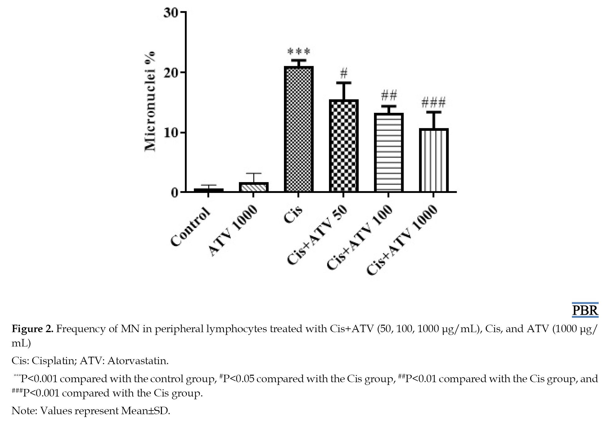
Based on the data presented in Figure 2, the percentage of micronuclei in lymphocytes treated with Cis (26.50%) was significantly higher than that in the control group (0.6%).
Full-Text: (26 Views)
Introduction
Cisplatin (Cis) was initially recognized for its inhibitory effects on the proliferation of Escherichia coli. Subsequent investigations have revealed its substantial antineoplastic and cytotoxic impacts on malignant cells [1].
Cis is a prominent chemotherapeutic agent with significant therapeutic value in the management of diverse malignancies, including leukemia, lymphoma, breast carcinoma, ovarian carcinoma, head and neck carcinoma, cervical carcinoma, sarcoma, and, especially, testicular carcinoma [1, 2]. Despite its broad therapeutic spectrum, some side effects of Cis limit its use. Nephrotoxicity, ototoxicity (cochlear damage), and neurotoxicity are the primary complications associated with Cis administration. These adverse effects restrict the broader use of Cis in clinical settings [3].
Alkylation is one of the most critical mechanisms of Cis’s action. Upon cellular entry, Cis dissociates its chloride ligand to form more stable bonds with cellular molecules, thereby exerting anticancer effects [4]. This molecular transformation results in the formation of DNA adducts, inhibiting DNA synthesis and impeding cellular proliferation [1].
Nevertheless, due to its alkylating properties, Cis can also harm normal cells [2]. This significant interaction with DNA initiates a sequence of cytotoxic reactions, such as genotoxicity [5].
Furthermore, oxidative stress plays a central role in Cis-induced genotoxicity [2]. Cis generates reactive oxygen species (ROS), including superoxide and hydroxyl free radicals. Research has indicated that Cis-based chemotherapy reduces plasma levels of intrinsic antioxidants, such as glutathione (GSH) [6]. Consequently, these events lead to apoptosis, necrosis, and genotoxicity [1, 2].
Recently, Cis-induced genotoxicity has garnered significant attention due to its capacity to induce profound DNA damage in non-tumor cells [1, 2]. Long-term use of Cis in patients can cause genetic abnormalities during chemotherapy and in subsequent years. Even a single dose of Cis increases the incidence of micronucleated polychromatic erythrocytes, indicating genetic damage and chromosomal abnormalities [7, 8].
Oxidative stress occurs when there is an imbalance between harmful reactive oxygen and nitrogen molecules and the body’s ability to neutralize them. This imbalance may result in various health issues, including tumors, heart problems, artery blockage, metabolic imbalances, and cancers [9]. The deleterious impact of antineoplastic agents on healthy tissues culminates in the development of secondary malignancies [6].
Therefore, antioxidant compounds offer an effective means to attenuate oxidative genotoxicity [10].
In recent years, increasing attention has been directed toward the antioxidant and pleiotropic properties of Atv [11, 12]. Atorvastatin (Atv), a potent inhibitor of 3-hydroxy-3-methylglutaryl coenzyme A reductase, exerts its primary pharmacological effect by suppressing endogenous cholesterol biosynthesis, thereby facilitating hypercholesterolemia management [13, 14]. Beyond its lipid-lowering capacity, a growing body of evidence demonstrates that Atv confers significant antioxidant effects, particularly in individuals with diabetes, where it has been shown to attenuate oxidative stress by modulating ROS, enhancing endothelial function, and downregulating pro-inflammatory pathways [12, 15-17]. Moreover, recent investigations suggest that Atv may mitigate or delay the progression of oxidative steatohepatitis in patients with diabetes, potentially through its integrated antioxidant, anti-inflammatory, and metabolic regulatory mechanisms [18, 19].
This study examined the genotoxic effects of Cis, a commonly used chemotherapeutic drug, emphasizing its role in promoting oxidative stress and DNA damage in non-cancerous cells. Additionally, it investigates the potential protective properties of antioxidant agents, particularly Atv, in alleviating Cis-induced genotoxicity and oxidative stress.
Materials and Methods
Chemicals
RPMI-1640 medium, fetal bovine serum (FBS), antibiotic/antimycotic solution, phytohemagglutinin (PHA), Cis, cytochalasin B, potassium chloride (KCl), methanol, glacial acetic acid, sucrose, magnesium chloride, disodium phosphate, tris-hydrochloride, phosphoric acid, thiobarbituric acid (TBA), n-butanol, tetramethoxypropane, ethylenediaminetetraacetic acid, trichloroacetic acid (TCA), tris-ammonium, Sodium acetate, dithionitro benzoic acid (DTNB), Atv, Cis and GSH were used in this study. These chemicals were purchased from Sigma–Aldrich (St. Louis, MI, USA) and Merck (Darmstadt, Germany).
Cell preparation
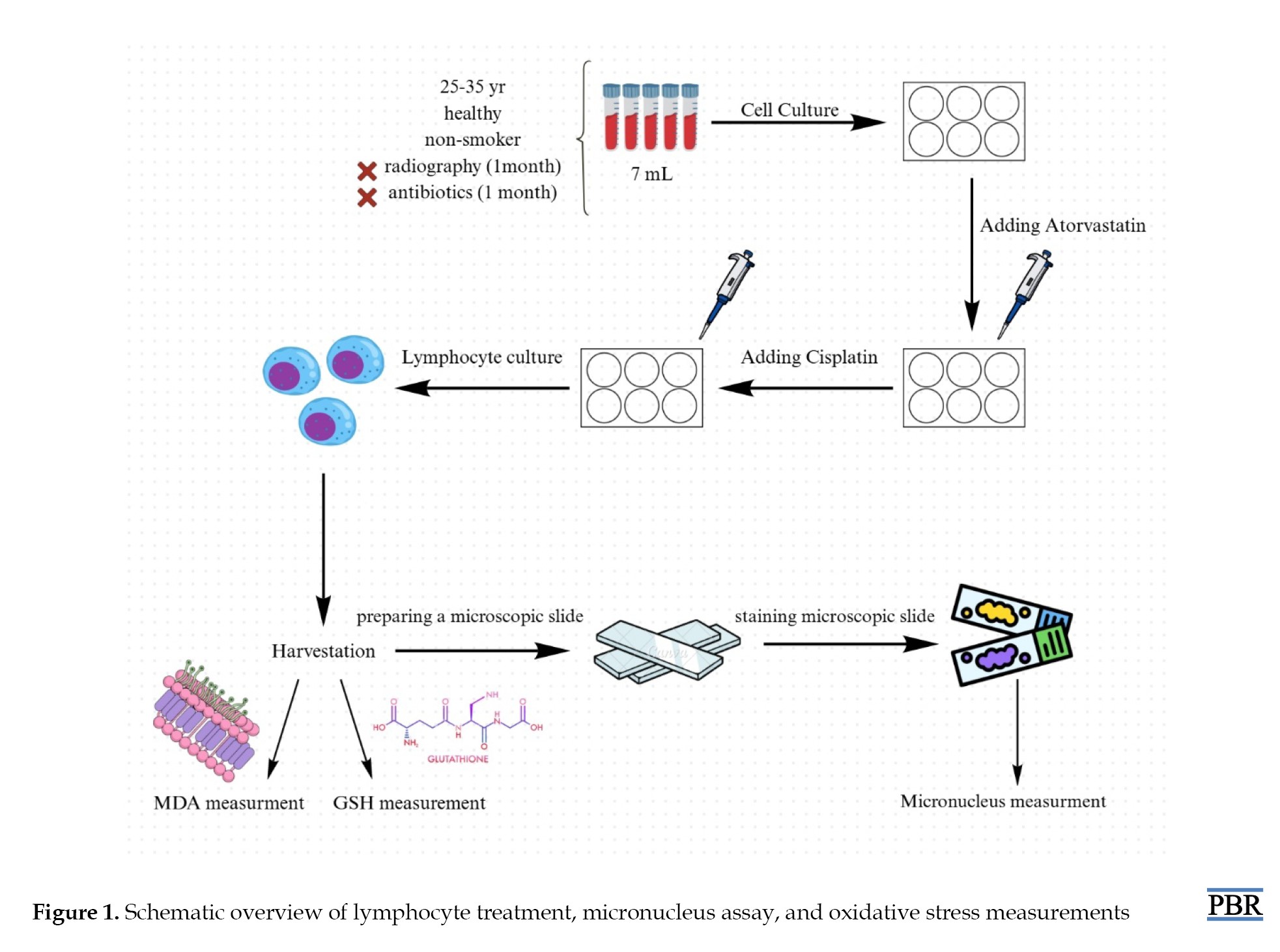
Blood samples (7 mL) were collected from five volunteers in accordance. Subsequently, the samples were cultured in 6-well plates containing RPMI 1640 medium, 10% FBS, and an antibiotic/antimycotic solution. Cell division was stimulated by adding 100 μL PHA to each well. Following this, the cells were incubated at 37 °C with 5% CO2 for 24 hours. Subsequently, specific treatments were administered to the cells based on the experimental groups [20-23].
The experimental groups were outlined as follows:
Group 1 (control): Peripheral blood lymphocytes with no treatment (RPMI-1640 Medium); group 2 (positive control): Cells treated with Cis (12 μM); group 3: Cells pre-treated with Atv (50 μM)+Cis; group 4: Cells pre-treated with Atv (100 μM)+Cis; group 5: Cells pre-treated with Atv (1000 μM)+Cis. The concentration of Atv deemed effective was established based on pretests and comparable studies. Group 6: Atv (1000 μM) without Cis. All procedures are shown in Figure 1.
Cisplatin (Cis) was initially recognized for its inhibitory effects on the proliferation of Escherichia coli. Subsequent investigations have revealed its substantial antineoplastic and cytotoxic impacts on malignant cells [1].
Cis is a prominent chemotherapeutic agent with significant therapeutic value in the management of diverse malignancies, including leukemia, lymphoma, breast carcinoma, ovarian carcinoma, head and neck carcinoma, cervical carcinoma, sarcoma, and, especially, testicular carcinoma [1, 2]. Despite its broad therapeutic spectrum, some side effects of Cis limit its use. Nephrotoxicity, ototoxicity (cochlear damage), and neurotoxicity are the primary complications associated with Cis administration. These adverse effects restrict the broader use of Cis in clinical settings [3].
Alkylation is one of the most critical mechanisms of Cis’s action. Upon cellular entry, Cis dissociates its chloride ligand to form more stable bonds with cellular molecules, thereby exerting anticancer effects [4]. This molecular transformation results in the formation of DNA adducts, inhibiting DNA synthesis and impeding cellular proliferation [1].
Nevertheless, due to its alkylating properties, Cis can also harm normal cells [2]. This significant interaction with DNA initiates a sequence of cytotoxic reactions, such as genotoxicity [5].
Furthermore, oxidative stress plays a central role in Cis-induced genotoxicity [2]. Cis generates reactive oxygen species (ROS), including superoxide and hydroxyl free radicals. Research has indicated that Cis-based chemotherapy reduces plasma levels of intrinsic antioxidants, such as glutathione (GSH) [6]. Consequently, these events lead to apoptosis, necrosis, and genotoxicity [1, 2].
Recently, Cis-induced genotoxicity has garnered significant attention due to its capacity to induce profound DNA damage in non-tumor cells [1, 2]. Long-term use of Cis in patients can cause genetic abnormalities during chemotherapy and in subsequent years. Even a single dose of Cis increases the incidence of micronucleated polychromatic erythrocytes, indicating genetic damage and chromosomal abnormalities [7, 8].
Oxidative stress occurs when there is an imbalance between harmful reactive oxygen and nitrogen molecules and the body’s ability to neutralize them. This imbalance may result in various health issues, including tumors, heart problems, artery blockage, metabolic imbalances, and cancers [9]. The deleterious impact of antineoplastic agents on healthy tissues culminates in the development of secondary malignancies [6].
Therefore, antioxidant compounds offer an effective means to attenuate oxidative genotoxicity [10].
In recent years, increasing attention has been directed toward the antioxidant and pleiotropic properties of Atv [11, 12]. Atorvastatin (Atv), a potent inhibitor of 3-hydroxy-3-methylglutaryl coenzyme A reductase, exerts its primary pharmacological effect by suppressing endogenous cholesterol biosynthesis, thereby facilitating hypercholesterolemia management [13, 14]. Beyond its lipid-lowering capacity, a growing body of evidence demonstrates that Atv confers significant antioxidant effects, particularly in individuals with diabetes, where it has been shown to attenuate oxidative stress by modulating ROS, enhancing endothelial function, and downregulating pro-inflammatory pathways [12, 15-17]. Moreover, recent investigations suggest that Atv may mitigate or delay the progression of oxidative steatohepatitis in patients with diabetes, potentially through its integrated antioxidant, anti-inflammatory, and metabolic regulatory mechanisms [18, 19].
This study examined the genotoxic effects of Cis, a commonly used chemotherapeutic drug, emphasizing its role in promoting oxidative stress and DNA damage in non-cancerous cells. Additionally, it investigates the potential protective properties of antioxidant agents, particularly Atv, in alleviating Cis-induced genotoxicity and oxidative stress.
Materials and Methods
Chemicals
RPMI-1640 medium, fetal bovine serum (FBS), antibiotic/antimycotic solution, phytohemagglutinin (PHA), Cis, cytochalasin B, potassium chloride (KCl), methanol, glacial acetic acid, sucrose, magnesium chloride, disodium phosphate, tris-hydrochloride, phosphoric acid, thiobarbituric acid (TBA), n-butanol, tetramethoxypropane, ethylenediaminetetraacetic acid, trichloroacetic acid (TCA), tris-ammonium, Sodium acetate, dithionitro benzoic acid (DTNB), Atv, Cis and GSH were used in this study. These chemicals were purchased from Sigma–Aldrich (St. Louis, MI, USA) and Merck (Darmstadt, Germany).
Cell preparation
Blood samples (7 mL) were collected from five volunteers in accordance. Subsequently, the samples were cultured in 6-well plates containing RPMI 1640 medium, 10% FBS, and an antibiotic/antimycotic solution. Cell division was stimulated by adding 100 μL PHA to each well. Following this, the cells were incubated at 37 °C with 5% CO2 for 24 hours. Subsequently, specific treatments were administered to the cells based on the experimental groups [20-23].
The experimental groups were outlined as follows:
Group 1 (control): Peripheral blood lymphocytes with no treatment (RPMI-1640 Medium); group 2 (positive control): Cells treated with Cis (12 μM); group 3: Cells pre-treated with Atv (50 μM)+Cis; group 4: Cells pre-treated with Atv (100 μM)+Cis; group 5: Cells pre-treated with Atv (1000 μM)+Cis. The concentration of Atv deemed effective was established based on pretests and comparable studies. Group 6: Atv (1000 μM) without Cis. All procedures are shown in Figure 1.
Micronucleus (MN) assay
MN assay was performed to assess genotoxicity in human lymphocytes, as previously described [24].
Twenty hours after exposure, cytochalasin B (5 mg/mL) was added to the cells to arrest the cell cycle in cytokinesis. The cell culture medium was harvested and centrifuged after 28 hours. Next, the supernatant was gently removed, and the cells were treated with a mild hypotonic solution (0.075 M potassium chloride [KCL] for 5 minutes). After fixing the samples with glacial acetic acid, they were refrigerated for 24 hours. The slides were transferred to a freezer and kept until frozen. Next, three drops of the cell suspension were placed on each microscopic slide from a distance of 10 cm, then the drops were smeared. The microscopic slides were air-dried at room temperature. In the next step, the slides were stained with Giemsa stain. First, a 2% Giemsa solution was prepared using phosphate buffer. The slides were then incubated in this solution for 10 min. Next, the slides were rinsed with distilled water for 10-15 s to remove any possible staining sediment. The samples were air-dried and prepared for microscopic analysis. The slides were analyzed at 40x magnification (100x if more detail was required). The frequency of micronuclei and nucleoplasmic bridges per 1000 cells was calculated and reported as a percentage. The MN frequency was calculated per 1000 binucleated cells and reported as a percentage.
Quantification of ROS level
ROS levels were measured using dichlorodihydrofluorescein diacetate (DCFH-DA) as an indicator. Following exposure, DCFH-DA was added at a final concentration of 10 mM and incubated for 10 minutes. ROS generation in isolated human lymphocytes was subsequently determined using a fluorescence spectrophotometer (Shimadzu RF5000U) with excitation at 485 nm and emission at 520 nm.
Lipid peroxidation (LPO) measurement
The concentration of malondialdehyde (MDA), a byproduct of LPO, was assessed using TBA, which reacts with MDA. To elaborate, 0.25 mL of 0.05 M phosphoric acid was added to the cellular suspension from each test group, followed by the addition of 25 µM TBA reagent into the microtubes.
The microtubes were incubated for 30 minutes in a boiling water bath at 95 °C, then immersed in an ice bath. Subsequently, 4 mL of n-butanol was added to each microtube and thoroughly mixed, after which centrifugation was conducted at 3500 rpm for 10 minutes. Approximately 150 µL of the resulting upper solution from the centrifugation process was carefully transferred to a microplate. MDA content was determined by measuring the absorbance at 535 nm [20]. A standard curve was generated using tetramethoxypropane, and the MDA concentrations were expressed as micromoles per litre (µM).
GSH content measurement
The GSH content was assessed using DTNB, which produces a yellow colour upon reaction with sulfhydryl-containing compounds. Each sample (1 mL) was combined with 1 mL of 10% TCA, mixed for 2 minutes using a shaker, and then centrifuged at 3500 RPM for 15 minutes. Subsequently, 2.5 mL of Tris buffer (0.4 M, pH 8.9) and 0.5 mL of DTNB reagent were added to 1 mL of the supernatant. The resulting yellow colour was measured at 412 nm using a spectrophotometer, allowing for the determination of GSH concentration via a standard calibration curve [24].
Statistical analysis
The results are presented as Mean±SD. All statistical analyses were performed using GraphPad Prism software, version 6. Statistical significance was determined using one-way analysis of variance, followed by Tukey’s post hoc test. Statistical significance was set at P<0.05.
Results
Management of genotoxicity in human lymphocytes via MN assay
Results of MN assay
Based on the data presented in Figure 2, the percentage of micronuclei in lymphocytes treated with Cis (26.50%) was significantly higher than that in the control group (0.6%).
Groups treated with Cis+Atv 1000 exhibited a statistically significant decrease compared to the Cis group (P<0.001). Figure 2 demonstrates that Atv dose-dependently reduced the frequency of micronuclei, suggesting a dose-dependent effect on MN frequency.
Results of ROS production
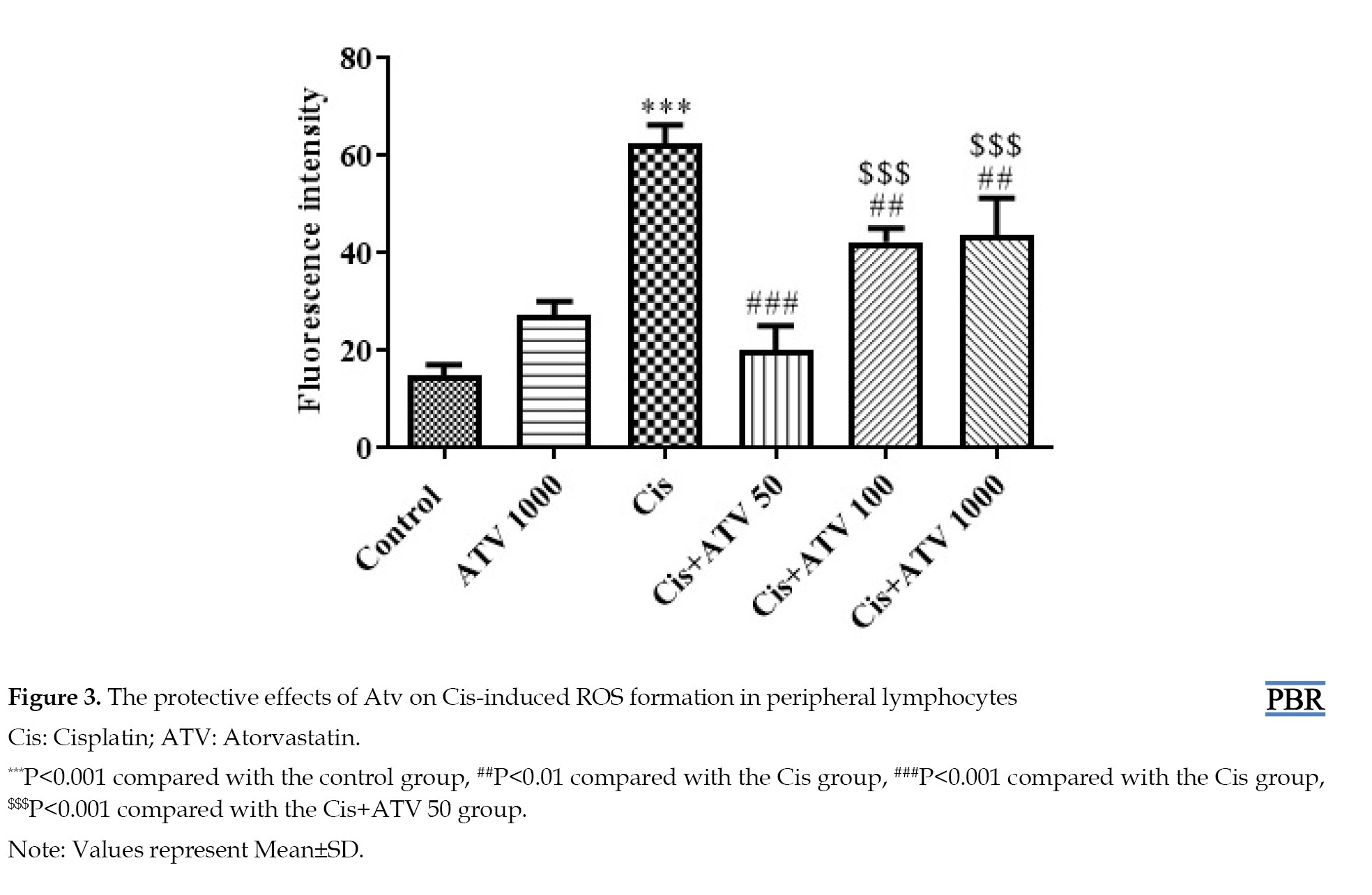
As shown in Figure 3, the Cis-treated group exhibited the highest ROS levels, while the control group showed the lowest (P<0.001).
Results of ROS production
As shown in Figure 3, the Cis-treated group exhibited the highest ROS levels, while the control group showed the lowest (P<0.001).
Atv administration notably reduced ROS levels following Cis exposure, particularly at 50 μM (P<0.001). Moreover, there was no significant difference in ROS levels between the Cis+ATV 100/1000 groups (P<0.01).
Results of LPO measurement
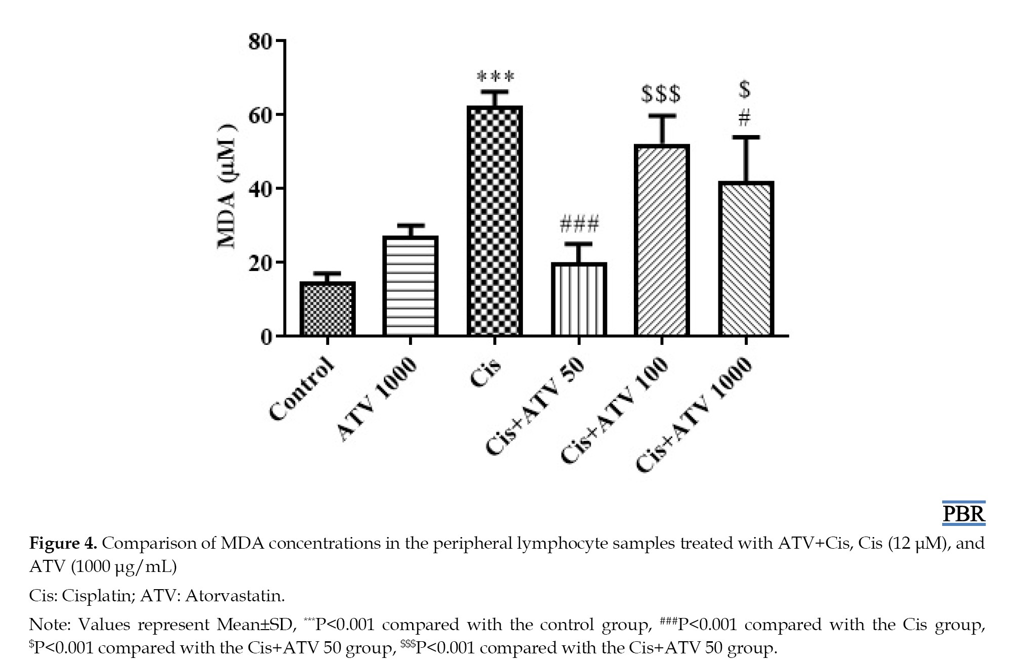
As shown in Figure 4, the Cis group had the highest LPO levels, while the control group had the lowest (P<0.001).
Results of LPO measurement
As shown in Figure 4, the Cis group had the highest LPO levels, while the control group had the lowest (P<0.001).
Furthermore, compared to the Cis group, the Cis+ATV50 group demonstrated even more significant differences compared to the Cis+Atv100 group (P<0.001). Moreover, the most significant reduction in LPO peroxidation was observed in the Cis+Atv50 group.
Results of GSH content measurement
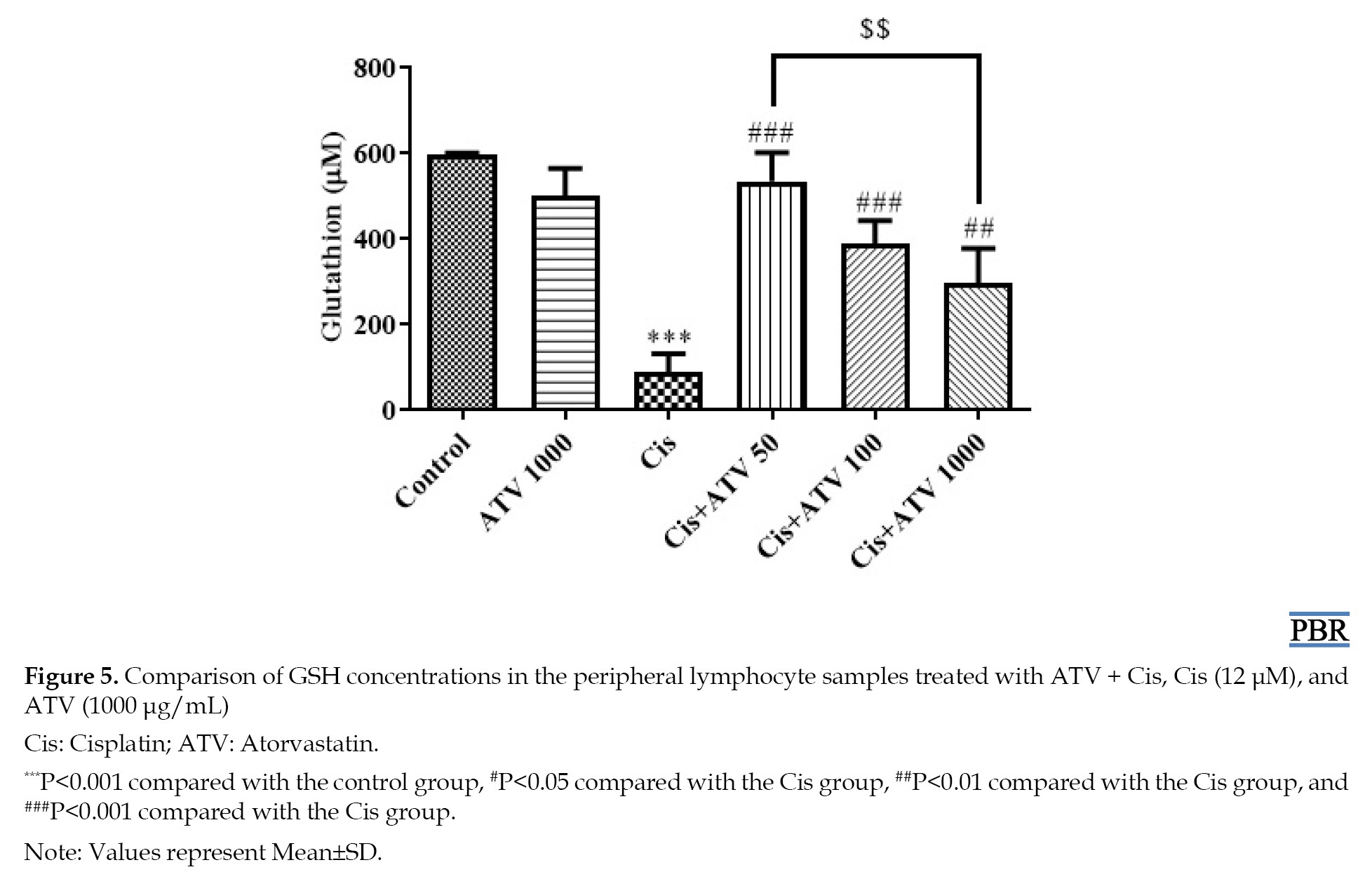
As shown in Figure 5, the control group exhibited the highest GSH levels.
Results of GSH content measurement
As shown in Figure 5, the control group exhibited the highest GSH levels.
Remarkably, there was a minimal disparity between the control and Cis+Atv50 groups. Conversely, the group receiving Cis showed the lowest GSH levels among all groups, with a statistically significant reduction compared to the control group (P<0.001). Atv led to a higher level of GSH than in the Cis group. However, the maximum effect and increase were observed in the Cis+Atv 50 group (P<0.05).
Discussion
Cis is an antineoplastic agent that is widely used in the treatment of cancers, such as head and neck, ovarian, cervical, and lung cancers through platinum-based chemotherapy [25]. Numerous studies have explored the genotoxicity of Cis. For example, researchers have observed that treating peripheral blood lymphocytes with Cis results in significant increases in chromosomal breaks, exchanges, and gaps. Additionally, there was a marked elevation in sister chromatid exchanges (SCEs) [26]. Furthermore, in another study at a concentration of 0.05 μg/mL, Cis induced increased chromosomal damage, chromosomal abnormalities, sister SCEs, and alterations in the mitotic index [27]. These findings align with the observations made in the present study regarding Cis’s genotoxic impact on human lymphocytes. It is important to note that our study employed a higher concentration (12 µM). The current study also revealed an increase in the percentage of micronuclei and elevated ROS formation in Cis-treated human lymphocytes. These differences arise from variations in the tested cell types, consumption methods, platinum salt concentrations, and laboratory conditions used in the different studies.
However, despite these findings, the authors of that study noted the need for further research to conclusively confirm Cis’s genotoxic effects. This study aimed to evaluate the genoprotective impact of Atv on Cis-induced genotoxicity in peripheral blood lymphocytes.
Alkylation plays a crucial role in Cis’s mode of action. When Cis enters a cell, it undergoes a process in which it replaces its chloride ligand with stronger bonds to cellular constituents. This transformation leads to the formation of DNA adducts, ultimately inhibiting DNA synthesis and the proliferation of affected cells [28]. Exposure to Cis is frequently associated with increased free radical production, leading to various forms of damage to cellular genetic material [29]. This damage can result in subsequent malignancies in normal cells [6].
To this end, MDA and GSH concentrations were assessed in human peripheral lymphocyte cultures. The findings revealed that lymphocytes treated with 12 µg/mL Cis showed a marked increase in MDA levels and a notable reduction in GSH levels. In conclusion, a final concentration of 12 µg/mL Cis induced oxidative stress in human peripheral blood lymphocytes, as manifested by increased LPO and decreased GSH levels. A study conducted by Sami et al. revealed that the administration of Cis, a well-established chemotherapy agent, leads to increased levels of renal ROS and MDA, as well as nitric oxide, and to the activation of nuclear factor kappa B (NF-κB) p65, inducible nitric oxide synthase, and pro-inflammatory cytokines. These findings indicate that Cis treatment may contribute to oxidative stress and inflammatory responses within different tissues [30].
Extensive research has been conducted to elucidate the impact of Cis-induced oxidative stress [31, 32]. Numerous studies have consistently demonstrated that Cis can induce oxidative damage, particularly when administered over extended periods. Hu et al. found that exposure of human embryonic kidney (HEK-293) cells to Cis led to a significant decrease in GSH, superoxide dismutase (SOD), and catalase (CAT) levels, along with a notable increase in MDA levels compared to the control group [33].
In another study, Cis exposure significantly decreased superoxide dismutase (SOD) and catalase (CAT) levels in intestinal homogenates. Additionally, as an end product of LPO, MDA levels dramatically increased in the intestines of Cis-exposed mice [34]. Cis exposure is frequently associated with increased free radicals and, subsequently, different types of damage to cellular genetic material, which can lead to malignancies in normal cells [35]. Since Cis can cause genotoxicity by inducing oxidative stress, reducing oxidative stress can mitigate genotoxicity [36].
The use of antioxidants is effective in achieving this objective. Numerous studies have provided evidence that antioxidants can mitigate both oxidative stress and genotoxicity [37-39].
Recently, Atv has gained attention for its antioxidant properties. Atv has been shown to have an antioxidant effect, and numerous studies have demonstrated its protective effect against oxidative damage [11].
For example, a study examined the impact of Atv at varying concentrations (50, 100, and 1000 µM) on the genotoxic effects of ionizing radiation in peripheral blood lymphocytes and found a significant reduction in the frequency of MN in lymphocytes treated with ionizing radiation. This implies that Atv exhibited a strong ability to mitigate DNA damage induced by ionizing radiation [40]. The study conducted by Shaghaghi et al. also demonstrated a significant reduction in the frequency of micronuclei with the use of Atv in cancer therapy [41].
Numerous in vivo studies have examined Atv’s effects on oxidative stress, consistently demonstrating its antioxidant properties [11, 42, 43]. For example, one study found a significant increase in MDA levels and a decrease in GSH content in the CP-treated group compared to the control group. The results of this assessment clearly demonstrated that the addition of 50 µM Atv to Cis-treated lymphocytes substantially reduced the frequency of micronuclei. In addition to the reduction in the MN assay, Atv also reduced oxidative damage, as measured by MDA levels and GSH concentrations. Amirhossein Sahebkar and Kadhim, Sahar S., demonstrated through their research that statin therapy significantly reduced MDA levels [44, 45]. In summary, Atv demonstrated a significant protective effect by reducing MDA levels and increasing GSH levels, with the effect being most pronounced at the lowest concentration (50 µM). Najah et al. also showed a significant increase in GSH levels following Atv administration in type 2 diabetic patients, as indicated by oxidative stress parameters and lipid profile [46].
Based on these findings, Atv appears to have a protective effect against oxidative stress, particularly by reducing LPO. Additionally, in the GSH concentration assay, lower Atv concentrations exhibited greater efficacy. These results collectively suggest that Atv exerts a protective influence against Cis-induced oxidative stress via diverse mechanisms.
Conclusion
This study demonstrates that Cis induces pronounced genotoxicity and oxidative stress in human lymphocytes, as evidenced by elevated MN frequency, LPO, and ROS, alongside diminished GSH levels. The co-administration of Atv significantly attenuated these effects, effectively reducing genotoxic and oxidative markers while restoring antioxidant capacity, particularly at lower concentrations. These findings underscore the potential of Atv as a protective adjunct in Cis-based chemotherapy, mitigating its deleterious effects on non-cancerous cells. Further investigations are warranted to elucidate the underlying mechanisms and evaluate their translational potential in clinical settings.
Ethical Considerations
Compliance with ethical guidelines
This study was approved by Ethics Committee of Mazandaran University of Medical Sciences, Sari, Iran (Code: IR.MAZUMS.REC.1396.574).
Funding
This study was extracted from PhD Dissertation of Mina Fasihbeiki, approved by Mazandaran University of Medical Sciences and was financially supported by the Research Council of Mazandaran University of Medical Sciences, Sari, Iran (Grant No.: 574).
Authors' contributions
Conceptualization, resources and supervision: Mohammad Shokrzadeh and Fatemeh Shaki; Validation, visualization and project administration: Mina Fasihbeikia, Mohammad Shokrzadeh, Mona Modanloo and Fatemeh Shaki; Writing and software: Mohammad Shokrzadeh, Ehsan Zamani, Mona Alinia and Fatemeh Shaki; Funding acquisition: Mohammad Shokrzadeh; Data collection, formal analysis, investigation and methodology: All authors.
Conflict of interest
The authors declared no conflict of interest.
Discussion
Cis is an antineoplastic agent that is widely used in the treatment of cancers, such as head and neck, ovarian, cervical, and lung cancers through platinum-based chemotherapy [25]. Numerous studies have explored the genotoxicity of Cis. For example, researchers have observed that treating peripheral blood lymphocytes with Cis results in significant increases in chromosomal breaks, exchanges, and gaps. Additionally, there was a marked elevation in sister chromatid exchanges (SCEs) [26]. Furthermore, in another study at a concentration of 0.05 μg/mL, Cis induced increased chromosomal damage, chromosomal abnormalities, sister SCEs, and alterations in the mitotic index [27]. These findings align with the observations made in the present study regarding Cis’s genotoxic impact on human lymphocytes. It is important to note that our study employed a higher concentration (12 µM). The current study also revealed an increase in the percentage of micronuclei and elevated ROS formation in Cis-treated human lymphocytes. These differences arise from variations in the tested cell types, consumption methods, platinum salt concentrations, and laboratory conditions used in the different studies.
However, despite these findings, the authors of that study noted the need for further research to conclusively confirm Cis’s genotoxic effects. This study aimed to evaluate the genoprotective impact of Atv on Cis-induced genotoxicity in peripheral blood lymphocytes.
Alkylation plays a crucial role in Cis’s mode of action. When Cis enters a cell, it undergoes a process in which it replaces its chloride ligand with stronger bonds to cellular constituents. This transformation leads to the formation of DNA adducts, ultimately inhibiting DNA synthesis and the proliferation of affected cells [28]. Exposure to Cis is frequently associated with increased free radical production, leading to various forms of damage to cellular genetic material [29]. This damage can result in subsequent malignancies in normal cells [6].
To this end, MDA and GSH concentrations were assessed in human peripheral lymphocyte cultures. The findings revealed that lymphocytes treated with 12 µg/mL Cis showed a marked increase in MDA levels and a notable reduction in GSH levels. In conclusion, a final concentration of 12 µg/mL Cis induced oxidative stress in human peripheral blood lymphocytes, as manifested by increased LPO and decreased GSH levels. A study conducted by Sami et al. revealed that the administration of Cis, a well-established chemotherapy agent, leads to increased levels of renal ROS and MDA, as well as nitric oxide, and to the activation of nuclear factor kappa B (NF-κB) p65, inducible nitric oxide synthase, and pro-inflammatory cytokines. These findings indicate that Cis treatment may contribute to oxidative stress and inflammatory responses within different tissues [30].
Extensive research has been conducted to elucidate the impact of Cis-induced oxidative stress [31, 32]. Numerous studies have consistently demonstrated that Cis can induce oxidative damage, particularly when administered over extended periods. Hu et al. found that exposure of human embryonic kidney (HEK-293) cells to Cis led to a significant decrease in GSH, superoxide dismutase (SOD), and catalase (CAT) levels, along with a notable increase in MDA levels compared to the control group [33].
In another study, Cis exposure significantly decreased superoxide dismutase (SOD) and catalase (CAT) levels in intestinal homogenates. Additionally, as an end product of LPO, MDA levels dramatically increased in the intestines of Cis-exposed mice [34]. Cis exposure is frequently associated with increased free radicals and, subsequently, different types of damage to cellular genetic material, which can lead to malignancies in normal cells [35]. Since Cis can cause genotoxicity by inducing oxidative stress, reducing oxidative stress can mitigate genotoxicity [36].
The use of antioxidants is effective in achieving this objective. Numerous studies have provided evidence that antioxidants can mitigate both oxidative stress and genotoxicity [37-39].
Recently, Atv has gained attention for its antioxidant properties. Atv has been shown to have an antioxidant effect, and numerous studies have demonstrated its protective effect against oxidative damage [11].
For example, a study examined the impact of Atv at varying concentrations (50, 100, and 1000 µM) on the genotoxic effects of ionizing radiation in peripheral blood lymphocytes and found a significant reduction in the frequency of MN in lymphocytes treated with ionizing radiation. This implies that Atv exhibited a strong ability to mitigate DNA damage induced by ionizing radiation [40]. The study conducted by Shaghaghi et al. also demonstrated a significant reduction in the frequency of micronuclei with the use of Atv in cancer therapy [41].
Numerous in vivo studies have examined Atv’s effects on oxidative stress, consistently demonstrating its antioxidant properties [11, 42, 43]. For example, one study found a significant increase in MDA levels and a decrease in GSH content in the CP-treated group compared to the control group. The results of this assessment clearly demonstrated that the addition of 50 µM Atv to Cis-treated lymphocytes substantially reduced the frequency of micronuclei. In addition to the reduction in the MN assay, Atv also reduced oxidative damage, as measured by MDA levels and GSH concentrations. Amirhossein Sahebkar and Kadhim, Sahar S., demonstrated through their research that statin therapy significantly reduced MDA levels [44, 45]. In summary, Atv demonstrated a significant protective effect by reducing MDA levels and increasing GSH levels, with the effect being most pronounced at the lowest concentration (50 µM). Najah et al. also showed a significant increase in GSH levels following Atv administration in type 2 diabetic patients, as indicated by oxidative stress parameters and lipid profile [46].
Based on these findings, Atv appears to have a protective effect against oxidative stress, particularly by reducing LPO. Additionally, in the GSH concentration assay, lower Atv concentrations exhibited greater efficacy. These results collectively suggest that Atv exerts a protective influence against Cis-induced oxidative stress via diverse mechanisms.
Conclusion
This study demonstrates that Cis induces pronounced genotoxicity and oxidative stress in human lymphocytes, as evidenced by elevated MN frequency, LPO, and ROS, alongside diminished GSH levels. The co-administration of Atv significantly attenuated these effects, effectively reducing genotoxic and oxidative markers while restoring antioxidant capacity, particularly at lower concentrations. These findings underscore the potential of Atv as a protective adjunct in Cis-based chemotherapy, mitigating its deleterious effects on non-cancerous cells. Further investigations are warranted to elucidate the underlying mechanisms and evaluate their translational potential in clinical settings.
Ethical Considerations
Compliance with ethical guidelines
This study was approved by Ethics Committee of Mazandaran University of Medical Sciences, Sari, Iran (Code: IR.MAZUMS.REC.1396.574).
Funding
This study was extracted from PhD Dissertation of Mina Fasihbeiki, approved by Mazandaran University of Medical Sciences and was financially supported by the Research Council of Mazandaran University of Medical Sciences, Sari, Iran (Grant No.: 574).
Authors' contributions
Conceptualization, resources and supervision: Mohammad Shokrzadeh and Fatemeh Shaki; Validation, visualization and project administration: Mina Fasihbeikia, Mohammad Shokrzadeh, Mona Modanloo and Fatemeh Shaki; Writing and software: Mohammad Shokrzadeh, Ehsan Zamani, Mona Alinia and Fatemeh Shaki; Funding acquisition: Mohammad Shokrzadeh; Data collection, formal analysis, investigation and methodology: All authors.
Conflict of interest
The authors declared no conflict of interest.
References
- Brown A, Kumar S, Tchounwou PB. Cisplatin-based chemotherapy of human cancers. J Cancer Sci Ther. 2019; 11(4):97. [PMID]
- Ranjbar A, Ghaseminejhad S, Takalu H, Baiaty A, Rahimi F, Abdollahi M. Anti oxidative stress potential of cinnamon (cinnamomum zeylanicum) in operating room personnel; a before/after cross sectional clinical trial. Int J Pharmacol. 2007; 3(6):482-86. [DOI:10.3923/ijp.2007.482.486]
- Cohen SM, Lippard SJ. Cisplatin: From DNA damage to cancer chemotherapy. Prog Nucleic Acid Res Mol Biol. 2001; 67:93-130. [DOI:10.1016/S0079-6603(01)67026-0] [PMID]
- Makovec T. Cisplatin and beyond: Molecular mechanisms of action and drug resistance development in cancer chemotherapy. Radiol Oncol. 2019; 53(2):148-58. [DOI:10.2478/raon-2019-0018] [PMID]
- Sancho-Martínez SM, Prieto-García L, Prieto M, López-Novoa JM, López-Hernández FJ. Subcellular targets of cisplatin cytotoxicity: An integrated view. Pharmacol Ther. 2012; 136(1):35-55. [DOI:10.1016/j.pharmthera.2012.07.003] [PMID]
- Dos Santos GC, Mendonça LM, Antonucci GA, Dos Santos AC, Antunes LM, Bianchi Mde L. Protective effect of bixin on cisplatin-induced genotoxicity in PC12 cells. Food Chem Toxicol. 2012; 50(2):335-40. [DOI:10.1016/j.fct.2011.10.033] [PMID]
- Erbas M, Sekerci H. Importance of free radicals and occurring during food processing. GIDA J Food. 2011; 36(6):1. [Link]
- Lobo V, Patil A, Phatak A, Chandra N. Free radicals, antioxidants and functional foods: Impact on human health. Pharmacogn Rev. 2010; 4(8):118-26. [DOI:10.4103/0973-7847.70902] [PMID]
- Ji LL. Oxidative stress during exercise: Implication of antioxidant nutrients. Free Radic Biol Med. 1995; 18(6):1079-86. [DOI:10.1016/0891-5849(94)00212-3] [PMID]
- Akbari B, Baghaei-Yazdi N, Bahmaie M, Mahdavi Abhari F. The role of plant-derived natural antioxidants in reduction of oxidative stress. Biofactors. 2022; 48(3):611-33. [DOI:10.1002/biof.1831] [PMID]
- Mansouri A, Reiner Ž, Ruscica M, Tedeschi-Reiner E, Radbakhsh S, Bagheri Ekta M, et al. Antioxidant effects of statins by modulating Nrf2 and Nrf2/HO-1 signaling in different diseases. J Clin Med. 2022; 11(5):1313. [DOI:10.3390/jcm11051313] [PMID]
- Clim A, Maranduca MA, Filip N, Tănase DM, Floria M, Pinzariu AC, et al. The Influence of Atorvastatin Treatment on Homocysteine Metabolism and Oxidative Stress in an Experimental Model of Diabetic Rats. Life. 2024; 14(11):1414. [DOI:10.3390/life14111414] [PMID]
- Voltan R, Secchiero P, Casciano F, Milani D, Zauli G, Tisato V. Redox signaling and oxidative stress: Cross talk with TNF-related apoptosis inducing ligand activity. Int J Biochem Cell Biol. 2016; 81(Pt B):364-74. [DOI:10.1016/j.biocel.2016.09.019] [PMID]
- Yuan W, Fan H, Yang H, Tang L, Liu Z, Ouyang F, et al. Effect and mechanism of HMG-CoA reductase inhibitor on the improvement of elderly essential hypertension-induced vascular endothelial function impairment based on the JAK/STAT pathway. Diagn Pathol. 2023;18(1):108. [DOI:10.1186/s13000-023-01393-x] [PMID]
- Stefanis L, Burke RE, Greene LA. Apoptosis in neurodegenerative disorders. Curr Opin Neurol. 1997; 10(4):299-305. [DOI:10.1097/00019052-199708000-00004] [PMID]
- Mayyas F. Short-term effect of atorvastatin on renal oxidative stress, inflammation, and fibrosis in a rat model of streptozotocin-induced diabetes. J Diabetes Metab Disord. 2024; 24(1):12. [DOI:10.1007/s40200-024-01514-3] [PMID]
- Zhang Y, Qu Y, Cai R, Gao J, Xu Q, Zhang L, et al. Atorvastatin ameliorates diabetic nephropathy through inhibiting oxidative stress and ferroptosis signaling. Eur J Pharmacol. 2024; 976:176699. [DOI:10.1016/j.ejphar.2024.176699] [PMID]
- Vance DE, Vance JE. Biochemistry of lipids, lipoproteins and membranes. Edinburgh: Elsevier; 1996. [Link]
- Inia JA, Stokman G, Pieterman EJ, Morrison MC, Menke AL, Verschuren L, et al. Atorvastatin attenuates diet-induced non-alcoholic steatohepatitis in APOE*3-leiden mice by reducing hepatic inflammation. Int J Mol Sci. 2023; 24(9):7818. [DOI:10.3390/ijms24097818] [PMID]
- Evazalipour M, Moayedi S, Safarzadeh Kozani P, Safarzadeh Kozani P. The protective effects of carvacrol on diphenhydramine-induced genotoxicity in human peripheral blood lymphocytes. Res J Pharmacognosy. 2021; 8(1):19-27. [Link]
- Yamamoto V, Wang B, Lee AS. Suppression of head and neck cancer cell survival and cisplatin resistance by GRP78 small molecule inhibitor YUM70. Front Oncol. 2023; 12:1044699. [DOI:10.3389/fonc.2022.1044699] [PMID]
- Marullo R, Werner E, Degtyareva N, Moore B, Altavilla G, Ramalingam SS, et al. Cisplatin induces a mitochondrial-ROS response that contributes to cytotoxicity depending on mitochondrial redox status and bioenergetic functions. Plos One. 2013; 8(11):e81162. [DOI:10.1371/journal.pone.0081162] [PMID]
- Araujo-Lima CF, Christoni LSA, Justo G, Soeiro MNC, Aiub CAF, Felzenszwalb I. Atorvastatin downregulates in vitro methyl methanesulfonate and cyclophosphamide alkylation-mediated cellular and DNA injuries. Oxid Med Cell Longev. 2018; 2018:7820890. [DOI:10.1155/2018/7820890] [PMID]
- Soufi BR, Evazalipour M, Motavallian A, Chakosari MH, Zamani E. The protective effect of melatonin on diazepam-induced genotoxicity in peripheral blood lymphocytes using micronucleus assay. J Rep Pharm Sci. 2022; 11(1):92-7. [DOI:10.4103/jrptps.JRPTPS_111_20]
- Ghosh S. Cisplatin: The first metal based anticancer drug. Bioorganic chemistry. 2019; 88:102925. [DOI:10.1016/j.bioorg.2019.102925] [PMID]
- Khabour OF, Alzoubi KH, Doa’a SM, Alasseiri M, Hasheesh TF. Tempol protects human lymphocytes from genotoxicity induced by cisplatin. Int J Clin Experiment Med. 2014 Apr 15;7(4):982. [PMID]
- Al-Eitan LN, Alzoubi KH, Al-Smadi LI, Khabour OF. Vitamin E protects against cisplatin-induced genotoxicity in human lymphocytes. Toxicol In Vitro. 2020; 62:104672. [DOI:10.1016/j.tiv.2019.104672] [PMID]
- Siddik ZH. Mechanisms of action of cancer chemotherapeutic agents: DNA-interactive alkylating agents and antitumour platinum-based drugs. In: Alison MR, editor. The Cancer Handbook 1st Edition. Hoboken: John Wiley & Sons, Ltd; 2002. [Link]
- Tchounwou PB, Dasari S, Noubissi FK, Ray P, Kumar S. Advances in our understanding of the molecular mechanisms of action of cisplatin in cancer therapy. J Exp Pharmacol. 2021; 13:303-28. [DOI:10.2147/JEP.S267383] [PMID]
- Sami DH, Soliman AS, Khowailed AA, Hassanein EHM, Kamel EM, Mahmoud AM. 7-hydroxycoumarin modulates Nrf2/HO-1 and microRNA-34a/SIRT1 signaling and prevents cisplatin-induced oxidative stress, inflammation, and kidney injury in rats. Life Sci. 2022; 310:121104. [DOI:10.1016/j.lfs.2022.121104] [PMID]
- Ghosh P, Roy SS, Chakraborty P, Ghosh S, Bhattacharya S. Effects of organoselenium compound 2-(5-selenocyanato-pentyl)-benzo[de]isoquinoline 1,3-dione on cisplatin induced nephrotoxicity and genotoxicity: An investigation of the influence of the compound on oxidative stress and antioxidant enzyme system. Biometals. 2013; 26(1):61-73. [DOI:10.1007/s10534-012-9594-y] [PMID]
- Brahmi D, Ayed Y, Hfaiedh M, Bouaziz C, Mansour HB, Zourgui L, et al. Protective effect of cactus cladode extract against cisplatin induced oxidative stress, genotoxicity and apoptosis in balb/c mice: Combination with phytochemical composition. BMC Complement Altern Med. 2012; 12:111. [DOI:10.1186/1472-6882-12-111] [PMID]
- Hu JN, Leng J, Shen Q, Liu Y, Li XD, Wang SH, et al. Platycodin D suppresses cisplatin-induced cytotoxicity by suppressing ROS-mediated oxidative damage, apoptosis, and inflammation in HEK-293 cells. J Biochem Mol Toxicol. 2021; 35(1):e22624. [DOI:10.1002/jbt.22624] [PMID]
- Hu JN, Yang JY, Jiang S, Zhang J, Liu Z, Hou JG, et al. Panax quinquefolium saponins protect against cisplatin evoked intestinal injury via ROS-mediated multiple mechanisms. Phytomedicine. 2021; 82:153446. [DOI:10.1016/j.phymed.2020.153446] [PMID]
- Nazari A, Mirian M, Aghaei M, Aliomrani M. 4-Hydroxyhalcone effects on cisplatin-induced genotoxicity model. Toxicol Res. 2021; 10(1):11-7. [DOI:10.1093/toxres/tfaa091] [PMID]
- Said Salem NI, Noshy MM, Said AA. Modulatory effect of curcumin against genotoxicity and oxidative stress induced by cisplatin and methotrexate in male mice. Food Chem Toxicol. 2017; 105:370-6. [DOI:10.1016/j.fct.2017.04.007] [PMID]
- Blasiak J, Trzeciak A, Malecka-Panas E, Drzewoski J, Wojewódzka M. In vitro genotoxicity of ethanol and acetaldehyde in human lymphocytes and the gastrointestinal tract mucosa cells. Toxicol In Vitro. 2000; 14(4):287-95. [DOI:10.1016/S0887-2333(00)00022-9] [PMID]
- Singh M, Kaur P, Sandhir R, Kiran R. Protective effects of vitamin E against atrazine-induced genotoxicity in rats. Mutat Res. 2008; 654(2):145-9. [DOI:10.1016/j.mrgentox.2008.05.010] [PMID]
- Anderson D, Yu TW, Phillips BJ, Schmezer P. The effect of various antioxidants and other modifying agents on oxygen-radical-generated DNA damage in human lymphocytes in the COMET assay. Mutat Res. 1994; 307(1):261-71. [DOI:10.1016/0027-5107(94)90300-X] [PMID]
- Hosseinimehr SJ, Izakmehri M, Ghasemi A. In vitro protective effect of atorvastatin against ionizing radiation induced genotoxicity in human lymphocytes. Cell Mol Biol. 2015; 61(1):68-71. [PMID]
- Shaghaghi Z, Alvandi M, Farzipour S, Dehbanpour MR, Nosrati S. A review of effects of atorvastatin in cancer therapy. Med Oncol. 2022; 40(1):27. [DOI:10.1007/s12032-022-01892-9] [PMID]
- Zhang XB, Cheng HJ, Yuan YT, Chen Y, Chen YY, Chiu KY, et al. Atorvastatin attenuates intermittent hypoxia-induced myocardial oxidative stress in a mouse obstructive sleep apnea model. Aging. 2021; 13(14):18870-8. [DOI:10.18632/aging.203339] [PMID]
- Ghoreshi ZA, Kabirifar R, Khodarahmi A, Karimollah A, Moradi A. The preventive effect of atorvastatin on liver fibrosis in the bile duct ligation rats via antioxidant activity and down-regulation of Rac1 and NOX1. Iran J Basic Med Sci. 2020; 23(1):30-5. [DOI:10.22038/IJBMS.2019.33663.8047] [PMID]
- Sahebkar A, Foroutan Z, Katsiki N, Jamialahmadi T, Mantzoros CS. Ferroptosis, a new pathogenetic mechanism in cardiometabolic diseases and cancer: Is there a role for statin therapy? Metabolism. 2023; 146:155659. [DOI:10.1016/j.metabol.2023.155659] [PMID]
- Kadhim SS, Al-Windy SA, Al-Nami MS, Al Kuraishy HM, Al Gareeb AI. Statins improve periodontal disease-induced inflammatory changes and associated lipid peroxidation in patients with dyslipidemia: Two birds by one stone. J Int Oral Health. 2020; 12(1):66-73. [DOI:10.4103/jioh.jioh_194_19]
- Najah R, Mohammad A, Ammar R. Effect of atorvastatin on oxidative stress parameters and lipid profile in type 2 diabetic patients. IIUM Med J Malaysia. 2008; 7(2):43-50. [DOI:10.31436/imjm.v7i2.783]
Type of Study: Original Research |
Subject:
Toxicology
| Rights and permissions | |
 |
This work is licensed under a Creative Commons Attribution-NonCommercial 4.0 International License. |


This is an open access article distributed under the terms of the Creative Commons Attribution License (CC-By-NC), which permits use, distribution, and reproduction in any medium, provided the original work is properly cited and is not used for commercial purposes.
Contact Information