

Volume 12, Issue 2 (2026)
Pharm Biomed Res 2026, 12(2): 119-132 |
Back to browse issues page


Download citation:
BibTeX | RIS | EndNote | Medlars | ProCite | Reference Manager | RefWorks
Send citation to:


BibTeX | RIS | EndNote | Medlars | ProCite | Reference Manager | RefWorks
Send citation to:
amini E, Mirahmadi M, Kaboli Farshchi H, Baniasadi F. Palliative Effects of Date Palm Pollen and Its Green Selenium Nanoparticles on FSH, LH, and Ovarian Histology in PCOS Mouse Model. Pharm Biomed Res 2026; 12 (2) :119-132
URL: http://pbr.mazums.ac.ir/article-1-749-en.html
URL: http://pbr.mazums.ac.ir/article-1-749-en.html


1- Department of Animal Biology, Faculty of Biological Sciences, Kharazmi University, Tehran, Iran.
2- Stem Cell and Regenerative Medicine Research Group, Iranian Academic Center for Education, Culture and Research (ACECR), Khorasan Razavi Branch, Mashhad, Iran. & Pars Takrokh Pharmaceutical Company, Bu Ali Research Institute, Science and Technology Park, Mashhad University of Medical Sciences, Mashhad, Iran.
3- Quantum Engineering and Photonics Technology Research Center, Sharif University of Technology, Tehran, Iran.
2- Stem Cell and Regenerative Medicine Research Group, Iranian Academic Center for Education, Culture and Research (ACECR), Khorasan Razavi Branch, Mashhad, Iran. & Pars Takrokh Pharmaceutical Company, Bu Ali Research Institute, Science and Technology Park, Mashhad University of Medical Sciences, Mashhad, Iran.
3- Quantum Engineering and Photonics Technology Research Center, Sharif University of Technology, Tehran, Iran.
Keywords: Selenium nanoparticles (SeNPs), Date palm pollen (DPP), Green synthesis, Folliculogenesis, Polycystic ovarian syndrome (PCOS)
Full-Text [PDF 2753 kb]
(91 Downloads)
| Abstract (HTML) (239 Views)
Full-Text: (37 Views)
Introduction
Polycystic ovarian syndrome (PCOS) is considered an endocrine disorder and the most common hormonal disorder in women, which is associated with symptoms, such as hyperandrogenism, polycystic ovarian morphology, chronic anovulation, and irregular gonadotropin secretion [1]. According to the literature, this disease affects 4–20% of women and is often associated with late diagnosis and metabolic dysfunction and problems such as obesity and insulin resistance [2]. Considering the symptoms of this disease, lifestyle modification, dietary adjustment, and pharmacological interventions are considered the most important strategies in the management of this disease [3]. Some of the most common medications and supplements used to manage PCOS include hormonal contraceptives, progestins, vitamin D, calcium, clomiphene citrate, and metformin [4, 5].
The most important herbs used to improve PCOS symptoms include cinnamon, aloe vera, flaxseed, licorice, evening primrose, and berberine [6-8]. Studies have shown that herbal compounds play an important role in improving fertility by increasing the number of ovarian follicles, reducing testosterone and androgen levels, mitigating blood lipid and glucose levels, and declining estrogen and hyperplasia [9].
Date palm pollen (DPP) from the date palm (Phoenix dactylifera L.) has been traditionally used to enhance sexual ability and fertility. In the past, it has been proven that DPP treats male infertility by improving the quality of sperm parameters [10]. To date, the presence of phenolic and flavonoid compounds with therapeutic effects, such as quercetin, gallic acid, rutin, catechin, ferulic acid, caffeic acid, apigenin, luteolin, and coumaric acid has been reported in DPP. In addition, DPP contains various bioactive compounds including fatty acids, amino acids, sterols, saponins, volatile unsaturated fatty acids, vitamins, and minerals [11].
Previous studies indicated that DPP can enhance fertility and sexual potency in both men and women through two main mechanisms, due to its antioxidant and gonadotropin-stimulating properties. Several animal studies have shown the effects of this herbal compound on testosterone, luteinizing hormone (LH), and follicle-stimulating hormone (FSH) [12-14].
A study has shown that reduced fertility in women is often associated with insufficient selenium levels in the body, and selenium supplementation is recommended in women with selenium deficiency to improve reproductive efficiency and increase pregnancy rates [15]. Studies have shown that selenium, helps protection of the developing oocyte from oxidative stress during folliculogenesis mainly via antioxidant function [16]. Previous studies have reported that selenium supplementation increased the gene expression levels of certain enzymes and improved lipid metabolism, and may have beneficial effects on patients with PCOS [17]. It has also been documented that co-administration of probiotics and selenium for 12 weeks in women with PCOS had beneficial effects on mental health parameters, total serum testosterone, hirsutism, total antioxidant capacity (TAC), glutathione GSH, and malondialdehyde (MDA) levels [18].
Studies have shown that selenium in the form of nanoparticles or SeNPs has higher bioavailability and lower excretion in animals, and has higher and better activity than selenium. In addition, SeNPs have lower toxicity than organic and inorganic selenium-based compounds, are biocompatible, and are capable of efficient delivery of the compounds to specific cells [19]. Given that oxidative stress can compromise egg quality and lead to infertility or poor outcomes in assisted reproductive technologies, SeNPs have been demonstrated to play a vital role in female reproduction via accelerating the maintenance of reproductive health, especially egg quality and protection against oxidative stress. They also play a role in regulating the menstrual cycle, ovulation, and potentially reducing the effects of diseases such as PCOS [20, 17]. The green synthesis is an environmentally friendly and relatively inexpensive method as compared to physical and chemical synthesis that does not require complex techniques and devices and involves the use of plant extracts or biocompounds to prepare nanoparticles as reducing and stabilizing agents [21]. Considering the advantages of green synthesis, including lower cost, less pollution, and improved safety for the environment and health, the present study aimed to investigate the effect of green-synthesized SeNPs on serum FSH and LH levels and ovarian tissue in mice model of PCOS.
Materials and Methods
Chemicals
Sodium selenite prepared from Sigma Company (code S5261), ascorbic acid (Sigma Company, code A92902), DPP (Liano Company, Bushehr), deionized water (Zalal Teb Chemi), estradiol valerate (Abo Raihan Pharmaceutical Company), hematoxylin from Sigma Company (code H3136), eosin Y from Sigma Company (code E6003), xylene (294780), ethanol (Razi Yeast Company), and toluene (code 244511) were used in this study.
Biosynthesis of SeNPs using aqueous extract of DPP
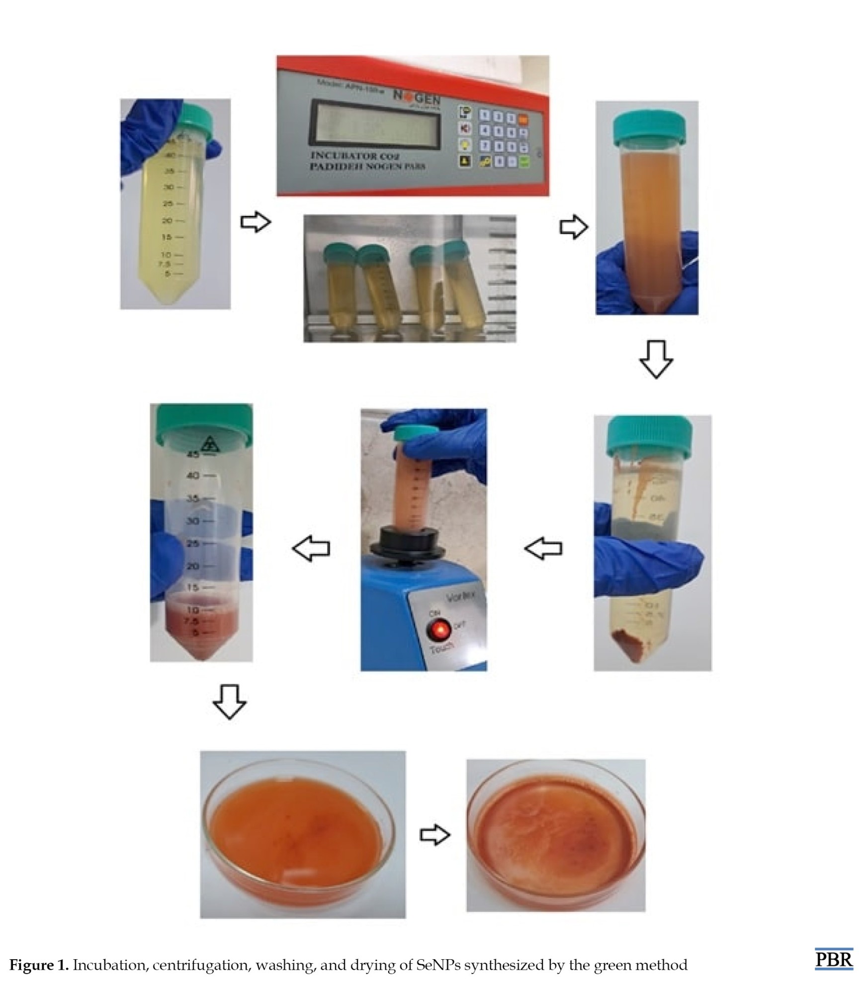
The synthesis of SeNPs was carried out based on Wakid et al. protocol. For this purpose, DPP was collected from Bushehr Province, Iran in March, transferred to the herbarium of Kharazmi University, identified, and confirmed ad herbarium code of 25556. Aqueous extract of DPP was used to prepare SeNPs. In this regard, the dried powder (10 g) was dissolved in deionized water (100 mL at room temperature), filtered after 30 min and evaporated at 55 to 60 °C [22]. At next step, 10 mmol/L ascorbic acid solution (10 mL), and DPP aqueous extract (10 mL) were added dropwise to selenium salt solution (10 mmol/L). After 24-hour incubation at 37 °C, serial centrifugation (2000 g, 10 min; NOGEN, Iran) performed using deionized water and the final precipitate are being collected as Se-NPs and dried to carry out biological assays [23] (Figure 1).
Characterization of SeNPs synthesized by green method
Dynamic light scattering (DLS), zeta potential, and transmission electron microscopy (TEM) analysis
To perform this method, the synthesized SeNPs sample was made into a suspension. To prepare a suspension of nanoparticles, SeNPs were dissolved in a suitable solvent (distilled water). To measure the particle size, a volume equivalent to 500 μL of SeNPs solution in 2 mL of deionized water was placed in a polystyrene cuvette and measured at 25 °C. The prepared sample was placed in the nanoparticle size analyzer (Nanotrac Wave II, Japan). Finally, the data collected by the device were analyzed using specific algorithms to calculate the particle size distribution. To evaluate the stability of the particles, the surface charge (zeta potential) was calculated in distilled water with pH 7 [24]. For TEM analysis, synthesized SeNPs was poured as a drop over carbon coated copper grids and maintain in room temperature to evaporate solvent. TEM analysis was conducted on a JEOL model 1200 EX instrument operated at an accelerating voltage at 80 kV.
Animal study
In this experimental study, 30 NMRI mice weighing between 22 and 24 g were used. The mice were obtained from the breeding and maintenance center of the Mashhad Bu Ali Research Institute and maintained under standard conditions (25 °C, 12 hours light/dark cycle). Animals’ intervention was carried out in accordance with the standards of the Kharazmi University Ethics Committee. The mice were divided into 6 groups of 5 mice. The control group received no treatment. PCOS group received estradiol valerate 4 mg/kg body weight in a volume of 100 μL intramuscularly dissolved in olive oil. The vaginal smears of mice were monitored for 60 days, until the time when abnormal estrus cycles and persistent vaginal cornification as the confirmation of PCOS induction [25]. DPP+PCOS aqueous extract groups received DPP at a dose of 200 and 20 mg/kg body weight for 14 days by gavage (after 8 weeks of estradiol valerate injection). SeNPs synthesized by the green method received SeNPs at a dose of 2 and 0.2 mg/kg body weight for 14 days by gavage (after 8 weeks of estradiol valerate injection). Blinding performed to reduce bias in research. For the overall study period, researchers were blind to the treatment allocation. The DPP dose was selected based on the research of Moshfegh and associates. In the case of SeNPs, the dose was selected first based on the research of Kandaparthi et al., who reported that SeNPs at a low dose of 1 to 2 mg/kg have less toxicity and higher antioxidant properties than high doses, and then the desired dose was experimentally investigated [14-26]. Based on previous studies, the LD50 for SeNPs was reported to be 14.6 mg/kg body weight and for DPP, about 5 g/kg body weight [27, 28].
Vaginal smear evaluation
This assay was performed to determine the regularity of the sexual cycle and to select mice in the estrus or estrous stage, which is characterized by the presence of epithelial cells and the absence of leukocytes. To do this, a sample was taken from the vagina of the mouse using a swab dipped in physiological serum and placed on a slide, and after staining with methylene blue, it was examined under a microscope. After the necessary examinations to ensure the regularity and problem-free reproductive cycle (at least 3 consecutive cycles) through smear test and observation under a microscope, after 12-14 days, only mice with a regular estrus cycle were randomly selected for this study.
FSH and LH analysis
One day after the last gavage, in order to collect blood from the hearts of control, PCOS, and PCOS mice treated with aqueous extract of DPP and SeNPs synthesized by DPP, they were first anesthetized by injection of ketamine (100 mg/kg)/xylazine (10 mg/kg) and then sacrificed by cervical dislocation. Blood was collected from the heart (1 mL per mouse) using an insulin syringe (Helma Teb). Then, the blood samples were placed at room temperature for about 30 to 40 min. Afterward, centrifugation was performed for 10 min at 800×g (NOGEN, Iran). The obtained serum was gently separated by 100 μL sampler and poured into a 1.5 mL microtube. Finally, the serum samples were transferred on ice to the Mashhad University Jahad Laboratory for analysis of LH and FSH hormone levels (Diazist, Iran). The samples were stored at -20 °C and the results of the hormone levels were compared between the groups [29].
Histology of ovarian tissue
To isolate ovarian tissue, after the treatment period, the mice were euthanized by cervical dislocation. The ovaries were removed and washed with physiological serum. After collection, the ovarian tissue was placed in 10% formalin fixative and transferred to the Pathobiology Department of Omid Hospital. To prepare the ovarian tissue, the tissue was first dehydrated using ascending grades of alcohol and placed in toluene for clarification. In the next step, the paraffin bath was used to cast the tissue and the tissue was embedded in paraffin blocks. Sectioning done using a 7-µ microtome (Hinotek, China), the sections were placed on coated slides. Hematoxylin and eosin staining was used for tissue staining.
Statistical analysis
Normality of data was assessed using the Kolmogorov-Smirnov test. After ensuring the normality of the data, parametric tests were used to analyze the data. SPSS software, version 22 was used to analyze the data. One-way analysis of variance was used to examine differences between groups and Tukey’s post hoc test was used to compare differences between groups. Experiments were repeated three times. Results were expressed as Mean±SD and P≤0.05 was considered significant. Excel software was used to draw graphs.
Results
DLS, zeta potential, andTEM analysis
The particle size distribution of SeNPs synthesized was evaluated using DLS (Figure 2A).
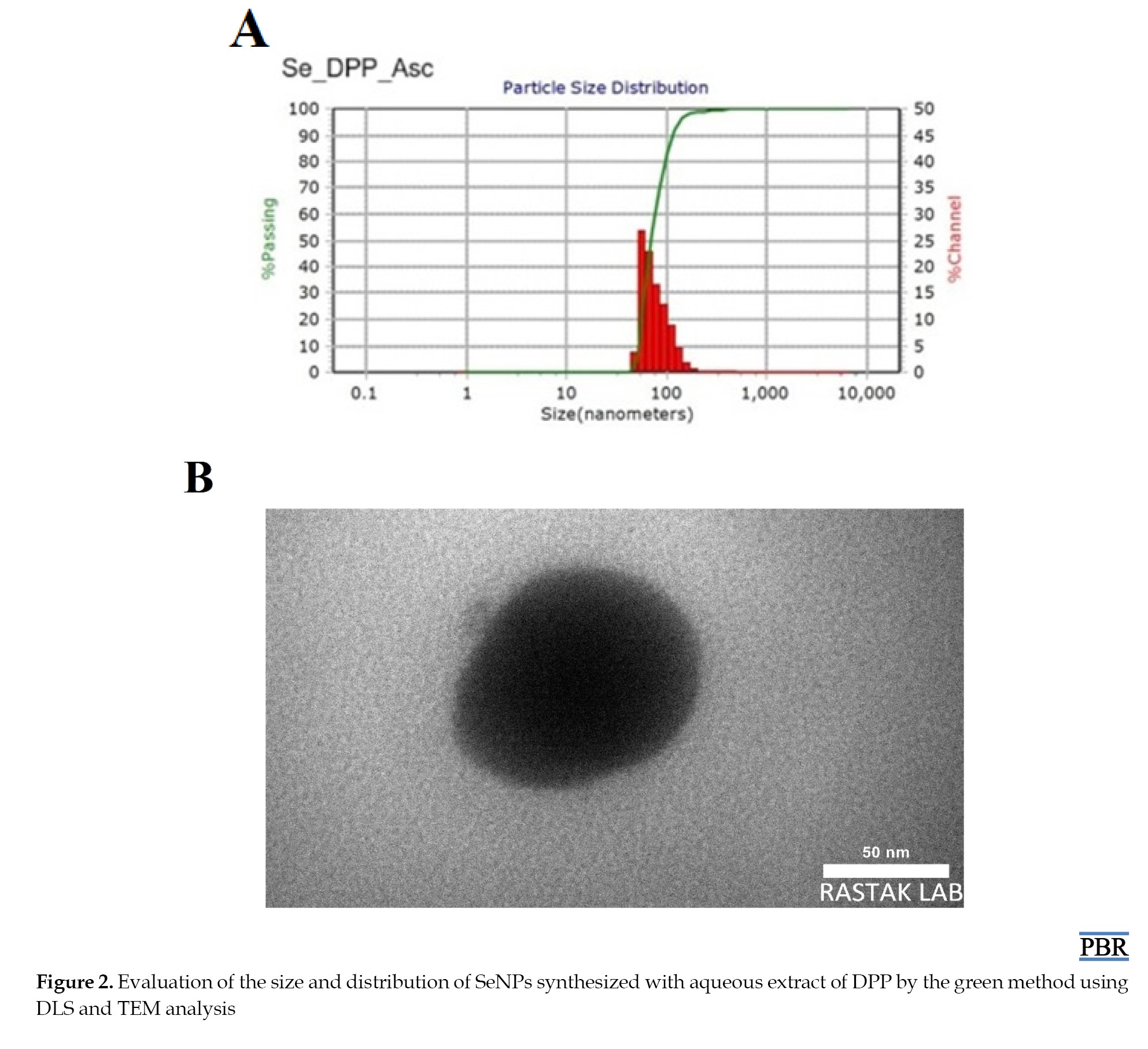 The results indicated that nanoparticles synthesized by DPP extract and ascorbic acid had a narrower and more symmetrical particle size distribution. The majority of the particles were in the range of 40 to 120 nm and the main peak was observed at about 70.02 nm. The zeta potential was measured to be -32.9 mV. The morphology and size of the synthesized SeNPs by DPP extract were characterized by TEM micrograph as shown in Figure 2B. The TEM micrograph confirms the particle size of green synthesized SeNPs ranging from 70 to 90 nm. SeNPs indicated predominantly spherical shape.
Serum FSH and LH levels
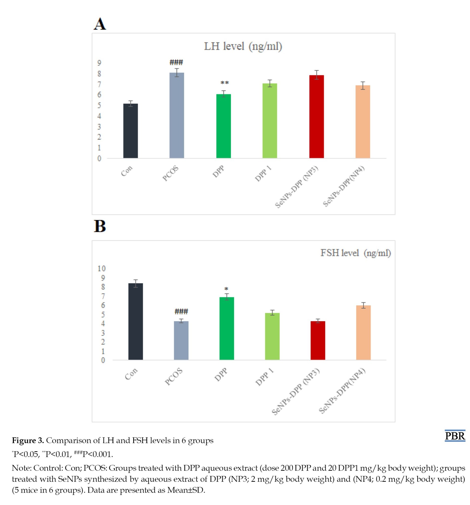
Data obtained from hormonal analysis performed by the Advia Centaur CP quantitative luminescence method showed that in PCOS mice induced by estradiol valerate, an increase in the LH/FSH ratio was observed compared to the control group (P<0.001). In the PCOS group, the FSH level was 4.3 compared to 8.4 in the control and the LH level was 8.1 compared to 5.2 in the control. In the group treated with DPP aqueous extract (dose 200 mg/kg), the LH hormone level (6.1) decreased significantly compared to the PCOS (8.1) (P<0.01) and the FSH hormone level (6.9) increased significantly compared to the PCOS (4.3) (P<0.05). However, no significant changes were observed in the levels of both hormones in the group treated with 20 mg/kg of DPP aqueous extract. Regarding the groups treated with SeNPs synthesized by the green method, in the group treated with 2 mg/kg of body weight of SeNPs, the LH hormone (7.9) decreased less compared to the PCOS (8.1), but the decrease was not significant. In the group treated with SeNPs (4.4), the level of FSH hormone did not show a significant increase compared to the PCOS (3.4). Under exposure with a dose of 0.2 mg/kg green synthesized SeNPs, the changes in LH and FSH hormones were not significant compared to the PCOS group, however, it had a greater effect on the levels of pituitary hormones FSH and LH (Figure 3).
Ovarian histopathological evaluation
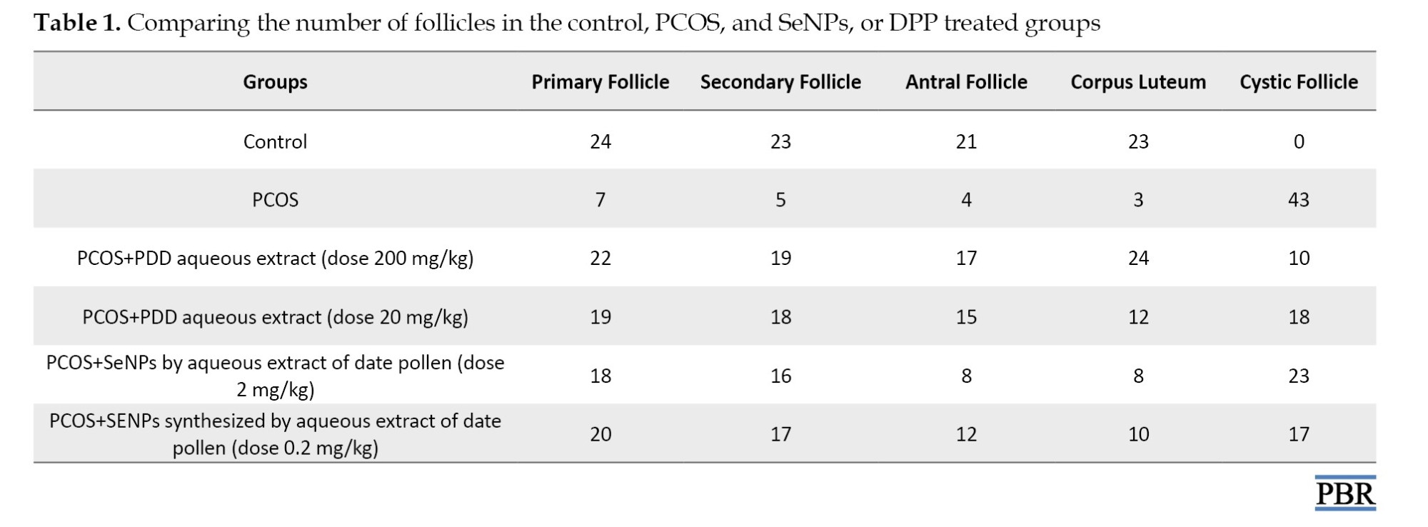
Histological analysis of the control group showed that the primary, secondary, and antral follicles were normal. Histological examination of the PCOS group induced by estradiol valerate showed that no primordial and primary follicles were observed in the germinal epithelium. Cystic follicles (CFs) were observed in different areas of the ovary with theca diameter of 44 µm and thin granulosa diameter of 32 µm. The cysts had a lining and were surrounded by dense fibrous tissue and were present in almost all areas. The unusual epithelial arrangement of the cyst was visible and, in the areas, marked with an asterisk, the lining of the cyst changed from cubic to cylindrical. In contrast, in the group treated with DPP aqueous extract (dose 200 mg/kg body weight), germinal epithelium is visible. In different areas of the ovary, growing follicles or secondary follicles and corpus luteum are visible. Antral or fully developed follicles are also observed and the cortical and central parts are completely distinguishable. In addition, treatment with a dose of 200 mg/kg body weight of DPP had better effects in ovarian tissue compared to its dose of 20 mg/kg. In the group treated with SeNPs synthesized by the green method (doses of 2 and 0.2 mg/kg body weight), primary, secondary, and antral follicles were visible and a number of atretic or atrophied follicles were also observed. In the comparison between groups, treatment with a dose of 0.2 mg/kg body weight of SeNPs synthesized by aqueous extract of DPP showed a significantly higher number of primary, secondary and antral follicles compared to the PCOS group (Figure 4).
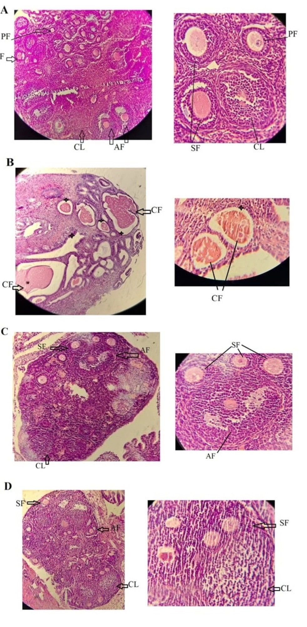
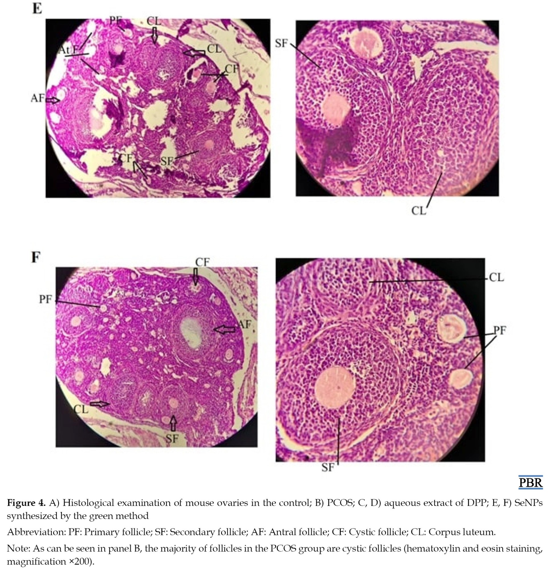 Table 1 lists the numerical data related with different types of follicles in the control, PCOS, and PCOS treated with DPP, and SeNPs.
Discussion
In this study, the effect of green-synthesized SeNPs using an aqueous extract of DPP was investigated in comparison with DPP aqueous extract in NMRI mice with polycystic ovary syndrome. Regarding the results obtained from DLS, zeta potential bioactivity, and TEM micrograph, the green synthesis of SeNPs were confirmed. It has been shown that DPP and ascorbic acid play an important role in the symmetry, particle size and size uniformity of SeNPs. DPP extract contains bioactive compounds, including flavonoids, polyphenols, and proteins that can cover the surface of nanoparticles with negatively charged functional groups such as –OH and –COOH. Ascorbic acid also has negative functional groups, such as -COO-, which further reduce the zeta potential by increasing the negative charge density on the nanoparticle surface. Despite the numerical decrease in zeta potential, this compound has led to increased colloidal stability of nanoparticles in aqueous media, since a greater distance from the isothermal point (close to zero) is usually associated with greater electrostatic repulsion and, consequently, higher stability.
The findings of this study regarding the synthesis of SeNPs showed that the aqueous extract of DPP is capable of synthesizing SeNPs via the presence of bioactive compounds as a reducing and stabilizing agent along with ascorbic acid. Regarding the use of DPP, previous studies have shown the potential of this extract in the synthesis of iron oxide, gold, and silver nanoparticles [30, 31]. In 2023, Wakid et al. investigated the efficacy of DPP extract loaded without the presence of ascorbic acid on SeNPs synthesis and showed that nanoparticles coated with DPP extract are potent agents against Toxoplasma gondii infections in a mouse model and can be suggested as a drug for the treatment of these infections in humans [23].
Various studies have shown that nano-based drug delivery system has occurred as a novel healing against PCOS. The synthetic NPs, including Ag-NPs, Cu-NPs/Fe3O4-NPs, superparamagnetic iron oxide NPs, and selenium-NPs are used for PCOS treatment and showed beneficial effects against PCOS [32]. Abhari et al. investigated the effects of curcumin-loaded super-paramagnetic iron oxide (Fe3O4) nanoparticles on histological parameters in an experimental mouse model of PCOS and found that curcumin-loaded Fe3O4 mitigated the ovarian volume and elevated a total number of primary, secondary, antral, and primordial follicles in comparison with the PCOS group [33]. Alwan et al, indicated that silver nanoparticles biofabricated from Cinnamomum zeylanicum improved PCOS signs by anti-inflammatory mechanism indicating via attenuation of inflammatory cytokines, such as tumor necrosis factor-α, Interleukin-6 and interleukin-18 [34]. The findings of the present study showed that DPP aqueous extract and SeNPs synthesized by green method at the tissue level can improve the symptoms of PCOS in a mouse model. However, hormonal analysis showed that aqueous extract of DPP at a dose of 200 mg/kg body weight had a better and significant effect on modulating the levels of FSH and LH hormones in mice with PCOS. In contrast, SeNPs at dose of 0.2 mg/kg revealed medium improvement and dose dependent toxicity which has been observed after exposure with 2 mg/kg indicating hormesis effect.
Previously, it demonstrated that selenium deficiency has been associated with an increased risk of PCOS, a condition that can significantly affect fertility [35]. Although caution is advised in the daily use of selenium in various organic, inorganic, and nanoparticle forms (intakes above 3 μg/kg body weight), low doses may have beneficial biological effects [36].
Zhao et al. (2023) reported that selenium supplementation improves follicle quality by increasing insulin sensitivity, reducing lipid peroxidation, and inflammation in PCOS patients. The results of a meta-analysis showed that selenium supplementation could only induce positive effects in patients with very poor follicle quality or who were forced to undergo in vitro fertilization (IVF) [35]. Zadeh Modarres et al. (2022) evaluated the effect of selenium supplementation on the metabolic profile in infertile women with PCOS. They exhibited that selenium supplementation for 8 weeks had beneficial effects on glycemic control and MDA levels in infertile women with PCOS undergoing IVF, but had no effect on pregnancy rate, lipid profile, TAC, and glutathione levels [37]. Mansouri Nejad et al. (2019) reported that selenium alone is not able to combat oxidative stress. Rather, the use of vitamin E, C, and selenium supplements synergistically improves the symptoms of PCOS in a rat model by reducing oxidative stress. They indicated that in the case of individual treatment, a better improvement was observed in the group receiving vitamin E compared to the group receiving vitamin C and selenium [38].
Regarding the reason for SeNPs administration in the present study, studies have elucidated that selenium in the form of nanoparticles or SeNPs is more available and less excreted in animals and has more and better activity than selenium. SeNPs are less toxic than organic and inorganic selenium-based compounds. In addition, SeNPs are biocompatible and are able to effectively deliver the compounds to specific cells [19]. Studies have proved that SeNPs are promising in improving metabolic and reproductive functions in PCOS models. SeNPs act as antioxidants and help neutralize harmful free radicals that can damage reproductive tissues and cells [39].
Studies have shown that higher levels of selenium, including SeNPs, may be associated with better egg quality and higher fertilization rates in IVF treatments [40]. SeNPs have been investigated as a potential therapeutic strategy for PCOS and other reproductive disorders due to their ability to regulate steroidogenesis and improve metabolic and reproductive functions. Studies have exhibited that SeNPs modulate steroidogenesis-related genes and improve ovarian function by regulating androgen receptor expression in a rat model of PCOS [17, 41].
Husseini et al. synthesized SeNPs using fenugreek and watercress extracts and showed that the synthesized nanoparticles could improve the function of antioxidant enzymes and symptoms of PCOS [42, 43]. In addition, Akintola et al. reported that SeNPs synthesized by green method from the leaves of Corchorus olitorius. L could modulate estrogen and progesterone levels and increase FSH and aromatase in rats with PCOS [48]. The findings of the present study on SeNPs synthesized by green method using aqueous extract of DPP showed that among the doses used (2 and 0.2 mg/kg body weight), the nanoparticles at a dose of 0.2 had a better effect on the number of primary, secondary and antral follicles. However, the best response was in the group treated with aqueous extract of DPP (dose 200 mg/kg).
Moshfegh et al. investigated the effect of DPP at a dose of 100 and 200 mg/kg body weight on Balb/C mice and showed that DPP, with its steroid compounds, can promote oogenesis and maintain fertility in female mice by increasing estrogen and progesterone levels [14]. The positive effects of DPP extract on fertility potential and the folliculogenesis process in females have been reported in several studies [44, 45]. Previous studies have shown that estrogenic compounds present in ethanolic extract of DPP improve the levels of glutathione and gonadotropin hormones and balance the levels of FSH and LH hormones in rats [46]. In addition, it has been reported that the protective effects of DPP may be related to flavonoids, glycosides, saponins, estradiol, and various types of vitamins such as vitamin A, vitamin E, and minerals, such as manganese, zinc, and selenium in DPP, which can result in improved reproductive performance [47, 48].
In 2019, 68 women in Khalkhal City were randomly assigned to one of two groups: Placebo group (n=35) and palm pollen group (n=35) and received one capsule of starch or palm pollen (300 mg/day) for 35 days, respectively. The female sexual function index (FSFI) was used to assess women’s sexual function. Results showed that DPP (300 mg supplement for 35 days) in premenopausal women can improve the FSFI libido domain [44]. Studies have shown that a daily dose of 6 g of DPP dry powder in two separate doses (3 g every 12 hours) orally administered to 30 subjects for 3 months showed that serum testosterone levels increased and serum LH levels decreased with increasing DPP. However, DPP had no significant effect on serum FSH levels. Karimi Jashani et al. (2016) reported that DPP extract can improve the symptoms of PCOS by reducing the number of CFs and increasing the number of corpus luteum, which probably indicate the resumption of the ovulation process [49]. Sena et al. evaluated the effect of DPP and its extract on PCOS-induced mice. Their results showed that treatment with DPP significantly improved the hormonal profile, liver and kidney function, and reduced oxidative stress compared to the PCOS group by modulating the levels of alanine transaminase, aspartate transaminase, alkaline phosphatase, bilirubin, albumin, and globulin [50].
Regarding the mechanism of action, a balanced level of free radicals is crucial for follicular development and oocyte maturation. Studies have shown that an imbalance between these two and an increase in free radicals leads to oxidative stress and damage to the reproductive system. Among the most important damages are oocyte immaturity due to abnormal extracellular matrix, increased free radicals in follicular fluid, increased cumulus ovum, and consequently damage to mtDNA, DNA strand breaks, induction of apoptosis, and anovulation [51].
In addition, polycystic ovary syndrome, as one of the causes of infertility, is associated with insulin resistance, which leads to lipid peroxidation, and together with hyperglycemia, leads to enhanced oxidative stress, and ultimately has an adverse effect on steroidogenesis and follicular growth. Therefore, the use of antioxidant supplements, including SeNPs, considering greater availability, lower excretion, and greater and better activity than selenium supplements, can play an important role in eliminating damage caused by the accumulation of free radicals, reducing hydrogen peroxide and lipid peroxides, and preventing insulin resistance resulting from PCOS induction by estradiol valerate [52]. On the other hand, DPP also contains many polyphenolic and flavonoid compounds, including gallic acid, caffeic acid, epicatechin, vanillic acid, coumarin, quercetin, and rutin, which have strong antioxidant activity. As a natural supplement carrying a variety of antioxidant and anti-inflammatory compounds, DPP plays an important role in preventing oxidative stress damage induced by estradiol valerate in the ovaries of PCOS model mice [53].
Conclusion
The data of the present study showed that the aqueous extract of DPP at a dose of 200 mg/kg compared to SeNPs synthesized by DPP (doses of 2 and 0.2 mg/kg) played a better role in improving the symptoms of polycystic ovary syndrome, including a decrease in serum LH levels and an increase in FSH in mice with PCOS. In addition, treatment with the aqueous extract of DPP compared to SeNPs enhanced folliculogenesis and increased ovulation by reducing the number of CFs and increasing the number of healthy follicles and corpus luteum. The main reason for the superior performance of DPP over SeNPs synthesized by this compound is the presence of bioactive compounds with antioxidant and anti-inflammatory effects in DPP. Therefore, the findings of the present study suggest the use of aqueous extract of DPP as a suitable herbal compound for the synthesis of SeNPs with antioxidant properties and improvement of symptoms of polycystic ovary syndrome.
Ethical Considerations
Compliance with ethical guidelines
This study was approved by Research Ethics Committee of Kharazmi University, Tehran, Iran (Code: IR.KHU.REC.1403.182).
Funding
This research did not receive any grant from funding agencies in the public, commercial, or non-profit sectors.
Authors' contributions
Investigation: Elaheh Amini, Mahdi Mirahmadi and Helaleh Kaboli Farshchi; Methodology, software, and validation: Farzaneh Baniasadi; Conceptualization, project administration, and writing the original draft: Elaheh Amini; Review and editing: Mahdi Mirahmadi and Helaleh Kaboli Farshchi.
Conflict of interest
The authors declared no conflict of interest.
Acknowledgments
The authors would like to thank all those who collaborated in this research, especially Engineer Mirahmadi at Pars Tek Rokh Pharmaceutical Company, Mashhad, Iran.
References
Polycystic ovarian syndrome (PCOS) is considered an endocrine disorder and the most common hormonal disorder in women, which is associated with symptoms, such as hyperandrogenism, polycystic ovarian morphology, chronic anovulation, and irregular gonadotropin secretion [1]. According to the literature, this disease affects 4–20% of women and is often associated with late diagnosis and metabolic dysfunction and problems such as obesity and insulin resistance [2]. Considering the symptoms of this disease, lifestyle modification, dietary adjustment, and pharmacological interventions are considered the most important strategies in the management of this disease [3]. Some of the most common medications and supplements used to manage PCOS include hormonal contraceptives, progestins, vitamin D, calcium, clomiphene citrate, and metformin [4, 5].
The most important herbs used to improve PCOS symptoms include cinnamon, aloe vera, flaxseed, licorice, evening primrose, and berberine [6-8]. Studies have shown that herbal compounds play an important role in improving fertility by increasing the number of ovarian follicles, reducing testosterone and androgen levels, mitigating blood lipid and glucose levels, and declining estrogen and hyperplasia [9].
Date palm pollen (DPP) from the date palm (Phoenix dactylifera L.) has been traditionally used to enhance sexual ability and fertility. In the past, it has been proven that DPP treats male infertility by improving the quality of sperm parameters [10]. To date, the presence of phenolic and flavonoid compounds with therapeutic effects, such as quercetin, gallic acid, rutin, catechin, ferulic acid, caffeic acid, apigenin, luteolin, and coumaric acid has been reported in DPP. In addition, DPP contains various bioactive compounds including fatty acids, amino acids, sterols, saponins, volatile unsaturated fatty acids, vitamins, and minerals [11].
Previous studies indicated that DPP can enhance fertility and sexual potency in both men and women through two main mechanisms, due to its antioxidant and gonadotropin-stimulating properties. Several animal studies have shown the effects of this herbal compound on testosterone, luteinizing hormone (LH), and follicle-stimulating hormone (FSH) [12-14].
A study has shown that reduced fertility in women is often associated with insufficient selenium levels in the body, and selenium supplementation is recommended in women with selenium deficiency to improve reproductive efficiency and increase pregnancy rates [15]. Studies have shown that selenium, helps protection of the developing oocyte from oxidative stress during folliculogenesis mainly via antioxidant function [16]. Previous studies have reported that selenium supplementation increased the gene expression levels of certain enzymes and improved lipid metabolism, and may have beneficial effects on patients with PCOS [17]. It has also been documented that co-administration of probiotics and selenium for 12 weeks in women with PCOS had beneficial effects on mental health parameters, total serum testosterone, hirsutism, total antioxidant capacity (TAC), glutathione GSH, and malondialdehyde (MDA) levels [18].
Studies have shown that selenium in the form of nanoparticles or SeNPs has higher bioavailability and lower excretion in animals, and has higher and better activity than selenium. In addition, SeNPs have lower toxicity than organic and inorganic selenium-based compounds, are biocompatible, and are capable of efficient delivery of the compounds to specific cells [19]. Given that oxidative stress can compromise egg quality and lead to infertility or poor outcomes in assisted reproductive technologies, SeNPs have been demonstrated to play a vital role in female reproduction via accelerating the maintenance of reproductive health, especially egg quality and protection against oxidative stress. They also play a role in regulating the menstrual cycle, ovulation, and potentially reducing the effects of diseases such as PCOS [20, 17]. The green synthesis is an environmentally friendly and relatively inexpensive method as compared to physical and chemical synthesis that does not require complex techniques and devices and involves the use of plant extracts or biocompounds to prepare nanoparticles as reducing and stabilizing agents [21]. Considering the advantages of green synthesis, including lower cost, less pollution, and improved safety for the environment and health, the present study aimed to investigate the effect of green-synthesized SeNPs on serum FSH and LH levels and ovarian tissue in mice model of PCOS.
Materials and Methods
Chemicals
Sodium selenite prepared from Sigma Company (code S5261), ascorbic acid (Sigma Company, code A92902), DPP (Liano Company, Bushehr), deionized water (Zalal Teb Chemi), estradiol valerate (Abo Raihan Pharmaceutical Company), hematoxylin from Sigma Company (code H3136), eosin Y from Sigma Company (code E6003), xylene (294780), ethanol (Razi Yeast Company), and toluene (code 244511) were used in this study.
Biosynthesis of SeNPs using aqueous extract of DPP
The synthesis of SeNPs was carried out based on Wakid et al. protocol. For this purpose, DPP was collected from Bushehr Province, Iran in March, transferred to the herbarium of Kharazmi University, identified, and confirmed ad herbarium code of 25556. Aqueous extract of DPP was used to prepare SeNPs. In this regard, the dried powder (10 g) was dissolved in deionized water (100 mL at room temperature), filtered after 30 min and evaporated at 55 to 60 °C [22]. At next step, 10 mmol/L ascorbic acid solution (10 mL), and DPP aqueous extract (10 mL) were added dropwise to selenium salt solution (10 mmol/L). After 24-hour incubation at 37 °C, serial centrifugation (2000 g, 10 min; NOGEN, Iran) performed using deionized water and the final precipitate are being collected as Se-NPs and dried to carry out biological assays [23] (Figure 1).
Characterization of SeNPs synthesized by green method
Dynamic light scattering (DLS), zeta potential, and transmission electron microscopy (TEM) analysis
To perform this method, the synthesized SeNPs sample was made into a suspension. To prepare a suspension of nanoparticles, SeNPs were dissolved in a suitable solvent (distilled water). To measure the particle size, a volume equivalent to 500 μL of SeNPs solution in 2 mL of deionized water was placed in a polystyrene cuvette and measured at 25 °C. The prepared sample was placed in the nanoparticle size analyzer (Nanotrac Wave II, Japan). Finally, the data collected by the device were analyzed using specific algorithms to calculate the particle size distribution. To evaluate the stability of the particles, the surface charge (zeta potential) was calculated in distilled water with pH 7 [24]. For TEM analysis, synthesized SeNPs was poured as a drop over carbon coated copper grids and maintain in room temperature to evaporate solvent. TEM analysis was conducted on a JEOL model 1200 EX instrument operated at an accelerating voltage at 80 kV.
Animal study
In this experimental study, 30 NMRI mice weighing between 22 and 24 g were used. The mice were obtained from the breeding and maintenance center of the Mashhad Bu Ali Research Institute and maintained under standard conditions (25 °C, 12 hours light/dark cycle). Animals’ intervention was carried out in accordance with the standards of the Kharazmi University Ethics Committee. The mice were divided into 6 groups of 5 mice. The control group received no treatment. PCOS group received estradiol valerate 4 mg/kg body weight in a volume of 100 μL intramuscularly dissolved in olive oil. The vaginal smears of mice were monitored for 60 days, until the time when abnormal estrus cycles and persistent vaginal cornification as the confirmation of PCOS induction [25]. DPP+PCOS aqueous extract groups received DPP at a dose of 200 and 20 mg/kg body weight for 14 days by gavage (after 8 weeks of estradiol valerate injection). SeNPs synthesized by the green method received SeNPs at a dose of 2 and 0.2 mg/kg body weight for 14 days by gavage (after 8 weeks of estradiol valerate injection). Blinding performed to reduce bias in research. For the overall study period, researchers were blind to the treatment allocation. The DPP dose was selected based on the research of Moshfegh and associates. In the case of SeNPs, the dose was selected first based on the research of Kandaparthi et al., who reported that SeNPs at a low dose of 1 to 2 mg/kg have less toxicity and higher antioxidant properties than high doses, and then the desired dose was experimentally investigated [14-26]. Based on previous studies, the LD50 for SeNPs was reported to be 14.6 mg/kg body weight and for DPP, about 5 g/kg body weight [27, 28].
Vaginal smear evaluation
This assay was performed to determine the regularity of the sexual cycle and to select mice in the estrus or estrous stage, which is characterized by the presence of epithelial cells and the absence of leukocytes. To do this, a sample was taken from the vagina of the mouse using a swab dipped in physiological serum and placed on a slide, and after staining with methylene blue, it was examined under a microscope. After the necessary examinations to ensure the regularity and problem-free reproductive cycle (at least 3 consecutive cycles) through smear test and observation under a microscope, after 12-14 days, only mice with a regular estrus cycle were randomly selected for this study.
FSH and LH analysis
One day after the last gavage, in order to collect blood from the hearts of control, PCOS, and PCOS mice treated with aqueous extract of DPP and SeNPs synthesized by DPP, they were first anesthetized by injection of ketamine (100 mg/kg)/xylazine (10 mg/kg) and then sacrificed by cervical dislocation. Blood was collected from the heart (1 mL per mouse) using an insulin syringe (Helma Teb). Then, the blood samples were placed at room temperature for about 30 to 40 min. Afterward, centrifugation was performed for 10 min at 800×g (NOGEN, Iran). The obtained serum was gently separated by 100 μL sampler and poured into a 1.5 mL microtube. Finally, the serum samples were transferred on ice to the Mashhad University Jahad Laboratory for analysis of LH and FSH hormone levels (Diazist, Iran). The samples were stored at -20 °C and the results of the hormone levels were compared between the groups [29].
Histology of ovarian tissue
To isolate ovarian tissue, after the treatment period, the mice were euthanized by cervical dislocation. The ovaries were removed and washed with physiological serum. After collection, the ovarian tissue was placed in 10% formalin fixative and transferred to the Pathobiology Department of Omid Hospital. To prepare the ovarian tissue, the tissue was first dehydrated using ascending grades of alcohol and placed in toluene for clarification. In the next step, the paraffin bath was used to cast the tissue and the tissue was embedded in paraffin blocks. Sectioning done using a 7-µ microtome (Hinotek, China), the sections were placed on coated slides. Hematoxylin and eosin staining was used for tissue staining.
Statistical analysis
Normality of data was assessed using the Kolmogorov-Smirnov test. After ensuring the normality of the data, parametric tests were used to analyze the data. SPSS software, version 22 was used to analyze the data. One-way analysis of variance was used to examine differences between groups and Tukey’s post hoc test was used to compare differences between groups. Experiments were repeated three times. Results were expressed as Mean±SD and P≤0.05 was considered significant. Excel software was used to draw graphs.
Results
DLS, zeta potential, andTEM analysis
The particle size distribution of SeNPs synthesized was evaluated using DLS (Figure 2A).
Serum FSH and LH levels
Data obtained from hormonal analysis performed by the Advia Centaur CP quantitative luminescence method showed that in PCOS mice induced by estradiol valerate, an increase in the LH/FSH ratio was observed compared to the control group (P<0.001). In the PCOS group, the FSH level was 4.3 compared to 8.4 in the control and the LH level was 8.1 compared to 5.2 in the control. In the group treated with DPP aqueous extract (dose 200 mg/kg), the LH hormone level (6.1) decreased significantly compared to the PCOS (8.1) (P<0.01) and the FSH hormone level (6.9) increased significantly compared to the PCOS (4.3) (P<0.05). However, no significant changes were observed in the levels of both hormones in the group treated with 20 mg/kg of DPP aqueous extract. Regarding the groups treated with SeNPs synthesized by the green method, in the group treated with 2 mg/kg of body weight of SeNPs, the LH hormone (7.9) decreased less compared to the PCOS (8.1), but the decrease was not significant. In the group treated with SeNPs (4.4), the level of FSH hormone did not show a significant increase compared to the PCOS (3.4). Under exposure with a dose of 0.2 mg/kg green synthesized SeNPs, the changes in LH and FSH hormones were not significant compared to the PCOS group, however, it had a greater effect on the levels of pituitary hormones FSH and LH (Figure 3).
Ovarian histopathological evaluation
Histological analysis of the control group showed that the primary, secondary, and antral follicles were normal. Histological examination of the PCOS group induced by estradiol valerate showed that no primordial and primary follicles were observed in the germinal epithelium. Cystic follicles (CFs) were observed in different areas of the ovary with theca diameter of 44 µm and thin granulosa diameter of 32 µm. The cysts had a lining and were surrounded by dense fibrous tissue and were present in almost all areas. The unusual epithelial arrangement of the cyst was visible and, in the areas, marked with an asterisk, the lining of the cyst changed from cubic to cylindrical. In contrast, in the group treated with DPP aqueous extract (dose 200 mg/kg body weight), germinal epithelium is visible. In different areas of the ovary, growing follicles or secondary follicles and corpus luteum are visible. Antral or fully developed follicles are also observed and the cortical and central parts are completely distinguishable. In addition, treatment with a dose of 200 mg/kg body weight of DPP had better effects in ovarian tissue compared to its dose of 20 mg/kg. In the group treated with SeNPs synthesized by the green method (doses of 2 and 0.2 mg/kg body weight), primary, secondary, and antral follicles were visible and a number of atretic or atrophied follicles were also observed. In the comparison between groups, treatment with a dose of 0.2 mg/kg body weight of SeNPs synthesized by aqueous extract of DPP showed a significantly higher number of primary, secondary and antral follicles compared to the PCOS group (Figure 4).
Discussion
In this study, the effect of green-synthesized SeNPs using an aqueous extract of DPP was investigated in comparison with DPP aqueous extract in NMRI mice with polycystic ovary syndrome. Regarding the results obtained from DLS, zeta potential bioactivity, and TEM micrograph, the green synthesis of SeNPs were confirmed. It has been shown that DPP and ascorbic acid play an important role in the symmetry, particle size and size uniformity of SeNPs. DPP extract contains bioactive compounds, including flavonoids, polyphenols, and proteins that can cover the surface of nanoparticles with negatively charged functional groups such as –OH and –COOH. Ascorbic acid also has negative functional groups, such as -COO-, which further reduce the zeta potential by increasing the negative charge density on the nanoparticle surface. Despite the numerical decrease in zeta potential, this compound has led to increased colloidal stability of nanoparticles in aqueous media, since a greater distance from the isothermal point (close to zero) is usually associated with greater electrostatic repulsion and, consequently, higher stability.
The findings of this study regarding the synthesis of SeNPs showed that the aqueous extract of DPP is capable of synthesizing SeNPs via the presence of bioactive compounds as a reducing and stabilizing agent along with ascorbic acid. Regarding the use of DPP, previous studies have shown the potential of this extract in the synthesis of iron oxide, gold, and silver nanoparticles [30, 31]. In 2023, Wakid et al. investigated the efficacy of DPP extract loaded without the presence of ascorbic acid on SeNPs synthesis and showed that nanoparticles coated with DPP extract are potent agents against Toxoplasma gondii infections in a mouse model and can be suggested as a drug for the treatment of these infections in humans [23].
Various studies have shown that nano-based drug delivery system has occurred as a novel healing against PCOS. The synthetic NPs, including Ag-NPs, Cu-NPs/Fe3O4-NPs, superparamagnetic iron oxide NPs, and selenium-NPs are used for PCOS treatment and showed beneficial effects against PCOS [32]. Abhari et al. investigated the effects of curcumin-loaded super-paramagnetic iron oxide (Fe3O4) nanoparticles on histological parameters in an experimental mouse model of PCOS and found that curcumin-loaded Fe3O4 mitigated the ovarian volume and elevated a total number of primary, secondary, antral, and primordial follicles in comparison with the PCOS group [33]. Alwan et al, indicated that silver nanoparticles biofabricated from Cinnamomum zeylanicum improved PCOS signs by anti-inflammatory mechanism indicating via attenuation of inflammatory cytokines, such as tumor necrosis factor-α, Interleukin-6 and interleukin-18 [34]. The findings of the present study showed that DPP aqueous extract and SeNPs synthesized by green method at the tissue level can improve the symptoms of PCOS in a mouse model. However, hormonal analysis showed that aqueous extract of DPP at a dose of 200 mg/kg body weight had a better and significant effect on modulating the levels of FSH and LH hormones in mice with PCOS. In contrast, SeNPs at dose of 0.2 mg/kg revealed medium improvement and dose dependent toxicity which has been observed after exposure with 2 mg/kg indicating hormesis effect.
Previously, it demonstrated that selenium deficiency has been associated with an increased risk of PCOS, a condition that can significantly affect fertility [35]. Although caution is advised in the daily use of selenium in various organic, inorganic, and nanoparticle forms (intakes above 3 μg/kg body weight), low doses may have beneficial biological effects [36].
Zhao et al. (2023) reported that selenium supplementation improves follicle quality by increasing insulin sensitivity, reducing lipid peroxidation, and inflammation in PCOS patients. The results of a meta-analysis showed that selenium supplementation could only induce positive effects in patients with very poor follicle quality or who were forced to undergo in vitro fertilization (IVF) [35]. Zadeh Modarres et al. (2022) evaluated the effect of selenium supplementation on the metabolic profile in infertile women with PCOS. They exhibited that selenium supplementation for 8 weeks had beneficial effects on glycemic control and MDA levels in infertile women with PCOS undergoing IVF, but had no effect on pregnancy rate, lipid profile, TAC, and glutathione levels [37]. Mansouri Nejad et al. (2019) reported that selenium alone is not able to combat oxidative stress. Rather, the use of vitamin E, C, and selenium supplements synergistically improves the symptoms of PCOS in a rat model by reducing oxidative stress. They indicated that in the case of individual treatment, a better improvement was observed in the group receiving vitamin E compared to the group receiving vitamin C and selenium [38].
Regarding the reason for SeNPs administration in the present study, studies have elucidated that selenium in the form of nanoparticles or SeNPs is more available and less excreted in animals and has more and better activity than selenium. SeNPs are less toxic than organic and inorganic selenium-based compounds. In addition, SeNPs are biocompatible and are able to effectively deliver the compounds to specific cells [19]. Studies have proved that SeNPs are promising in improving metabolic and reproductive functions in PCOS models. SeNPs act as antioxidants and help neutralize harmful free radicals that can damage reproductive tissues and cells [39].
Studies have shown that higher levels of selenium, including SeNPs, may be associated with better egg quality and higher fertilization rates in IVF treatments [40]. SeNPs have been investigated as a potential therapeutic strategy for PCOS and other reproductive disorders due to their ability to regulate steroidogenesis and improve metabolic and reproductive functions. Studies have exhibited that SeNPs modulate steroidogenesis-related genes and improve ovarian function by regulating androgen receptor expression in a rat model of PCOS [17, 41].
Husseini et al. synthesized SeNPs using fenugreek and watercress extracts and showed that the synthesized nanoparticles could improve the function of antioxidant enzymes and symptoms of PCOS [42, 43]. In addition, Akintola et al. reported that SeNPs synthesized by green method from the leaves of Corchorus olitorius. L could modulate estrogen and progesterone levels and increase FSH and aromatase in rats with PCOS [48]. The findings of the present study on SeNPs synthesized by green method using aqueous extract of DPP showed that among the doses used (2 and 0.2 mg/kg body weight), the nanoparticles at a dose of 0.2 had a better effect on the number of primary, secondary and antral follicles. However, the best response was in the group treated with aqueous extract of DPP (dose 200 mg/kg).
Moshfegh et al. investigated the effect of DPP at a dose of 100 and 200 mg/kg body weight on Balb/C mice and showed that DPP, with its steroid compounds, can promote oogenesis and maintain fertility in female mice by increasing estrogen and progesterone levels [14]. The positive effects of DPP extract on fertility potential and the folliculogenesis process in females have been reported in several studies [44, 45]. Previous studies have shown that estrogenic compounds present in ethanolic extract of DPP improve the levels of glutathione and gonadotropin hormones and balance the levels of FSH and LH hormones in rats [46]. In addition, it has been reported that the protective effects of DPP may be related to flavonoids, glycosides, saponins, estradiol, and various types of vitamins such as vitamin A, vitamin E, and minerals, such as manganese, zinc, and selenium in DPP, which can result in improved reproductive performance [47, 48].
In 2019, 68 women in Khalkhal City were randomly assigned to one of two groups: Placebo group (n=35) and palm pollen group (n=35) and received one capsule of starch or palm pollen (300 mg/day) for 35 days, respectively. The female sexual function index (FSFI) was used to assess women’s sexual function. Results showed that DPP (300 mg supplement for 35 days) in premenopausal women can improve the FSFI libido domain [44]. Studies have shown that a daily dose of 6 g of DPP dry powder in two separate doses (3 g every 12 hours) orally administered to 30 subjects for 3 months showed that serum testosterone levels increased and serum LH levels decreased with increasing DPP. However, DPP had no significant effect on serum FSH levels. Karimi Jashani et al. (2016) reported that DPP extract can improve the symptoms of PCOS by reducing the number of CFs and increasing the number of corpus luteum, which probably indicate the resumption of the ovulation process [49]. Sena et al. evaluated the effect of DPP and its extract on PCOS-induced mice. Their results showed that treatment with DPP significantly improved the hormonal profile, liver and kidney function, and reduced oxidative stress compared to the PCOS group by modulating the levels of alanine transaminase, aspartate transaminase, alkaline phosphatase, bilirubin, albumin, and globulin [50].
Regarding the mechanism of action, a balanced level of free radicals is crucial for follicular development and oocyte maturation. Studies have shown that an imbalance between these two and an increase in free radicals leads to oxidative stress and damage to the reproductive system. Among the most important damages are oocyte immaturity due to abnormal extracellular matrix, increased free radicals in follicular fluid, increased cumulus ovum, and consequently damage to mtDNA, DNA strand breaks, induction of apoptosis, and anovulation [51].
In addition, polycystic ovary syndrome, as one of the causes of infertility, is associated with insulin resistance, which leads to lipid peroxidation, and together with hyperglycemia, leads to enhanced oxidative stress, and ultimately has an adverse effect on steroidogenesis and follicular growth. Therefore, the use of antioxidant supplements, including SeNPs, considering greater availability, lower excretion, and greater and better activity than selenium supplements, can play an important role in eliminating damage caused by the accumulation of free radicals, reducing hydrogen peroxide and lipid peroxides, and preventing insulin resistance resulting from PCOS induction by estradiol valerate [52]. On the other hand, DPP also contains many polyphenolic and flavonoid compounds, including gallic acid, caffeic acid, epicatechin, vanillic acid, coumarin, quercetin, and rutin, which have strong antioxidant activity. As a natural supplement carrying a variety of antioxidant and anti-inflammatory compounds, DPP plays an important role in preventing oxidative stress damage induced by estradiol valerate in the ovaries of PCOS model mice [53].
Conclusion
The data of the present study showed that the aqueous extract of DPP at a dose of 200 mg/kg compared to SeNPs synthesized by DPP (doses of 2 and 0.2 mg/kg) played a better role in improving the symptoms of polycystic ovary syndrome, including a decrease in serum LH levels and an increase in FSH in mice with PCOS. In addition, treatment with the aqueous extract of DPP compared to SeNPs enhanced folliculogenesis and increased ovulation by reducing the number of CFs and increasing the number of healthy follicles and corpus luteum. The main reason for the superior performance of DPP over SeNPs synthesized by this compound is the presence of bioactive compounds with antioxidant and anti-inflammatory effects in DPP. Therefore, the findings of the present study suggest the use of aqueous extract of DPP as a suitable herbal compound for the synthesis of SeNPs with antioxidant properties and improvement of symptoms of polycystic ovary syndrome.
Ethical Considerations
Compliance with ethical guidelines
This study was approved by Research Ethics Committee of Kharazmi University, Tehran, Iran (Code: IR.KHU.REC.1403.182).
Funding
This research did not receive any grant from funding agencies in the public, commercial, or non-profit sectors.
Authors' contributions
Investigation: Elaheh Amini, Mahdi Mirahmadi and Helaleh Kaboli Farshchi; Methodology, software, and validation: Farzaneh Baniasadi; Conceptualization, project administration, and writing the original draft: Elaheh Amini; Review and editing: Mahdi Mirahmadi and Helaleh Kaboli Farshchi.
Conflict of interest
The authors declared no conflict of interest.
Acknowledgments
The authors would like to thank all those who collaborated in this research, especially Engineer Mirahmadi at Pars Tek Rokh Pharmaceutical Company, Mashhad, Iran.
References
- Yuan J, Li Z, Yu Y, Wang X, Zhao Y. Natural compounds in the management of polycystic ovary syndrome: A comprehensive review of hormonal regulation and therapeutic potential. Front Nutr. 2025; 12:1520695. [DOI:10.3389/fnut.2025.1520695] [PMID]
- Ghafari A, Maftoohi M, Eslami Samarin M, Barani S, Banimohammad M, Samie R. The last update on polycystic ovary syndrome(PCOS), diagnosis criteria, and novel treatment. Endocrine Metabol Sci. 2025; 17:100228. [DOI:10.1016/j.endmts.2025.100228]
- Singh S, Pal N, Shubham S, Sarma DK, Verma V, Marotta F, et al. Polycystic Ovary Syndrome: Etiology, Current Management, and Future Therapeutics. J Clin Med. 2023; 12(4):1454. [DOI:10.3390/jcm12041454] [PMID]
- Akre S, Sharma K, Chakole S, Wanjari MB. Recent advances in the management of polycystic ovary syndrome: A review article. Cureus. 2022; 14(8):e27689. [DOI:10.7759/cureus.27689]
- Williams T, Mortada R, Porter S. Diagnosis and treatment of polycystic ovary syndrome. Am Fam Physician. 2016; 94(2):106-13. [Link]
- Hemayatkhah-Jahromi V, Rahmanian-Koushkaki M. [Effect of hydro-alcoholic extract of Aloe vera L. on polycystic ovary syndrome in rat (Persian)]. Feyz. 2016; 20(3):221-7. [Link]
- Ionescu OM, Frincu F, Mehedintu A, Plotogea M, Cirstoiu M, Petca A, et al. Berberine-A promising therapeutic approach to polycystic ovary syndrome in infertile/pregnant women. Life. 2023; 13(1):125. [DOI:10.3390/life13010125] [PMID]
- Maleki V, Faghfouri AH, Tabrizi FPF, Moludi J, Saleh-Ghadimi S, Jafari-Vayghan H, et al. Mechanistic and therapeutic insight into the effects of cinnamon in polycystic ovary syndrome: A systematic review. J Ovarian Res. 2021; 14(1):130. [DOI:10.1186/s13048-021-00870-5] [PMID]
- Manouchehri A, Abbaszadeh S, Ahmadi M, Nejad FK, Bahmani M, Dastyar N. Polycystic ovaries and herbal remedies: A systematic review. JBRA Assist Reprod. 2023; 27(1):85-91. [DOI:10.5935/1518-0557.20220024]
- Chhoud R, El-Ghoul Y, Alminderej FM, Majdoub H. Phytochemical and technological insights into the fertility-enhancing potential of date palm pollen (Phoenix dactylifera L.). Appl Food Res. 2025; 5(2):101410. [DOI:10.1016/j.afres.2025.101410.]
- Salhi S, Chentouf M, Harrak H, Rahim A, Çakir C, Çam D, et al. Assessment of physicochemical parameters, bioactive compounds, biological activities, and nutritional value of the most two commercialized pollen types of date palm (Phoenix dactylifera L.) in Morocco. Food Sci Technol Int. 2024; 30(8):788-98. [DOI:10.1177/10820132231168914] [PMID]
- Abdi F, Roozbeh N, Mortazavian AM. Effects of date palm pollen on fertility: Research proposal for a systematic review. BMC Res Notes. 2017; 10(1):363. [DOI:10.1186/s13104-017-2697-3] [PMID]
- Falahati AM, Fallahi S, Allamehzadeh Z, Izadi Raieni M, Malekzadeh K. Effects of date palm pollen supplementations on the expression of PRDX1 and PRDX6 genes in infertile men: A controlled clinical trial. Int J Fertil Steril. 2023; 17(3):201-207. [DOI:10.22074/ijfs.2022.549291.1264] [PMID]
- Moshfegh F, Baharara J, Namvar F, Zafar-Balanezhad S, Amini E, Jafarzadeh L. Effects of date palm pollen on fertility and development of reproductive system in female Balb/C mice. J HerbMed Pharmacol. 2016; 5(1):23-8. [Link]
- Lima LG, Santos AAMD, Gueiber TD, Gomes RZ, Martins CM, Chaikoski AC. Relation between selenium and female fertility: A systematic review. Rev Bras Ginecol Obstet. 2022; 44(7):701-709. [DOI:10.1055/s-0042-1744288] [PMID]
- Safiyeh FD, Mojgan M, Parviz S, Sakineh MA, Behnaz SO. The effect of selenium and vitamin E supplementation on anti-Mullerian hormone and antral follicle count in infertile women with occult premature ovarian insufficiency: A randomized controlled clinical trial. Complement Ther Med. 2021; 56:102533. [DOI:10.1016/j.ctim.2020.102533] [PMID]
- Abdallah ABE, El-Ghannam MA, Hasan AA, Mohammad LG, Mesalam NM, Alsayed RM. Selenium nanoparticles modulate steroidogenesis-related genes and improve ovarian functions via regulating androgen receptors expression in polycystic ovary syndrome rat model. Biol Trace Elem Res. 2023; 201(12):5721-33. [DOI:10.1007/s12011-023-03616-0] [PMID]
- Jamilian M, Mansury S, Bahmani F, Heidar Z, Amirani E, Asemi Z. The effects of probiotic and selenium co-supplementation on parameters of mental health, hormonal profiles, and biomarkers of inflammation and oxidative stress in women with polycystic ovary syndrome. J Ovarian Res. 2018; 11(1):80. [DOI:10.1186/s13048-018-0457-1] [PMID]
- Khalil WA, El-Harairy MA, Zeidan AEB, Hassan MAE. Impact of selenium nano-particles in semen extender on bull sperm quality after cryopreservation. Theriogenology. 2019; 126:121-7. [DOI:10.1016/j.theriogenology.2018.12.017] [PMID]
- El-Dahhar AA, Khalifa GA, El-Zaeem SY, Abdel-Rahim MM, Mourad MM. Impact of organic and nano selenium on sexual, maturation, egg, and offspring quality of European sea bass (Dicentrarchus labrax) broodstock. Sci Rep. 2026; 16(1):1342. [DOI:10.1038/s41598-025-29108-w] [PMID]
- Mikhailova EO. Selenium Nanoparticles: Green Synthesis and Biomedical Application. Molecules. 2023; 28(24):8125. [DOI:10.3390/molecules28248125] [PMID]
- Lak Mazaheri E, Niknejad A, Amini E, Nabiuni M. Phoenix dactylifera L. pollen and fluvoxamine maleate protect PC12 cells against H2O2-induced oxidative stress by involvement of Nrf2 and SIGMAR1 gene expression. Jundishapur J Nat Pharm Prod. 2023; 19(1):e141738. [DOI:10.5812/jjnpp-141738]
- Wakid MH, Alsulami MN, Farid M, El Kholy WA. Potential anti-toxoplasmosis efficiency of phoenix dactylifera extracts loaded on selenium nanoparticles. Infect Drug Resist. 2023; 16:7743-58. [DOI:10.2147/IDR.S443047] [PMID]
- Vahdati M, Tohidi Moghadam T. Synthesis and characterization of selenium nanoparticles-lysozyme nanohybrid system with synergistic antibacterial properties. Sci Rep. 2020; 10(1):510. [DOI:10.1038/s41598-019-57333-7] [PMID]
- Gao J, Huang Y, Song Y, Huang X, Zhang D, Hou Z. Therapeutic effects of althaea officinalis L. and metformin on estradiol-induced polycystic ovary syndrome in rats: Insights into The PI3K/AKT pathway, inflammation, and oxidative stress. Cell J. 2024; 26(8):473-86. [DOI:10.22074/cellj.2024.2026329.1550]
- Kondaparthi P, Deore M, Naqvi S, Flora SJS. Dose-dependent hepatic toxicity and oxidative stress on exposure to nano and bulk selenium in mice. Environ Sci Pollut Res Int. 2021; 28(38):53034-44. [DOI:10.1007/s11356-021-14400-9] [PMID]
- Bano I, Skalickova S, Arbab S, Urbankova L, Horky P. Toxicological effects of nanoselenium in animals. J Anim Sci Biotechnol. 2022; 13(1):72. [DOI:10.1186/s40104-022-00722-2] [PMID]
- Elblehi SS, El-Sayed YS, Soliman MM, Shukry M. Date palm pollen extract avert doxorubicin-induced cardiomyopathy fibrosis and associated oxidative/nitrosative stress, inflammatory cascade, and apoptosis-targeting Bax/Bcl-2 and caspase-3 signaling pathways. Animals. 2021; 11(3):886. [DOI:10.3390/ani11030886] [PMID]
- Siavashi M, Ahmadi R. [The effect of oil paint vapor inhalation on FSH, LH, DHEAS, and testosterone serum levels in male rats (Persian)]. Feyz Med Sci J. 2015; 19(4):271-7. [Link]
- Nejadali Chaleshtari S, Amini E, Baniasadi F, Tavana S, Ghalamboran M. Oocyte maturation, fertilization, and embryo development in vitro by green and chemical iron oxide nanoparticles: A comparative study. Sci Rep. 2024; 14(1):14157. [DOI:10.1038/s41598-024-65121-1] [PMID]
- Turunc E, Kahraman O, Binzet R. Green synthesis of silver nanoparticles using pollen extract: Characterization, assessment of their electrochemical and antioxidant activities. Anal Biochem. 2021; 621:114123. [DOI:10.1016/j.ab.2021.114123.] [PMID]
- Mesgin RM, Nejati V, Talatapeh SP, Imani Z, Rezaie J. Nanoparticles for polycystic ovary syndrome (PCOS) therapy: Exosomes and synthetic nanoparticles, challenges and opportunities. Cell Biochem Funct. 2025; 43(9):e70114. [DOI:10.1002/cbf.70114] [PMID]
- Fatemi Abhari SM, Khanbabaei R, Hayati Roodbari N, Parivar K, Yaghmaei P. Curcumin-loaded super-paramagnetic iron oxide nanoparticle affects on apoptotic factors expression and histological changes in a prepubertal mouse model of polycystic ovary syndrome-induced by dehydroepiandrosterone-A molecular and stereological study. Life Sci. 2020; 249:117515. [DOI:10.1016/j.lfs.2020.117515] [PMID]
- Alwan SH, Al-Saeed MH. Silver nanoparticles biofabricated from cinnamomum zeylanicum reduce IL-6, IL-18, and tnf-ɑ in female rats with polycystic ovarian syndrome. Int J Fertil Steril. 2023; 17(1):80-84. [DOI:10.22074/ijfs.2022.539396.1189] [PMID]
- Zhao J, Dong L, Lin Z, Sui X, Wang Y, Li L, et al. Effects of selenium supplementation on polycystic ovarian syndrome: A systematic review and meta-analysis on randomized clinical trials. BMC Endocr Disord. 2023; 23(1):33. [DOI:10.1186/s12902-023-01286-6] [PMID]
- Hadrup N, Ravn-Haren G. Toxicity of repeated oral intake of organic selenium, inorganic selenium, and selenium nanoparticles: A review. J Trace Elem Med Biol. 2023; 79:127235. [DOI:10.1016/j.jtemb.2023.127235] [PMID]
- Zadeh Modarres S, Asemi Z, Heidar Z. The effects of selenium supplementation on glycemic control, serum lipoproteins and biomarkers of oxidative stress in infertile women diagnosed with polycystic ovary syndrome undergoing in vitro fertilization: A randomized, double-blind, placebo-controlled trial. Clin Nutr ESPEN. 2022; 51:92-6. [DOI:10.1016/j.clnesp.2022.07.017] [PMID]
- Mansouri Nejad F, Naghipour Hamzekolaei M, Jafarisani M, Sadeghi I. [Supplementary containing Vit E, C and Se reduced the PCOs induced oxidative stress in rat model (Persian)]. J Knowl Health Basic Med Sci. 2019; 14(2):47-52. [DOI:10.22100/jkh.v14i2.2260]
- Sentkowska A, Pyrzyńska K. Antioxidant properties of selenium nanoparticles synthesized using tea and herb water extracts. Appl Sci. 2023; 13(2):1071. [DOI:10.3390/app13021071]
- Asri-Rezaei S, Nourian A, Shalizar-Jalali A, Najafi G, Nazarizadeh A, Koohestani M, et al. Selenium supplementation in the form of selenium nanoparticles and selenite sodium improves mature male mice reproductive performances. Iran J Basic Med Sci. 2018; 21(6):577-85. [DOI:10.22038/IJBMS.2018.26023.6397] [PMID]
- Butt MA, Shafique HM, Mustafa M, Moghul NB, Munir A, Shamas U, et al. Therapeutic potential of selenium nanoparticles on letrozole-induced polycystic ovarian syndrome in female wistar rats. Biol Trace Elem Res. 2023; 201(11):5213-29. [DOI:10.1007/s12011-023-03579-2] [PMID]
- Husseini H, Zainulabdeen JA. The effect of selenium nanoparticles with fenugreek extract on oxidative stress related to polycystic ovary syndrome. J Med Pharm Chem Res. 2023; 5(4):371-81. [DOI:10.22034/ecc.2023.369594.1551]
- Husseini HH, Zainulabdeen JA. Effect of selenium nanoparticles with cress extract on polycystic ovary syndrome (PCOS). J Nanostruct. 2023; 13(4):1080-90. [DOI:10.22052/JNS.2023.04.016]
- Salmani R, Nasiri K, Javadzadeh Y, Salmani R, Clark CCT, Aghamohammadi V. Effect of date palm pollen supplementation on female sexual function in non-menopausal women: A double blind randomized clinical trial. Chin Herb Med. 2022; 14(4):643-8. [DOI:10.1016/j.chmed.2022.02.004] [PMID]
- Jamali S, Javadnoori M, Rahmanian V, Zahedian M, Mohammadi S. The effects of date palm on women's sexual function: A systemic review and meta-analysis of randomized controlled trials. Adv Integra Med. 2025; 12(3):100498. [DOI:10.1016/j.aimed.2025.100498]
- Jiheel M, Arrak J. Effect of different doses of ethanolic extract of date palm pollen grains on serum gonadotropin and total Glutathione in mature female rats. Kufa J Vet Med Sci. 2015; 6(2):109-16. [DOI:10.36326/kjvs/2015/v6i23992]
- Pujari RR, Vyawahare NS, Kagathara VG. Evaluation of antioxidant and neuroprotective effect of date palm (Phoenix dactylifera L.) against bilateral common carotid artery occlusion in rats. Indian J Exp Biol. 2011; 49(8):627-33. [Link]
- Hassanpoor M, Nabiuni M, Amini E, Salehghamari E. Assessment of date palm (Phoenix dactylifera L) pollen therapeutic potential on citalopram-induced testicular damage: In vitro and in vivo toxicity. Andrologia. 2026; 2026(1):2302087. [DOI:10.1155/and/2302087]
- Karimi Jashni H, Kargar Jahromi H, Bagheri Z. The effect of palm pollen extract on polycystic ovary syndrome (POS) in rats. Int JMed Res Health Sci. 2016; 5, 5(S):317-21. [Link]
- Sana M, Arshad M, Siddique F, Muhammad Irfan H, Hussain Asim M, Khalid W, et al. Therapeutic potential of Phoenix dactylifera pulp and seed extracts in mitigating oxidative stress and organ dysfunction in testosterone-induced polycystic ovary syndrome. J Func Food. 2025; 127:106743. [DOI:10.1016/j.jff.2025.106743]
- Akintola TE, Akintunde JK, Eteng OE, Thomas FC, Adeleke JT. Selenium nanoparticles from Corchorus olitorius corrects polycystic ovarian syndrome by inhibition of hormonal imbalance, aromatase and cellular inflammation via hypothalamic-ovarian axis in female rat. Endocr Metab Sci. 2024; 16:100202. [DOI:10.1016/j.endmts.2024.100202]
- Immediata V, Ronchetti C, Spadaro D, Cirillo F, Levi-Setti PE. Oxidative stress and human ovarian response-from somatic ovarian cells to oocytes damage: A clinical comprehensive narrative review. Antioxidants. 2022; 11(7):1335. [DOI:10.3390/antiox11071335] [PMID]
- Al-Habsi N. Date palm (Phoenix dactylifera L.) fruit: Strategic crop for food security, nutritional benefits, postharvest quality, and valorization into emerging functional products. Sustainability. 2025; 17(16):7491. [DOI:10.3390/su17167491]
Type of Study: Original Research |
Subject:
Cellular and Molecular Biology
| Rights and permissions | |
 |
This work is licensed under a Creative Commons Attribution-NonCommercial 4.0 International License. |


This is an open access article distributed under the terms of the Creative Commons Attribution License (CC-By-NC), which permits use, distribution, and reproduction in any medium, provided the original work is properly cited and is not used for commercial purposes.
Contact Information