

Volume 12, Issue 2 (2026)
Pharm Biomed Res 2026, 12(2): 87-98 |
Back to browse issues page


Download citation:
BibTeX | RIS | EndNote | Medlars | ProCite | Reference Manager | RefWorks
Send citation to:


BibTeX | RIS | EndNote | Medlars | ProCite | Reference Manager | RefWorks
Send citation to:
SarveAhrabi Y, Aqa Abedi S, Ahmadirad M, Zarrabi Ahrabi N. Anticancer 1,3-thiazole Derivatives: In Vitro Evaluation and in Silico Tubulin/Lipoxygenase Inhibition. Pharm Biomed Res 2026; 12 (2) :87-98
URL: http://pbr.mazums.ac.ir/article-1-679-en.html
URL: http://pbr.mazums.ac.ir/article-1-679-en.html


1- Department of Biology, CT.C., Islamic Azad University, Tehran, Iran.
Full-Text [PDF 2529 kb]
(83 Downloads)
| Abstract (HTML) (238 Views)
Full-Text: (42 Views)
Introduction
Drug resistance in cancer is a major barrier to effective treatment, often resulting in treatment failure and disease recurrence [1]. The incidence of treatment resistance varies significantly across cancer types and is influenced by multiple factors, including genetic mutations, the tumor microenvironment, and epigenetic modifications. Research indicates that approximately 90% of patients with advanced cancer may encounter some form of treatment resistance during the course of their illness [2]. Drug resistance can be categorized as intrinsic or acquired. Intrinsic resistance refers to the inherent ability of cancer cells to withstand a drug’s effects from the outset, often due to pre-existing genetic alterations or the expression of specific efflux pumps that actively remove the drug from the cell [3]. In contrast, acquired resistance develops over time as cancer cells adapt to therapeutic pressures. This adaptation can occur through various mechanisms, such as gene amplification (duplicating the target gene, leading to overexpression of the target protein), mutations in drug targets that alter binding sites, or the activation of alternative signaling pathways that bypass the inhibited pathway [4].
Moreover, the tumor microenvironment plays a crucial role in drug resistance. Factors, such as hypoxia, acidity, and the presence of stromal cells, can influence treatment efficacy and contribute to the development of a resistant phenotype [5]. Epigenetic changes, including DNA methylation and histone modifications, can also alter gene expression patterns, promoting resistance. Understanding the complex interplay among these factors is essential for developing novel therapeutic strategies to overcome drug resistance and improve patient outcomes in cancer therapy. Advanced approaches, such as combination therapies and personalized medicine, are being explored to target the specific mechanisms of resistance in individual tumors, thereby enhancing the effectiveness of cancer therapy [6]. Lung cancer, particularly non-small cell lung cancer (NSCLC), exemplifies the significant challenges associated with treatment resistance. Mutations in the epidermal growth factor receptor (EGFR) are prevalent in NSCLC, and although targeted therapies, such as tyrosine kinase inhibitors, have improved patient outcomes, resistance often develops [7]. The mechanisms underlying this resistance include secondary mutations in the EGFR gene, such as the T790M mutation; histological transformation to small cell lung cancer; and activation of alternative signaling pathways, including MET and HER2 amplification. Additionally, the tumor microenvironment significantly influences resistance, with factors, such as hypoxia and the presence of cancer-associated fibroblasts, playing critical roles. As a result, research is focusing on combinatorial treatment strategies and the development of novel therapeutic agents to overcome these resistance mechanisms [8]. Colorectal cancer (CRC) presents a distinct array of drug resistance challenges. Resistance to chemotherapeutic agents, such as fluorouracil, oxaliplatin, and irinotecan, is common, often arising from changes in drug metabolism, cellular efflux mechanisms, and the activation of anti-apoptotic pathways [9]. Furthermore, mutations in critical oncogenes and tumor suppressor genes, including KRAS and p53, are associated with treatment failure. Approximately 50% of patients with CRC harbor mutations in the KRAS oncogene, rendering them insensitive to anti-EGFR therapies. The capacity of tumor cells to enter a dormant state further complicates CRC treatment, as these dormant cells can evade standard therapies and contribute to disease recurrence. To address these resistance mechanisms and enhance treatment efficacy, new therapeutic strategies, including targeted agents and immunotherapy, are currently being investigated [10]. Among promising novel therapeutic agents for cancer treatment, 1,3-thiazole compounds have attracted significant interest due to their diverse biological activities. It is well established that both 1,3-thiazole and 1,3,4-thiadiazole derivatives exhibit promising anticancer activities, particularly when combined with heterocycles, such as 1,3,4-oxadiazole [11].
The mechanisms underlying their action often involve the modulation of various signaling pathways, particularly those associated with pro-apoptotic and anti-apoptotic proteins. Research has shown that thiazole derivatives exhibit activity against multiple cancer types, including breast, lung, and CRC [12]. Investigating the structure-activity relationships (SAR) of these compounds is essential for optimizing the efficacy and selectivity. In addition to their direct anticancer properties, thiazole derivatives may also serve as scaffolds for the development of multitarget drugs, potentially addressing resistance mechanisms in combination therapies [13]. Tubulin, a fundamental protein in the cytoskeleton, is essential for cell division and intracellular transport. The dynamic equilibrium of tubulin polymerization into microtubules is crucial for mitosis, making it a prime target for cancer therapies. Inhibiting tubulin polymerization disrupts mitotic spindle formation, resulting in cell cycle arrest and subsequent apoptosis in rapidly dividing cancer cells. Various classes of drugs, such as taxanes (e.g. paclitaxel) and vinca alkaloids (e.g. vincristine), directly target tubulin and have demonstrated efficacy across a range of malignancies. However, the development of resistance to these tubulin-targeting agents poses a significant challenge, often arising from the expression of drug efflux transporters or mutations in tubulin isotypes. A comprehensive understanding of tubulin dynamics, along with the development of novel tubulin inhibitors or combination therapies, holds great promise for enhancing the effectiveness of cancer treatments [14]. On the other hand, lipoxygenases (LOXs) are enzymes that catalyze the oxidation of polyunsaturated fatty acids, leading to the production of leukotrienes that play a role in inflammatory processes. Elevated expression of LOXs has been linked to various cancers, including breast, prostate, and CRC, where they contribute to tumor growth, metastasis, and the establishment of an inflammatory tumor microenvironment. Inhibiting LOXs has emerged as a potential therapeutic strategy due to their involvement in cancer progression. LOX inhibitors suppress cancer cell proliferation, induce apoptosis, and inhibit angiogenesis, thereby offering a multifaceted approach to combat cancer. Furthermore, ongoing research on selective LOX isoform inhibitors aims to reduce side effects while maximizing targeted therapeutic efficacy. As our understanding of LOX pathways in tumor biology expands, these inhibitors may serve as promising adjunctive therapies in comprehensive cancer treatment regimens [15].
Objectives
This study aimed to investigate the in vitro anticancer effects of 1,3-thiazole derivatives against A549 and HT-29 cancer cell lines and to evaluate the compounds in silico as inhibitors of tubulin and lipoxygenase enzymes.
Materials and Methods
This experimental research was conducted in the Microbiology Laboratory of Islamic Azad University, Tehran Branch, in 2023-2024. Starting materials, solvents, and culture media (RPMI 1640) were purchased from Merck (Germany). The cell lines were obtained from the Pasteur Institute of Iran (Cell Line Collections).
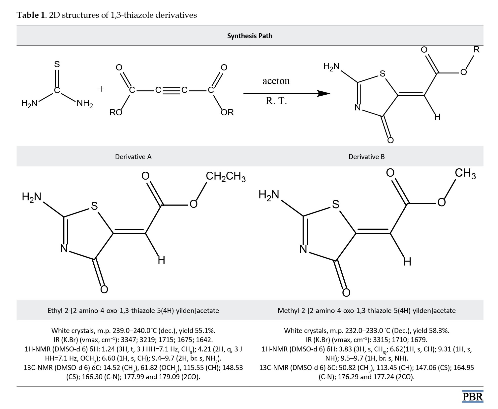
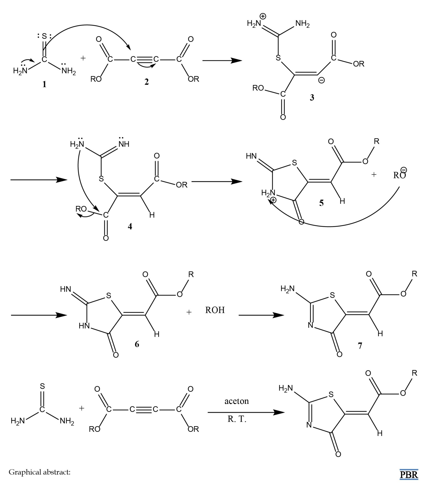
In vitro: All derivatives of 1,3-thiazole from our previous research were resynthesized, as shown in Table 1 [16].
The concentrations of 250, 500, and 1000 µg/mL were selected based on preliminary solubility and cytotoxicity screening of the compounds in dimethyl sulfoxide (DMSO), ensuring complete dissolution while spanning a wide therapeutic window. These doses align with standard ranges used in MTT assays for heterocyclic compounds [17] and allowed direct comparison with the positive control.
The new compounds were solubilized using DMSO at concentrations of 250, 500, and 1000 µg/mL to assess their anticancer properties. Additionally, Doxorubicin, a pure antibiotic powder obtained from Sigma, was prepared and employed as a control at the same concentrations.
Doxorubicin was chosen as the positive control due to its well-established clinical efficacy against both lung and CRC, its broad-spectrum cytotoxic mechanism (DNA intercalation and topoisomerase II inhibition), and its frequent use as a benchmark in 3-(4,5-dimethylthiazol-2-yl)-2,5-diphenyltetrazolium bromide (MTT)-based anticancer screening studies.
A549 and HT-29 cancer cell lines were cultured in RPMI 1640 medium supplemented with 10% fetal bovine serum and antibiotics, specifically penicillin and streptomycin, both at 100 µg/mL.
A549 is a human lung adenocarcinoma cell line (non-small cell lung cancer), while HT-29 is a human colorectal adenocarcinoma cell line, representing two of the most lethal malignancies worldwide.
This culture was maintained in an incubator at 37 ºC with 5% carbon dioxide (CO2) and saturated humidity in 25 cm flasks. The culture medium was refreshed every one to two days, and when the cells achieved 80% confluence, they were subcultured. Cell viability was assessed using the MTT assay. This colorimetric technique relies on the reduction of yellow tetrazolium crystals by the succinate dehydrogenase enzyme, yielding blue insoluble formazan crystals. During incubation, MTT is reduced by succinate dehydrogenase, an enzyme integral to mitochondrial respiration. The reduction process leads to the formation of recognizable blue formazan crystals under a microscope, with the color intensity directly correlating with the number of viable cells. To evaluate the effects of the tested compounds on cell growth and survival, 5×10⁴ cells were plated in 96-well plates. These cells were then exposed to different concentrations of the compounds for periods of 24, 48, and 72 hours. The yellow MTT powder was dissolved in 1 mL of PBS buffer shielded from light and subsequently filtered through a 0.2-micron filter for sterilization. After the incubation duration, the medium was aspirated, and MTT solution was introduced to each well. The plates were incubated for 4 hours at 37 ºC to facilitate the formation of formazan crystals. DMSO was added post-incubation to dissolve the insoluble formazan crystals. The optical density of each well’s solution was assessed using an ELISA reader at 570 nm. The optical density reflects the amount of MTT converted to formazan under these conditions. Cell viability was calculated using the Equation 1:
1. Percentage survival=(mean absorbance of treated cells/mean absorbance of the control cells)×100.
In silico: The 2D representations of the derivatives were produced with ChemDraw software, version 12.0.2.1076. Subsequently, the structures were normalized via the MM2 method in Chem3D Pro version 12.0.2.1076. Crystallographic structures of tubulin (PDB ID: 3HKB) [18] and lipoxygenase (PDB ID: 2IUJ) [19] were sourced from the Protein Data Bank, which provided resolutions of 3.65 Å for 3HKB and 2.40 Å for 2IUJ. Gasteiger charges and polar hydrogens were incorporated using AutoDockTools (version 1.5.6). All water molecules and ligands were eliminated from the protein structures. The grid dimensions were set to 48×48×48 for the 3HKB structure and 56×56×56 for the 2IUJ structure, with 3 Å spacing. The docking of the derivatives into the binding sites of 3HKB and 2IUJ was performed using AutoDock Vina and Discovery Studio 4.5. The interactions between the ligands and the binding sites were evaluated using Discovery Studio Client software, version 4.5 [20].
Results
In vitro
A549 cancer cell line: As shown in Figure 1, compound A demonstrated significant efficacy in reducing cancer cells, achieving a 50% reduction over 72 hours at a concentration of 500 µg/mL.
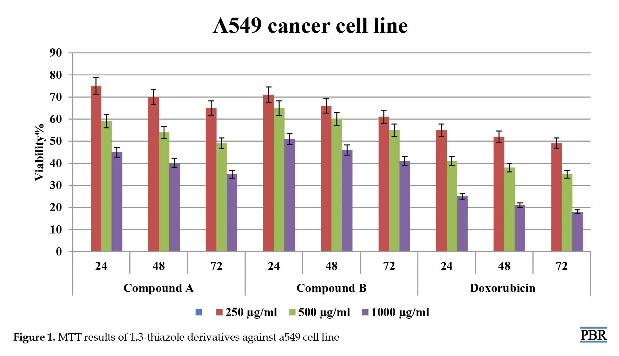 In comparison, compound B inhibited 50% of cancer cells within the first 24 hours at a higher concentration of 1000 µg/mL. Both compounds showed promising inhibitory effects, with greater efficacy at higher concentrations and longer exposure times. Notably, compound A outperformed compound B, suggesting stronger anticancer properties. Interestingly, the control sample also inhibited 50% of cancer cells at 250 µg/mL, demonstrating its effectiveness, albeit less potent than compound A. Moreover, results revealed that both compounds continued to suppress cancer cell viability at increased concentrations, illustrating a dose-dependent response. Overall, these data suggest that both compounds may be viable candidates for further investigation in cancer treatment, with compound A leading in efficacy.
HT-29 cancer cell line: As shown in Figure 2, compound A at 500 µg/mL eliminated approximately 50% of cancer cells within 24 hours. Notably, its effectiveness varied significantly over time and with changes in concentration.
 In comparison, compound B (500 µg/mL) achieved a similar 50% reduction in cancer cells but exhibited relatively stronger effects than compound A. The results indicated that compound B’s efficacy was also significantly influenced by time and concentration variations. Additionally, the control sample inhibited 50% of cancer cells at the lower concentration of 250 µg/mL, demonstrating its effectiveness even at lower levels. Overall, these findings suggest that both compounds possess notable anticancer properties, with compound B showing superior potency.
In silico
According to Table 2, all affinities were calculated, and the best affinity for each receptor with low ΔGbind (-ΔGbind) was selected for Auto dock interactions.
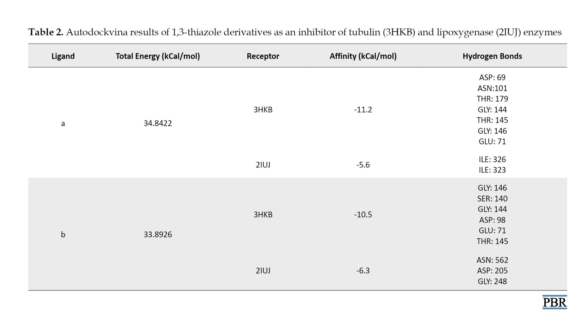
Table 2 and Figure 3 show ligand interactions within the active site of 3HKB and 2IUJ.
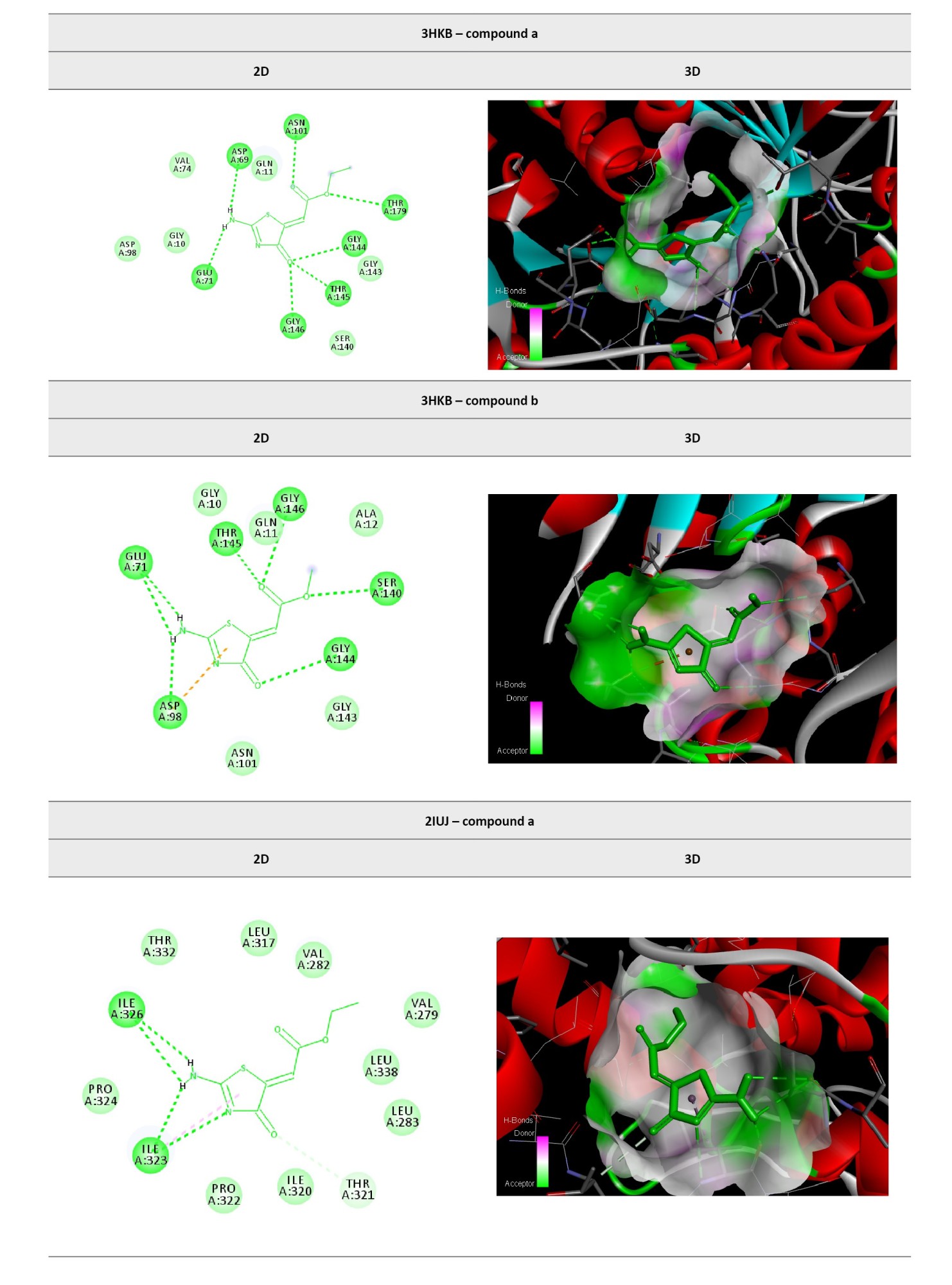
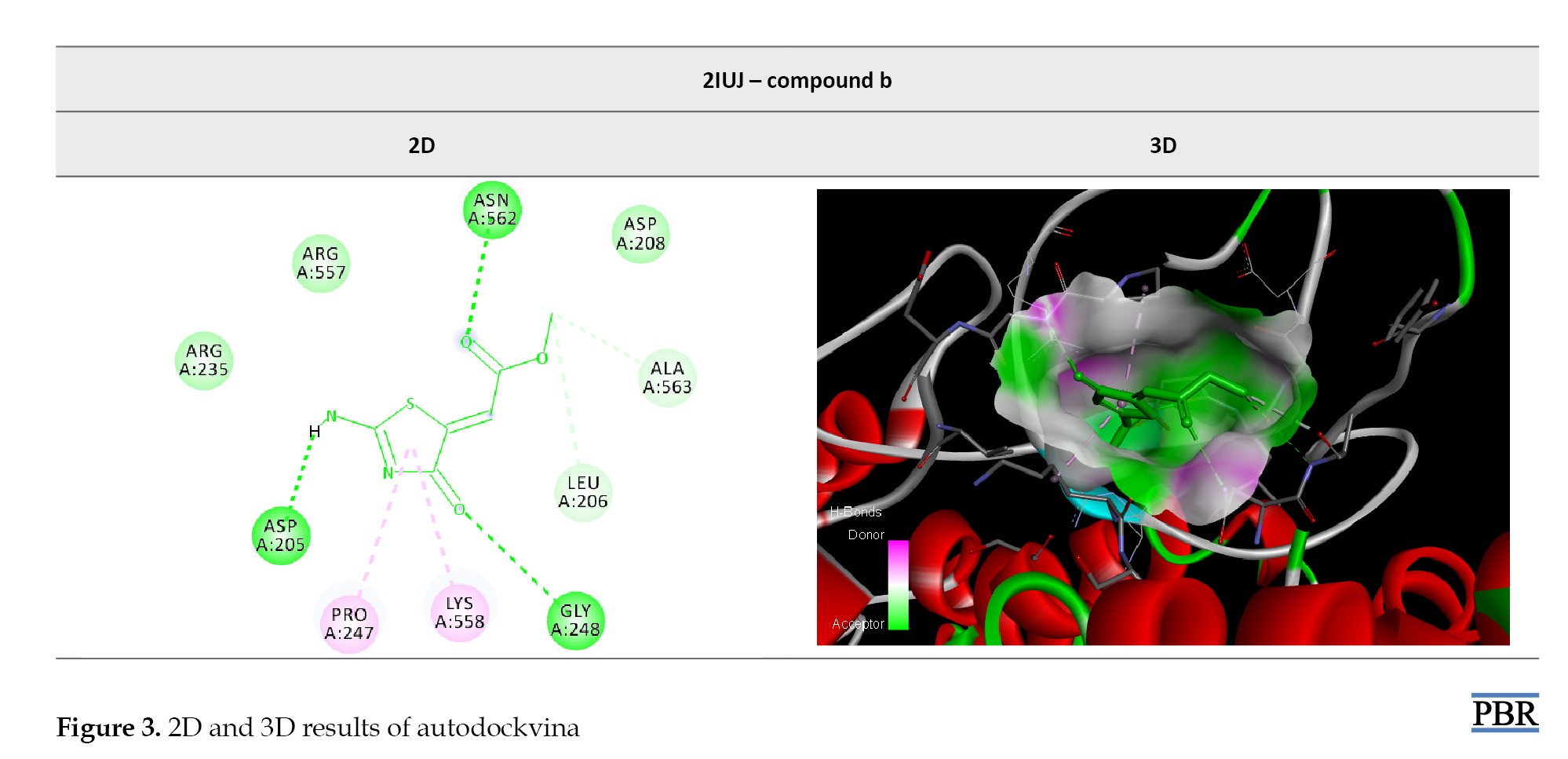 Compound A (ethyl-2-[2-amino-4-oxo-1,3-thiazole-5(4H)-yilden]acetate): Ligand a formed hydrogen bonds with the amino acids aspartic acid (69 hydrogen bonds), asparagine (101 hydrogen bonds), threonine (179 and 145 hydrogen bonds), glycine (144 and 146 hydrogen bonds) and glutamic acid (71 hydrogen bonds) to inhibit 3HKB receptor. This ligand formed hydrogen bonds with the amino acids isoleucine (326 and 323 hydrogen bonds) to inhibit the 2IUJ receptor. These results suggest that compound A may effectively inhibit tubulin. Compound B (methyl-2-[2-amino-4-oxo-1,3-thiazole-5(4H)-yilden]acetate): Ligand b formed hydrogen bonds with the amino acids glycine (146 and 144 hydrogen bonds), serine (140 hydrogen bonds), aspartic acid (98 hydrogen bonds), glutamic acid (71 hydrogen bonds), and threonine (145 hydrogen bonds) to inhibit the 3HKB receptor. This ligand also formed hydrogen bonds with the amino acids asparagine (562 hydrogen bonds), aspartic acid (205 hydrogen bonds), and glycine (248 hydrogen bonds) to inhibit the 2IUJ receptor. Compound B appears highly effective at inhibiting the tubulin enzyme. The results indicate that ligand A, which contains an ethyl group, exhibits significant inhibitory potential against the tubulin enzyme, while ligand B, featuring a methyl group, demonstrates considerable inhibitory effects against the lipoxygenase enzyme. Both ligands offer promising opportunities for the development of alternative drug structures for cancer treatment in future studies. Furthermore, these compounds may serve as valuable leads for the development of targeted therapies that could enhance efficacy and reduce side effects in cancer patients. Continued investigations into their mechanisms of action and SAR are essential for optimizing their therapeutic applications.
Discussion
Gaining insights into lung and CRC is essential, given their widespread occurrence and high mortality rates worldwide. Lung cancer has consistently been the leading cause of cancer-related deaths, primarily associated with smoking and environmental factors. Early detection and advancements in treatment approaches could play a vital role in diminishing mortality rates. CRC, on the other hand, is among the most prevalent cancers, with lifestyle and hereditary factors contributing significantly to its onset. Investigating these cancers is key to uncovering potential biomarkers for early detection, novel treatment targets, and effective preventive measures that could save many lives. Furthermore, research in these fields sheds light on the fundamental processes underlying tumor development, metastasis, and the challenges posed by therapy resistance [21]. In this context, the Global Cancer Observatory (2020) published an analysis of worldwide cancer statistics, confirming that lung cancer is responsible for approximately 19% of all cancer fatalities globally. Moreover, CRC is the second most frequently diagnosed cancer, indicating an urgent necessity for superior prevention and treatment strategies [22].
A different investigation by Sharma et al. (2020) examined the escalating rates of lung and CRC. Their findings illustrated a concerning uptrend in CRC among younger adults, while lung cancer statistics remained consistently high among older demographics [23]. These results highlight the need for targeted screening and proactive intervention initiatives to effectively address these trends. Recent innovations in cancer therapeutics have led to the discovery of new agents targeting specific biological pathways involved in lung and CRC progression [24]. For example, investigational agents that inhibit critical signaling routes, such as the MET pathway and BRAF mutations, have emerged as promising treatment avenues for lung cancer. In CRC, research has focused on the efficacy of monoclonal antibodies targeting PD-1 and novel immune modulators [25]. Additionally, advancements in nanotechnology-based drug delivery have the potential to enhance the targeting efficiency and effectiveness of chemotherapeutic agents, thereby reducing adverse effects and improving patient care. Ongoing inquiries into these new treatment modalities are crucial for addressing resistance challenges and improving patient survival outcomes [26]. Likewise, Patel et al. (2017) investigated targeting the MET pathway in lung cancer treatment, demonstrating that novel inhibitors significantly prolonged progression-free survival in patients with advanced lung cancer, underscoring the need for personalized therapeutic approaches [27]. Similarly, Johnson et al. (2019) focused on developing novel immunotherapies for CRC and identified several promising monoclonal antibodies that enhance the efficacy of existing treatments, particularly in metastatic patients, underscoring the growing significance of precision medicine in oncology [28]. Tubulin, a fundamental protein involved in microtubule assembly, is vital for processes, such as cell division and intracellular transport, making it a key target in the fight against cancer. Disruption of tubulin polymerization can interfere with mitotic spindle formation, thereby triggering programmed cell death in rapidly proliferating cancer cells [29]. Additionally, enzymes, such as cyclooxygenase, are integral to arachidonic acid metabolism, leading to the production of prostaglandins that facilitate inflammation and tumor development. Inhibiting these enzymes may slow cancer cell growth and enhance the efficacy of existing chemotherapy drugs. Thus, elucidating the functions of these enzymes and developing targeted inhibitors represents a promising path to the innovation of cancer therapies [30]. For instance, Čermák and Dostál (2020) emphasized the significance of microtubule-targeting agents in cancer treatment. Their research demonstrated that compounds that inhibit tubulin can halt the cell cycle and induce cell death in various cancer cell lines, reinforcing the idea that modulating microtubule dynamics is a viable strategy against a range of malignancies [31]. In a different study, Saadh et al. (2025) examined the effects of cyclooxygenase inhibitors on cancer cells. Their results indicated that specific cyclooxygenase inhibitors markedly reduced tumor growth in both laboratory and animal models, demonstrating their potential as a novel class of cancer treatment options [32]. The 1,3-thiazole framework is becoming increasingly prominent in medicinal chemistry due to its diverse bioactivity and promising anticancer properties. Molecules featuring this heterocyclic design have exhibited a broad spectrum of pharmacological effects, including anti-inflammatory, anti-microbial, and anticancer properties. The inclusion of the thiazole structure in synthesized compounds is associated with the inhibition of numerous cancer cell lines by disrupting cellular signaling pathways and inducing programmed cell death. Current research efforts are focused on refining these compounds to enhance their effectiveness and specificity against specific types of cancer. This highlights the critical need to investigate new synthetic derivatives that can enhance current cancer treatment strategies and improve therapeutic outcomes [33]. In line with this, Patel et al. (2019) examined the anticancer activities of thiazole-based compounds. Their findings demonstrated that derivatives of 1,3-thiazole exhibited potent cytotoxic effects across multiple cancer cell lines, indicating their potential as viable agents for cancer therapy [34]. Furthermore, Kumar and Singh (2020) focused on the development and assessment of thiazole derivatives. Their research revealed that certain thiazole compounds effectively inhibited cancer cell growth by modulating apoptotic pathways, reinforcing the therapeutic promise of these synthetic molecules in the fight against cancer [34]. In this study, the results obtained from the A549 and HT-29 cancer cell lines provide compelling evidence for the anticancer potential of compounds A and B. In the A549 cell line, compound A demonstrated a remarkable ability to reduce cancer cell viability by 50% over 72 hours at a concentration of 500 µg/mL. This finding aligns with the study of Zhang et al. (2019), who reported that a similar thiazole derivative exhibited significant cytotoxicity against lung cancer cells, reinforcing the notion that compounds targeting microtubule dynamics can effectively induce cell death in aggressive cancer types [35]. In contrast, compound B achieved a comparable 50% reduction in cancer cells within 24 hours at a higher concentration of 1000 µg/mL, suggesting that, while both compounds are effective, compound A may possess superior potency. Furthermore, in the HT-29 cell line, compound A demonstrated efficacy again, achieving a 50% reduction in cancer cells at 500 µg/mL within 24 hours. However, compound B showed a greater potency at the same concentration, indicating its potential for faster action against CRC. This observation is consistent with the findings of Lee et al. (2020), who reported that certain thiazole derivatives showed rapid cytotoxic effects on CRC cells, suggesting that time-dependent efficacy is a critical factor in therapeutic outcomes [36].
The differential anticancer activity observed between the ethyl (compound A) and methyl (compound B) derivatives highlights the critical role of alkyl chain length at the ester moiety. The ethyl group in compound A likely enhances lipophilicity (logP~0.3 units higher than that of the methyl group, calculated using ChemDraw), facilitating better membrane penetration and accumulation in A549 lung cancer cells. In contrast, the methyl group in compound B may form stronger hydrogen bonds with lipoxygenase residues (e.g. Asn562), as evidenced by the docking score and interaction profile, contributing to its superior activity in HT-29 colorectal cells. These findings suggest that minor structural modifications in the ester side chain can dramatically alter cellular uptake, target affinity, and therapeutic specificity.
From an In Silico perspective, the molecular docking results provide valuable insights into the mechanisms of action of these compounds. For instance, compound A (ethyl-2-[2-amino-4-oxo-1,3-thiazole-5(4H)-yilden]acetate) formed multiple hydrogen bonds with key amino acids in the active site of the 3HKB receptor, indicating a strong interaction that likely contributes to its inhibitory effects on tubulin. This finding is supported by Kumar et al. (2021), who demonstrated that similar compounds can bind to tubulin, disrupting its polymerization and enhancing apoptosis in cancer cells [37]. On the other hand, compound B (methyl-2-[2-amino-4-oxo-1,3-thiazole-5(4H)-yilden]acetate) also exhibited significant binding interactions with the 3HKB receptor. However, its interaction with the lipoxygenase enzyme highlights its potential as a dual-action therapeutic agent. is consistent with previous findings suggesting that compounds targeting both tubulin and lipoxygenase could synergistically enhance anticancer effects while minimizing side effects [38]. However, the novelty of the present study lies in the one-step synthesis of ethyl and methyl 2-[2-amino-4-oxo-1,3-thiazol-5(4H)-ylidene]acetate derivatives and their cell line-specific efficacy profiles: the ethyl derivative (compound A) showed superior activity against lung adenocarcinoma (A549), while the methyl derivative (compound B) was more potent against colorectal adenocarcinoma (HT-29). To the best of our knowledge, this structure-dependent selectivity has not been previously reported for this scaffold and may guide the design of tissue-specific thiazole-based therapeutics.
Conclusion
This study established that compounds A and B exhibit significant anticancer activity against A549 and HT-29 cell lines, with compound A showing greater potency in the A549 line and compound B demonstrating rapid efficacy in the HT-29 line. Molecular docking analysis revealed that both compounds effectively interact with critical receptors involved in cell proliferation, suggesting distinct mechanisms of action. These findings indicate the potential of these compounds as candidates for developing targeted cancer therapies, warranting further investigation into their SAR and in vivo efficacy.
Ethical Considerations
Compliance with ethical guidelines
There were no ethical considerations to be considered in this research.
Funding
This research was financially supported by the Central Tehran Branch, Islamic Azad University, Tehran, Iran (Grand No.: 2023-2024).
Authors' contributions
Study design: Nakisa Zarrabi Ahrabi and Yasin SarveAhrabi; Statistical analysis and writing: Nakisa Zarrabi Ahrabi and Yasin SarveAhrabi; Experiments and final approval: All authors.
Conflict of interest
The authors declared no conflict of interest.
Acknowledgments
The authors appreciate all professors who participated voluntarily in this investigation.
References
Drug resistance in cancer is a major barrier to effective treatment, often resulting in treatment failure and disease recurrence [1]. The incidence of treatment resistance varies significantly across cancer types and is influenced by multiple factors, including genetic mutations, the tumor microenvironment, and epigenetic modifications. Research indicates that approximately 90% of patients with advanced cancer may encounter some form of treatment resistance during the course of their illness [2]. Drug resistance can be categorized as intrinsic or acquired. Intrinsic resistance refers to the inherent ability of cancer cells to withstand a drug’s effects from the outset, often due to pre-existing genetic alterations or the expression of specific efflux pumps that actively remove the drug from the cell [3]. In contrast, acquired resistance develops over time as cancer cells adapt to therapeutic pressures. This adaptation can occur through various mechanisms, such as gene amplification (duplicating the target gene, leading to overexpression of the target protein), mutations in drug targets that alter binding sites, or the activation of alternative signaling pathways that bypass the inhibited pathway [4].
Moreover, the tumor microenvironment plays a crucial role in drug resistance. Factors, such as hypoxia, acidity, and the presence of stromal cells, can influence treatment efficacy and contribute to the development of a resistant phenotype [5]. Epigenetic changes, including DNA methylation and histone modifications, can also alter gene expression patterns, promoting resistance. Understanding the complex interplay among these factors is essential for developing novel therapeutic strategies to overcome drug resistance and improve patient outcomes in cancer therapy. Advanced approaches, such as combination therapies and personalized medicine, are being explored to target the specific mechanisms of resistance in individual tumors, thereby enhancing the effectiveness of cancer therapy [6]. Lung cancer, particularly non-small cell lung cancer (NSCLC), exemplifies the significant challenges associated with treatment resistance. Mutations in the epidermal growth factor receptor (EGFR) are prevalent in NSCLC, and although targeted therapies, such as tyrosine kinase inhibitors, have improved patient outcomes, resistance often develops [7]. The mechanisms underlying this resistance include secondary mutations in the EGFR gene, such as the T790M mutation; histological transformation to small cell lung cancer; and activation of alternative signaling pathways, including MET and HER2 amplification. Additionally, the tumor microenvironment significantly influences resistance, with factors, such as hypoxia and the presence of cancer-associated fibroblasts, playing critical roles. As a result, research is focusing on combinatorial treatment strategies and the development of novel therapeutic agents to overcome these resistance mechanisms [8]. Colorectal cancer (CRC) presents a distinct array of drug resistance challenges. Resistance to chemotherapeutic agents, such as fluorouracil, oxaliplatin, and irinotecan, is common, often arising from changes in drug metabolism, cellular efflux mechanisms, and the activation of anti-apoptotic pathways [9]. Furthermore, mutations in critical oncogenes and tumor suppressor genes, including KRAS and p53, are associated with treatment failure. Approximately 50% of patients with CRC harbor mutations in the KRAS oncogene, rendering them insensitive to anti-EGFR therapies. The capacity of tumor cells to enter a dormant state further complicates CRC treatment, as these dormant cells can evade standard therapies and contribute to disease recurrence. To address these resistance mechanisms and enhance treatment efficacy, new therapeutic strategies, including targeted agents and immunotherapy, are currently being investigated [10]. Among promising novel therapeutic agents for cancer treatment, 1,3-thiazole compounds have attracted significant interest due to their diverse biological activities. It is well established that both 1,3-thiazole and 1,3,4-thiadiazole derivatives exhibit promising anticancer activities, particularly when combined with heterocycles, such as 1,3,4-oxadiazole [11].
The mechanisms underlying their action often involve the modulation of various signaling pathways, particularly those associated with pro-apoptotic and anti-apoptotic proteins. Research has shown that thiazole derivatives exhibit activity against multiple cancer types, including breast, lung, and CRC [12]. Investigating the structure-activity relationships (SAR) of these compounds is essential for optimizing the efficacy and selectivity. In addition to their direct anticancer properties, thiazole derivatives may also serve as scaffolds for the development of multitarget drugs, potentially addressing resistance mechanisms in combination therapies [13]. Tubulin, a fundamental protein in the cytoskeleton, is essential for cell division and intracellular transport. The dynamic equilibrium of tubulin polymerization into microtubules is crucial for mitosis, making it a prime target for cancer therapies. Inhibiting tubulin polymerization disrupts mitotic spindle formation, resulting in cell cycle arrest and subsequent apoptosis in rapidly dividing cancer cells. Various classes of drugs, such as taxanes (e.g. paclitaxel) and vinca alkaloids (e.g. vincristine), directly target tubulin and have demonstrated efficacy across a range of malignancies. However, the development of resistance to these tubulin-targeting agents poses a significant challenge, often arising from the expression of drug efflux transporters or mutations in tubulin isotypes. A comprehensive understanding of tubulin dynamics, along with the development of novel tubulin inhibitors or combination therapies, holds great promise for enhancing the effectiveness of cancer treatments [14]. On the other hand, lipoxygenases (LOXs) are enzymes that catalyze the oxidation of polyunsaturated fatty acids, leading to the production of leukotrienes that play a role in inflammatory processes. Elevated expression of LOXs has been linked to various cancers, including breast, prostate, and CRC, where they contribute to tumor growth, metastasis, and the establishment of an inflammatory tumor microenvironment. Inhibiting LOXs has emerged as a potential therapeutic strategy due to their involvement in cancer progression. LOX inhibitors suppress cancer cell proliferation, induce apoptosis, and inhibit angiogenesis, thereby offering a multifaceted approach to combat cancer. Furthermore, ongoing research on selective LOX isoform inhibitors aims to reduce side effects while maximizing targeted therapeutic efficacy. As our understanding of LOX pathways in tumor biology expands, these inhibitors may serve as promising adjunctive therapies in comprehensive cancer treatment regimens [15].
Objectives
This study aimed to investigate the in vitro anticancer effects of 1,3-thiazole derivatives against A549 and HT-29 cancer cell lines and to evaluate the compounds in silico as inhibitors of tubulin and lipoxygenase enzymes.
Materials and Methods
This experimental research was conducted in the Microbiology Laboratory of Islamic Azad University, Tehran Branch, in 2023-2024. Starting materials, solvents, and culture media (RPMI 1640) were purchased from Merck (Germany). The cell lines were obtained from the Pasteur Institute of Iran (Cell Line Collections).
In vitro: All derivatives of 1,3-thiazole from our previous research were resynthesized, as shown in Table 1 [16].
The concentrations of 250, 500, and 1000 µg/mL were selected based on preliminary solubility and cytotoxicity screening of the compounds in dimethyl sulfoxide (DMSO), ensuring complete dissolution while spanning a wide therapeutic window. These doses align with standard ranges used in MTT assays for heterocyclic compounds [17] and allowed direct comparison with the positive control.
The new compounds were solubilized using DMSO at concentrations of 250, 500, and 1000 µg/mL to assess their anticancer properties. Additionally, Doxorubicin, a pure antibiotic powder obtained from Sigma, was prepared and employed as a control at the same concentrations.
Doxorubicin was chosen as the positive control due to its well-established clinical efficacy against both lung and CRC, its broad-spectrum cytotoxic mechanism (DNA intercalation and topoisomerase II inhibition), and its frequent use as a benchmark in 3-(4,5-dimethylthiazol-2-yl)-2,5-diphenyltetrazolium bromide (MTT)-based anticancer screening studies.
A549 and HT-29 cancer cell lines were cultured in RPMI 1640 medium supplemented with 10% fetal bovine serum and antibiotics, specifically penicillin and streptomycin, both at 100 µg/mL.
A549 is a human lung adenocarcinoma cell line (non-small cell lung cancer), while HT-29 is a human colorectal adenocarcinoma cell line, representing two of the most lethal malignancies worldwide.
This culture was maintained in an incubator at 37 ºC with 5% carbon dioxide (CO2) and saturated humidity in 25 cm flasks. The culture medium was refreshed every one to two days, and when the cells achieved 80% confluence, they were subcultured. Cell viability was assessed using the MTT assay. This colorimetric technique relies on the reduction of yellow tetrazolium crystals by the succinate dehydrogenase enzyme, yielding blue insoluble formazan crystals. During incubation, MTT is reduced by succinate dehydrogenase, an enzyme integral to mitochondrial respiration. The reduction process leads to the formation of recognizable blue formazan crystals under a microscope, with the color intensity directly correlating with the number of viable cells. To evaluate the effects of the tested compounds on cell growth and survival, 5×10⁴ cells were plated in 96-well plates. These cells were then exposed to different concentrations of the compounds for periods of 24, 48, and 72 hours. The yellow MTT powder was dissolved in 1 mL of PBS buffer shielded from light and subsequently filtered through a 0.2-micron filter for sterilization. After the incubation duration, the medium was aspirated, and MTT solution was introduced to each well. The plates were incubated for 4 hours at 37 ºC to facilitate the formation of formazan crystals. DMSO was added post-incubation to dissolve the insoluble formazan crystals. The optical density of each well’s solution was assessed using an ELISA reader at 570 nm. The optical density reflects the amount of MTT converted to formazan under these conditions. Cell viability was calculated using the Equation 1:
1. Percentage survival=(mean absorbance of treated cells/mean absorbance of the control cells)×100.
In silico: The 2D representations of the derivatives were produced with ChemDraw software, version 12.0.2.1076. Subsequently, the structures were normalized via the MM2 method in Chem3D Pro version 12.0.2.1076. Crystallographic structures of tubulin (PDB ID: 3HKB) [18] and lipoxygenase (PDB ID: 2IUJ) [19] were sourced from the Protein Data Bank, which provided resolutions of 3.65 Å for 3HKB and 2.40 Å for 2IUJ. Gasteiger charges and polar hydrogens were incorporated using AutoDockTools (version 1.5.6). All water molecules and ligands were eliminated from the protein structures. The grid dimensions were set to 48×48×48 for the 3HKB structure and 56×56×56 for the 2IUJ structure, with 3 Å spacing. The docking of the derivatives into the binding sites of 3HKB and 2IUJ was performed using AutoDock Vina and Discovery Studio 4.5. The interactions between the ligands and the binding sites were evaluated using Discovery Studio Client software, version 4.5 [20].
Results
In vitro
A549 cancer cell line: As shown in Figure 1, compound A demonstrated significant efficacy in reducing cancer cells, achieving a 50% reduction over 72 hours at a concentration of 500 µg/mL.
HT-29 cancer cell line: As shown in Figure 2, compound A at 500 µg/mL eliminated approximately 50% of cancer cells within 24 hours. Notably, its effectiveness varied significantly over time and with changes in concentration.
In silico
According to Table 2, all affinities were calculated, and the best affinity for each receptor with low ΔGbind (-ΔGbind) was selected for Auto dock interactions.
Table 2 and Figure 3 show ligand interactions within the active site of 3HKB and 2IUJ.
Discussion
Gaining insights into lung and CRC is essential, given their widespread occurrence and high mortality rates worldwide. Lung cancer has consistently been the leading cause of cancer-related deaths, primarily associated with smoking and environmental factors. Early detection and advancements in treatment approaches could play a vital role in diminishing mortality rates. CRC, on the other hand, is among the most prevalent cancers, with lifestyle and hereditary factors contributing significantly to its onset. Investigating these cancers is key to uncovering potential biomarkers for early detection, novel treatment targets, and effective preventive measures that could save many lives. Furthermore, research in these fields sheds light on the fundamental processes underlying tumor development, metastasis, and the challenges posed by therapy resistance [21]. In this context, the Global Cancer Observatory (2020) published an analysis of worldwide cancer statistics, confirming that lung cancer is responsible for approximately 19% of all cancer fatalities globally. Moreover, CRC is the second most frequently diagnosed cancer, indicating an urgent necessity for superior prevention and treatment strategies [22].
A different investigation by Sharma et al. (2020) examined the escalating rates of lung and CRC. Their findings illustrated a concerning uptrend in CRC among younger adults, while lung cancer statistics remained consistently high among older demographics [23]. These results highlight the need for targeted screening and proactive intervention initiatives to effectively address these trends. Recent innovations in cancer therapeutics have led to the discovery of new agents targeting specific biological pathways involved in lung and CRC progression [24]. For example, investigational agents that inhibit critical signaling routes, such as the MET pathway and BRAF mutations, have emerged as promising treatment avenues for lung cancer. In CRC, research has focused on the efficacy of monoclonal antibodies targeting PD-1 and novel immune modulators [25]. Additionally, advancements in nanotechnology-based drug delivery have the potential to enhance the targeting efficiency and effectiveness of chemotherapeutic agents, thereby reducing adverse effects and improving patient care. Ongoing inquiries into these new treatment modalities are crucial for addressing resistance challenges and improving patient survival outcomes [26]. Likewise, Patel et al. (2017) investigated targeting the MET pathway in lung cancer treatment, demonstrating that novel inhibitors significantly prolonged progression-free survival in patients with advanced lung cancer, underscoring the need for personalized therapeutic approaches [27]. Similarly, Johnson et al. (2019) focused on developing novel immunotherapies for CRC and identified several promising monoclonal antibodies that enhance the efficacy of existing treatments, particularly in metastatic patients, underscoring the growing significance of precision medicine in oncology [28]. Tubulin, a fundamental protein involved in microtubule assembly, is vital for processes, such as cell division and intracellular transport, making it a key target in the fight against cancer. Disruption of tubulin polymerization can interfere with mitotic spindle formation, thereby triggering programmed cell death in rapidly proliferating cancer cells [29]. Additionally, enzymes, such as cyclooxygenase, are integral to arachidonic acid metabolism, leading to the production of prostaglandins that facilitate inflammation and tumor development. Inhibiting these enzymes may slow cancer cell growth and enhance the efficacy of existing chemotherapy drugs. Thus, elucidating the functions of these enzymes and developing targeted inhibitors represents a promising path to the innovation of cancer therapies [30]. For instance, Čermák and Dostál (2020) emphasized the significance of microtubule-targeting agents in cancer treatment. Their research demonstrated that compounds that inhibit tubulin can halt the cell cycle and induce cell death in various cancer cell lines, reinforcing the idea that modulating microtubule dynamics is a viable strategy against a range of malignancies [31]. In a different study, Saadh et al. (2025) examined the effects of cyclooxygenase inhibitors on cancer cells. Their results indicated that specific cyclooxygenase inhibitors markedly reduced tumor growth in both laboratory and animal models, demonstrating their potential as a novel class of cancer treatment options [32]. The 1,3-thiazole framework is becoming increasingly prominent in medicinal chemistry due to its diverse bioactivity and promising anticancer properties. Molecules featuring this heterocyclic design have exhibited a broad spectrum of pharmacological effects, including anti-inflammatory, anti-microbial, and anticancer properties. The inclusion of the thiazole structure in synthesized compounds is associated with the inhibition of numerous cancer cell lines by disrupting cellular signaling pathways and inducing programmed cell death. Current research efforts are focused on refining these compounds to enhance their effectiveness and specificity against specific types of cancer. This highlights the critical need to investigate new synthetic derivatives that can enhance current cancer treatment strategies and improve therapeutic outcomes [33]. In line with this, Patel et al. (2019) examined the anticancer activities of thiazole-based compounds. Their findings demonstrated that derivatives of 1,3-thiazole exhibited potent cytotoxic effects across multiple cancer cell lines, indicating their potential as viable agents for cancer therapy [34]. Furthermore, Kumar and Singh (2020) focused on the development and assessment of thiazole derivatives. Their research revealed that certain thiazole compounds effectively inhibited cancer cell growth by modulating apoptotic pathways, reinforcing the therapeutic promise of these synthetic molecules in the fight against cancer [34]. In this study, the results obtained from the A549 and HT-29 cancer cell lines provide compelling evidence for the anticancer potential of compounds A and B. In the A549 cell line, compound A demonstrated a remarkable ability to reduce cancer cell viability by 50% over 72 hours at a concentration of 500 µg/mL. This finding aligns with the study of Zhang et al. (2019), who reported that a similar thiazole derivative exhibited significant cytotoxicity against lung cancer cells, reinforcing the notion that compounds targeting microtubule dynamics can effectively induce cell death in aggressive cancer types [35]. In contrast, compound B achieved a comparable 50% reduction in cancer cells within 24 hours at a higher concentration of 1000 µg/mL, suggesting that, while both compounds are effective, compound A may possess superior potency. Furthermore, in the HT-29 cell line, compound A demonstrated efficacy again, achieving a 50% reduction in cancer cells at 500 µg/mL within 24 hours. However, compound B showed a greater potency at the same concentration, indicating its potential for faster action against CRC. This observation is consistent with the findings of Lee et al. (2020), who reported that certain thiazole derivatives showed rapid cytotoxic effects on CRC cells, suggesting that time-dependent efficacy is a critical factor in therapeutic outcomes [36].
The differential anticancer activity observed between the ethyl (compound A) and methyl (compound B) derivatives highlights the critical role of alkyl chain length at the ester moiety. The ethyl group in compound A likely enhances lipophilicity (logP~0.3 units higher than that of the methyl group, calculated using ChemDraw), facilitating better membrane penetration and accumulation in A549 lung cancer cells. In contrast, the methyl group in compound B may form stronger hydrogen bonds with lipoxygenase residues (e.g. Asn562), as evidenced by the docking score and interaction profile, contributing to its superior activity in HT-29 colorectal cells. These findings suggest that minor structural modifications in the ester side chain can dramatically alter cellular uptake, target affinity, and therapeutic specificity.
From an In Silico perspective, the molecular docking results provide valuable insights into the mechanisms of action of these compounds. For instance, compound A (ethyl-2-[2-amino-4-oxo-1,3-thiazole-5(4H)-yilden]acetate) formed multiple hydrogen bonds with key amino acids in the active site of the 3HKB receptor, indicating a strong interaction that likely contributes to its inhibitory effects on tubulin. This finding is supported by Kumar et al. (2021), who demonstrated that similar compounds can bind to tubulin, disrupting its polymerization and enhancing apoptosis in cancer cells [37]. On the other hand, compound B (methyl-2-[2-amino-4-oxo-1,3-thiazole-5(4H)-yilden]acetate) also exhibited significant binding interactions with the 3HKB receptor. However, its interaction with the lipoxygenase enzyme highlights its potential as a dual-action therapeutic agent. is consistent with previous findings suggesting that compounds targeting both tubulin and lipoxygenase could synergistically enhance anticancer effects while minimizing side effects [38]. However, the novelty of the present study lies in the one-step synthesis of ethyl and methyl 2-[2-amino-4-oxo-1,3-thiazol-5(4H)-ylidene]acetate derivatives and their cell line-specific efficacy profiles: the ethyl derivative (compound A) showed superior activity against lung adenocarcinoma (A549), while the methyl derivative (compound B) was more potent against colorectal adenocarcinoma (HT-29). To the best of our knowledge, this structure-dependent selectivity has not been previously reported for this scaffold and may guide the design of tissue-specific thiazole-based therapeutics.
Conclusion
This study established that compounds A and B exhibit significant anticancer activity against A549 and HT-29 cell lines, with compound A showing greater potency in the A549 line and compound B demonstrating rapid efficacy in the HT-29 line. Molecular docking analysis revealed that both compounds effectively interact with critical receptors involved in cell proliferation, suggesting distinct mechanisms of action. These findings indicate the potential of these compounds as candidates for developing targeted cancer therapies, warranting further investigation into their SAR and in vivo efficacy.
Ethical Considerations
Compliance with ethical guidelines
There were no ethical considerations to be considered in this research.
Funding
This research was financially supported by the Central Tehran Branch, Islamic Azad University, Tehran, Iran (Grand No.: 2023-2024).
Authors' contributions
Study design: Nakisa Zarrabi Ahrabi and Yasin SarveAhrabi; Statistical analysis and writing: Nakisa Zarrabi Ahrabi and Yasin SarveAhrabi; Experiments and final approval: All authors.
Conflict of interest
The authors declared no conflict of interest.
Acknowledgments
The authors appreciate all professors who participated voluntarily in this investigation.
References
- Rezayatmand H, Razmkhah M, Razeghian-Jahromi I. Drug resistance in cancer therapy: The Pandora's box of cancer stem cells. Stem Cell Res Ther. 2022; 13(1):181. [DOI:10.1186/s13287-022-02856-6] [PMID]
- Fu YC, Liang SB, Luo M, Wang XP. Intratumoral heterogeneity and drug resistance in cancer. Cancer Cell Int. 2025; 25(1):103. [DOI:10.1186/s12935-025-03734-w] [PMID]
- Mengistu BA, Tsegaw T, Demessie Y, Getnet K, Bitew AB, Kinde MZ, et al. Comprehensive review of drug resistance in mammalian cancer stem cells: Implications for cancer therapy. Cancer Cell Int. 2024; 24(1):406. [DOI:10.1186/s12935-024-03558-0] [PMID] [PMCID]
- Tao X, Ke X, Xu G. Mechanisms of circular RNA in drug resistance of lung cancer: Therapeutic targets, biomarkers, and future research directions. Discov Oncol. 2025; 16(1):896.[DOI:10.1007/s12672-025-02713-x] [PMID] [PMCID]
- Garimella SS, Gampa SC, Garimella S. Role of the tumor microenvironment in cancer therapy: Unveiling new targets to overcome drug resistance. Med Oncol. 2025; 42(6):202. [DOI:10.1007/s12032-025-02754-w] [PMID]
- Minopoli A, Evangelista D, Marras M, Perini G, Augello A, Palmieri V, et al. Enhancing photothermal therapy effectiveness via tartrazine-induced optical clearing of biological tissues. Sci Rep. 2026; 16(1):7553. [DOI:10.1038/s41598-026-38616-2] [PMID] [PMCID]
- Pramanik P, Das T, Pal U, Rehman I. Unravelling the molecular genetics and epigenetic regulation of lung cancer vis a vis non-small cell lung cancer (NSCLC) for a targeted approach towards novel therapeutic strategies: A review. Nucleus. 2025. [DOI:10.1007/s13237-025-00629-z]
- Wang Y, Hao Y, Ranieri M, Abramyan TM, Tsidilkovski L, Hollenberg M, et al. Deep mutational scanning reveals EGFR mutations conferring resistance to the 4th-generation EGFR tyrosine kinase inhibitor BLU-945. NPJ Precis Oncol. 2025; 9(1):294. [DOI:10.1038/s41698-025-01086-2] [PMID] [PMCID]
- Lin D, Luo Y, Chen J, Ma Z, Kang H, Wang X, et al. Single-Cell-Derived Tumor Organoid (STO) arrays on a microfluidic chip for personalized drug screening to address heterogeneity-induced drug resistance in colorectal cancer. Microsyst Nanoeng. 2025; 11(1):253. [DOI:10.1038/s41378-025-01068-1] [PMID] [PMCID]
- Karnwal A, Dutta J, Aqueel-Ur-Rehman, Al-Tawaha ARMS, Nesterova N. Genetic landscape of cancer: Mechanisms, key genes, and therapeutic implications. Clin Transl Oncol. 2026; 28(2):424-5. [DOI:10.1007/s12094-025-04019-4] [PMID]
- Raman APS, Aslam M, Awasthi A, Ansari A, Jain P, Lal K, et al. An updated review on 1,2,3-/1,2,4-triazoles: synthesis and diverse range of biological potential. Mol Divers. 2025; 29(1):899-964. [DOI:10.1007/s11030-024-10858-0] [PMID]
- Dasgupta A, Rajesh R, Das PK, Matada GS, Dhiwar PS, Paik A. Medicinal chemistry perspective of chalcone derivatives as anticancer agents: Synthetic strategy, biological activity, and structure-activity relationship. Mol Divers. 2026. [DOI:10.1007/s11030-025-11434-w]
- Lee H, Kim M, Jeon B. Boron-containing anticancer agents: A target-centric review of structure-activity relationships and clinical pipeline. Arch Pharm Res. 2025; 48(11-12):1253-98. [DOI:10.1007/s12272-025-01582-w] [PMID]
- Wang X, Fan Y, Wang Q, Shu X, Lin J, Guo J, et al. Tumor-infiltrating nerves: Unraveling the role of cancer neuroscience in tumorigenesis, disease progression, and emerging therapies. Discov Oncol. 2025; 16(1):1209. [DOI:10.1007/s12672-025-02827-2] [PMID]
- No Author. 33rd Annual Meeting & Pre-Conference Programs of the Society for Immunotherapy of Cancer (SITC 2018) : Washington, D.C., USA. 7-11 November 2018. J Immunother Cancer. 2018; 6(Suppl 1):114. [DOI:10.1186/s40425-018-0422-y] [PMID]
- Sarveahrabi Y, Shirinbeig S. [One-step synthesis of ethyl and methyl derivatives of ylidene-acetate bonded at position 5 of ring 1,3-thiazole and evaluation of their antibacterial activities (Persian)]. Navid No. 2020; 23(73):66-77. [DOI:10.22038/nnj.2020.45706.1197]
- Aljohani GF, Abolibda TZ, Alhilal M, AlHumaidi JY, Alhilal S, Ahmed HA, et al. Novel thiadiazolethiazole hybrids: synthesis, molecular docking, and cytotoxicity evaluation against liver cancer cell lines. J Taibah Univ Sci. 2022; 16(1):1005-15. [DOI:10.1080/16583655.2022.2135805]
- Dorleans A, Gigant B, Ravelli RBG, Mailliet P, Mikol V, Knossow M. Tubulin: RB3 Stathmin-like domain complex. New York: Protein Data Bank; 2009. [DOI:10.2210/pdb3HKB/pdb]
- Youn B, Sellhorn GE, Mirchel RJ, Gaffney BJ, Grimes HD, Kang C. Crystal Structure of Soybean Lipoxygenase-B. New York: Protein Data Bank; 2006. [DOI:10.2210/pdb2iuj/pdb]
- AlShemary RK, Mohapatra RK, Kumar M, Sarangi AK, Azam M, Tuli HS, et al. Synthesis, structural investigations, XRD, DFT, anticancer and molecular docking study of a series of thiazolebased Schiff base metal complexes. J Mol Struct. 2023; 1275:134676. [DOI:10.1016/j.molstruc.2022.134676]
- Xia C, Liu Y, Qing X. Trends in incidence and mortality of early-onset gastrointestinal cancers: A comprehensive study. BMC Gastroenterol. 2025; 25(1):424. [DOI:10.1186/s12876-025-04015-6] [PMID]
- Elwali NE, AlShareef SM, Khamis AH, Elhassan MMA. Pancreatic cancer in Saudi Arabia (2005-2020): Increasing trend. BMC Cancer. 2024; 24(1):653. [DOI:10.1186/s12885-024-12401-8] [PMID]
- Sharma A, Baker S, Duijm M, Oomen-de Hoop E, Cornelissen R, Verhoef C, et al. Prognostic factors for local control and survival for inoperable pulmonary colorectal oligometastases treated with stereotactic body radiotherapy. Radiother Oncol. 2020; 144:23-9. [DOI:10.1016/j.radonc.2019.10.004] [PMID]
- Nur A, Seruwagi G, Odwe G, Kisaakye P, Muthuri S, Habteyesus D, et al. Screening for sexual violence against children in humanitarian settings: A feasibility study of a parasocial workerled intervention in Uganda. Int J Humanitarian Action. 2025; 10:19. [DOI:10.1186/s41018-025-00185-w]
- Noorkhajavi G, Banakholdi A, Torabi A, Zoghi A, Iranijam E, Safarzadeh E. Recent clinical advances in nonconjugated antibodies and antibody-drug conjugates for colorectal cancer treatment. Cancer Cell Int. 2025; 25(1):395. [DOI:10.1186/s12935-025-04039-8] [PMID]
- Saripilli R, Sharma DK. Nanotechnologybased drug delivery system for the diagnosis and treatment of ovarian cancer. Discov Oncol. 2025; 16(1):422. [DOI:10.1007/s12672-025-02062-9] [PMID]
- Green C, Kong AP, Brysbaert M, Keogh K. Crowdsourced and AIgenerated ageofacquisition (AoA) norms for vocabulary in print: extending the Kuperman et al. (2012) norms. Behav Res Methods. 2025; 57(11):304. [DOI:10.3758/s13428-025-02843-8] [PMID]
- No Author. Proceedings of the World Molecular Imaging Congress 2019, Montreal, Quebec, Canada, September 4-7, 2019: General abstracts. Mol Imaging Biol. 2019; 21(Suppl 1):1-166. [DOI:10.1007/s11307-019-01454-y]
- Stathatos GG, Merriner DJ, O'Connor AE, Zenker J, Dunleavy JE, O'Bryan MK. Epsilon tubulin is an essential determinant of microtubule-based structures in male germ cells. EMBO Rep. 2024; 25(6):2722-42. [DOI:10.1038/s44319-024-00159-w] [PMID]
- Wang Y, Hsu P, Hu H, Lin F, Wei X. Role of arachidonic acid metabolism in osteosarcoma prognosis by integrating WGCNA and bioinformatics analysis. BMC Cancer. 2025; 25(1):445. [DOI:10.1186/s12885-024-13278-3] [PMID]
- Čermák V, Dostál V, Jelínek M, Libusová L, Kovář J, Rösel D, et al. Microtubule-targeting agents and their impact on cancer treatment. Eur J Cell Biol. 2020; 99(4):151075. [DOI:10.1016/j.ejcb.2020.151075] [PMID]
- Saadh MJ, Ahmed HH, Chandra M, Al-Hussainy AF, Hamid JA, Mishra A, et al. Therapeutic effects of quercetin in oral cancer therapy: A systematic review of preclinical evidence focused on oxidative damage, apoptosis and anti-metastasis. Cancer Cell Int. 2025; 25(1):66. [DOI:10.1186/s12935-025-03694-1] [PMID]
- No Author. Abstracts of the 82nd Annual Meeting of the German Society for Experimental and Clinical Pharmacology and Toxicology (DGPT) and the 18th Annual Meeting of the Network Clinical Pharmacology Germany (VKliPha) in cooperation with the Arbeitsgemeinschaft für Angewandte Humanpharmakologie e.V. (AGAH). Naunyn Schmiedebergs Arch Pharmacol. 2016; 389(Suppl 1):1-104. [DOI:10.1007/s00210-016-1213-y] [PMID]
- Abhale YK, Patel K, Patil M, Mhaske PC, Jabir M, Ghotekar S. Recent advancements in the synthesis of bithiazole and its derivatives for versatile medicinal applications. Chem Pap. 2025; 79:7269-98. [DOI:10.1007/s11696-025-04312-0]
- Paul A, Mishra SS, Maji A, Samanta A, Nahar S, Maity TK. Exploring the therapeutic potentials of cuminaldehyde: a comprehensive review of biological activities, mechanisms, and novel delivery systems. Phytochem Rev. 2025; 24:5207-38. [DOI:10.1007/s11101-025-10069-x]
- No Author. Proceedings of the World Molecular Imaging Congress 2020, October 7-9, 2020: General Abstracts. Mol Imaging Biol. 2021; 23(Suppl 1):1-862. [DOI:10.1007/s11307-021-01691-0]
- Sharma A, Suvedi D, Kumar A, Khanal S, Verma R, Kumar D, et al. Bioactive compounds of Ganoderma species: Molecular mechanisms and therapeutic potential in cancer and metabolic disorders. World J Microbiol Biotechnol. 2025; 41(12):497. [DOI:10.1007/s11274-025-04687-y] [PMID]
- No Author. Abstracts from the 56th European Society of Human Genetics (ESHG) Conference: Hybrid Posters. Eur J Hum Genet. 2024; 32(Suppl 1):349-795. [DOI:10.1038/s41431-023-01482-x]
Type of Study: Original Research |
Subject:
Cellular and Molecular Biology
| Rights and permissions | |
 |
This work is licensed under a Creative Commons Attribution-NonCommercial 4.0 International License. |


This is an open access article distributed under the terms of the Creative Commons Attribution License (CC-By-NC), which permits use, distribution, and reproduction in any medium, provided the original work is properly cited and is not used for commercial purposes.
Contact Information