

Volume 12, Issue 3 (2026)
Pharm Biomed Res 2026, 12(3): 195-206 |
Back to browse issues page



Download citation:
BibTeX | RIS | EndNote | Medlars | ProCite | Reference Manager | RefWorks
Send citation to:


BibTeX | RIS | EndNote | Medlars | ProCite | Reference Manager | RefWorks
Send citation to:
Koochaki R, Nabiuni M, Amini E. In Vitro and In Vivo Effects of Quillaja saponaria Bark Saponin Against Ovarian Cancer Cells. Pharm Biomed Res 2026; 12 (3) :195-206
URL: http://pbr.mazums.ac.ir/article-1-755-en.html
URL: http://pbr.mazums.ac.ir/article-1-755-en.html


1- Department of Cell & Molecular Biology, Faculty of Biological Sciences, Kharazmi University, Tehran, Iran.
2- Department of Animal Biology, Faculty of Biological Sciences, Kharazmi University, Tehran, Iran.
2- Department of Animal Biology, Faculty of Biological Sciences, Kharazmi University, Tehran, Iran.
Full-Text [PDF 3064 kb]
(83 Downloads)
| Abstract (HTML) (560 Views)
Results
Effects of Q. saponaria bark saponin on viability of SKOV3 and A2780 cells in a dose-dependent manner
In SKOV3 cells, QBS at concentrations of 20, 30, and 80 µg/mL reduced cell survival percentage to 78.9±0.4, 61.4±0.2, and 52.6±0.1, respectively. In A2780 cells, cell viability under exposure to QBS at concentrations of 20, 30, and 80 µg/mL was 75.1±0.09, 64.4±0.06, and 45.9±0.1 (Figure 2).
Full-Text: (78 Views)
Introduction
Ovarian cancer is a fatal gynecological cancer ranking eighth among the leading causes of ca)ncer–related mortality in women worldwide. Epithelial ovarian cancer (EOC) is the most widespread subtype, including more than 95% of all ovarian cancer cases [1]. Based on surveys, ovarian cancer cells are very invasive; therefore, suppression of their invasion is considered a therapeutic approach for oncological research. In this regard, , epithelial cells must undergo epithelial–mesenchymal transition (EMT) to gain metastatic potential, a crucial step in cancer progression during which cancer cells acquire the invasive abilities necessary to complete all steps of the metastatic cascade [2, 3]. Related to EMT, metastasis of malignant epithelial cells requires lysis of the ECM by matrix metalloproteinase (MMP) enzymes. MMPs are zinc-dependent enzymes that play an essential role in degrading the basement membrane and ECM proteins. MMP2 overexpression in malignant ovarian cancer is related to the metastasis of ovarian cancer [4]. MMP2, a key member of this family, digests type IV collagen and is overexpressed in EOC, which correlates with poor survival [5-7]. Integrins are adhesion molecules in the cell surface containing α and β subunits, which have different functions in cells. Notably, tumor cells can resist anoikis by expressing ITGA5. In addition, it was reported that high expression of ITGA5 in tumor cells is associated with cell survival attenuation [8-10]. Studies showed that ITGA5 overexpression initiates transformed cells towards a more malignant phenotype and metastasis in malignant ovarian cells [11]. On the other hand, CDH1, as an epithelial marker and Ca-dependent glycoprotein, is involved in cellular connections and controls metastatic progression. Studies showed that when CDH1 is overexpressed, cancer cells are less likely to invade [12, 13]. In this regard, zinc finger E-box binding homeobox 1 (ZEB1) is a major transcription factor that plays a central role in EOC progression and stimulates EMT by downregulating epithelial genes, including CDH1, and increasing the metastatic ability of EOC [14, 15].
With the progression of ovarian cancer stage, the conventional therapeutic strategies, including surgery, chemotherapy, immunotherapy, and radiotherapy have indicated minimal success [16]. Meanwhile, natural compounds are considered promising sources of anticancer agents [17].
Saponins are triterpenoids found in many plant species, such as sunflower, chickpea, soybean, sugar beet, ginseng, and oats, and exhibit cytotoxic activity and play a protective role in plants by defending against predators. Many biological effects of saponins, such as anti-inflammatory and anti-microbial effects, cell membrane permeabilization, immune system stimulation, and cytotoxic attributes, have also been reported [18-20].
According to the literature, saponins have exhibited many pharmacological effects; however, their application is limited due to their in vitro hemolytic activity. Nevertheless, this hemolytic property is not the same in all saponins. Quillaja saponaria bark saponin (QBS), in particular, exhibits minimal hemolytic effects, so it was previously approved as a vaccine adjuvant [21]. Previous studies have demonstrated that saponin compounds can exert anti-cancer effects via modulation of EMT and invasion of ovarian and other cancer cells [19, 20]. Despite extensive evidence of the anti-proliferation and anti-invasion effects of saponins, the anti-migratory effects of QBS in ovarian cancer have not been fully elucidated. In the present study, SKOV3 and A2780 cell lines were used as ovarian cancer cell lines. A2780 cells are more sensitive than SKOV3 cells to treatment with fluorouracil, oxaliplatin, paclitaxel, etc. Furthermore, to provide more evidence supporting the potential anti-cancer properties of QBS, its effects were also tested in the Wistar rat model, which induced ovarian cancer with 7,12-Dimethylbenz[a]anthracene (DMBA) as carcinogen [19-22]. The DMBA is commonly used in animal models to induce breast cancer, skin cancer, and ovarian cancer. Studies have shown that DMBA-induced ovarian cancer resembles human ovarian cancer in terms of histology, molecular, and dissemination patterns. Thus, a DMBA-induced ovarian cancer model can be useful in human ovarian cancer studies [23, 24]. The SKOV3 cell line is a human ovarian adenocarcinoma derived from ascitic fluid, and the A2780 cell line is known as a human ovarian carcinoma isolated from tumor tissue. Besides, A2780 cells are more sensitive than SKOV3 cells to oxaliplatin, fluorouracil, gemcitabine, paclitaxel, etc. [25]. Furthermore, ITGA5, MMP2, ZEB1, and CDH1 genes were selected because of their well-documented involvement in EMT and their recognized roles in tumor progression and metastatic dissemination. In this study, we aimed to evaluate the effects of QBS on EMT-related genes in A2780 and SKOV3 human ovarian cancer cell lines.
Materials and Methods
Chemicals and reagents
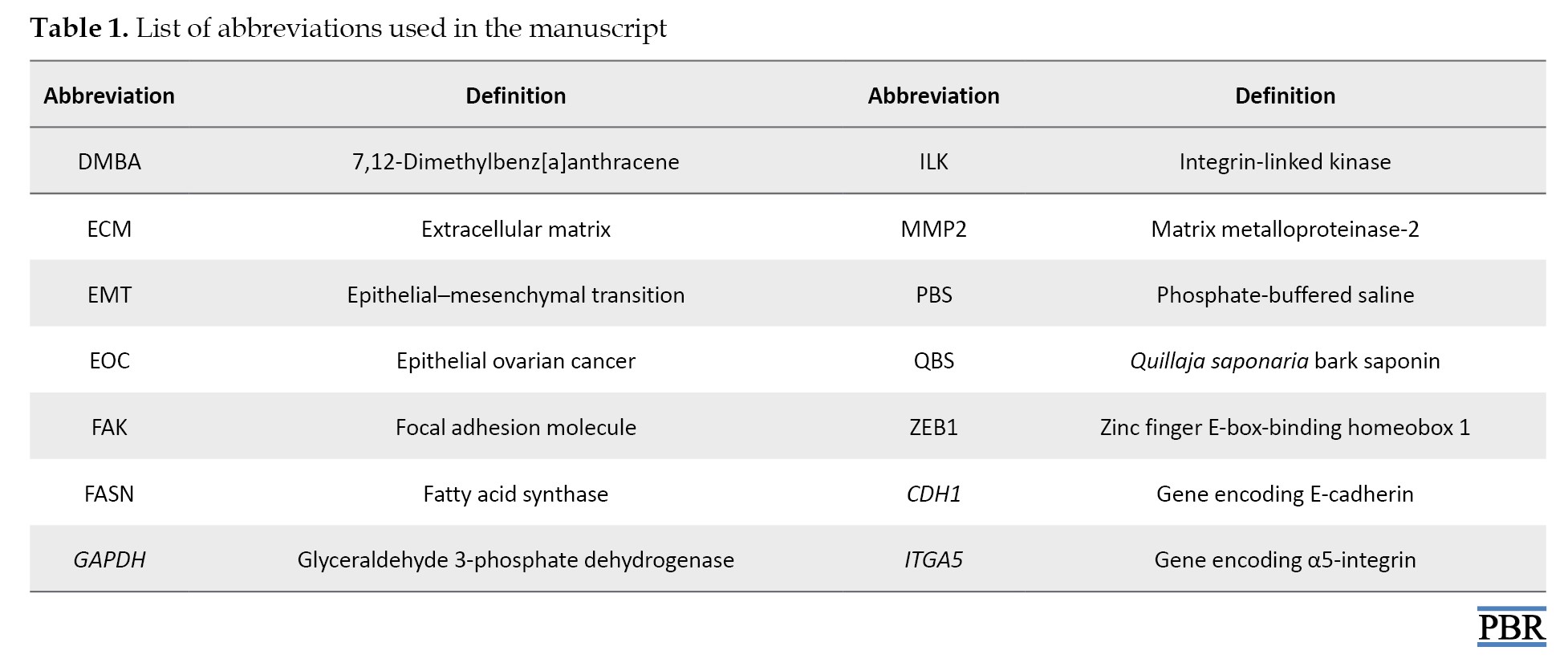
QBS powder (Cat No: S4521) was purchased from Sigma-Aldrich (USA). According to the manufacturer´s specifications, the product contained 25-35% sapogenins. A2780 and SKOV3 human ovarian cancer cell lines were obtained from the Gene-Iran laboratory (Tehran, Iran), and the NIH3T3 normal fibroblast cell line was obtained from National Cell Bank of Iran (NCBI). Roswell Park Memorial Institute (RPMI)-1640 and DMEM, fetal bovine serum (FBS), penicillin/streptomycin, and trypsin/EDTA were purchased from Bioidea (Tehran, Iran). 3-(4,5-dimethylthiazol-2-yl)-2,5-diphenyltetrazolium bromide (MTT) and DMBA solutions were prepared from Merck (Germany). RNA isolation (Cat No: A101231), complementary DNA (cDNA) synthesis (Cat No: A101161), and SYBR Green master mix (Cat No: C101051, C101052) were purchased from Parstous (Tehran, Iran). The abbreviations used in this study are presented in Table 1.
Preparation of Q. saponaria bark saponin stock and working solutions
A stock solution was prepared by dissolving QBS in sterile phosphate-buffered saline. The solution was sterilized by filtration through a 0.22-µm syringe filter and stored at 4 °C until use. Immediately before each experiment, the stock solution was diluted with complete culture medium to prepare working concentrations of 20, 30, and 80 µg/mL.
Cell culture
The study selected two ovarian cancer cell lines (A2780 and SKOV3) to assess the effects of QBS. Also, NIH3T3, a normal fibroblast cell line, was also cultured to assess the selectivity of QBS against ovarian cancer cells. A2780 and SKOV3 human ovarian cancer cells were cultured in RPMI-1640 medium with 1% antibiotics and 10% FBS, and NIH3T3 cells were cultured in DMEM under standard conditions (37 °C, 5% CO2 incubator).
Cell viability assay
The MTT assay relies on the capacity of live cells to convert the yellow MTT tetrazolium salt into purple formazan crystals. MTT passes through the cell membrane and the mitochondrial inner membrane of viable cells, and it is reduced to purple formazan crystals that produce a quantifiable colorimetric signal [26]. A2780, SKOV3, and NIH3T3 cells were cultured (104 cells/well) in a 96-well plate to determine the effect of QBS on cell survival. After 24 h, NIH3T3, SKOV3, and A2780 cells were treated with QBS at 20, 30, and 80 μg/mL, or without QBS (control), for 24 hours, then 40 µL of MTT solution (5 mg/mL) was added to each well. Following 3 hours of incubation, 80 of DMSO was added to each well. Cell viability was determined. Four hours later, DMSO was added to each well to dissolve the formazan crystals, and then the absorbance of each well was recorded at 570 nm using an ELISA reader, a microplate reader that measures the absorbance of solubilized formazan, indicating cell viability. The optimal wavelength for measuring formazan is 570 nm. The MTT assay was performed in three independent biological experiments, with each experimental concentration analyzed in triplicate.
In vitro scratch wound assay
Cell migration ability was assessed using a scratch wound-healing assay. A2780 and SKOV3 cells (2×10⁵) were cultured in a 6-well plate in RPMI-1640 medium with 1% FBS; when the cell density reached 70%, a wound was created with a sterile pipette tip. Detached cells were removed, and the wound was checked after 24 h of treatment with IC50 and sub-IC50 values of 20 and 30 µg/mL. Finally, cell invasion into the wound area was assessed by inverted microscopy, and images were captured. Then, the distance of cells from the scratch area was measured using ImageJ. The average scraped width per well was measured and subtracted from the pre-treatment measurement. The change in the width for each experimental condition was compared with the control.
RNA extraction and quantitative real-time polymerase chain reaction
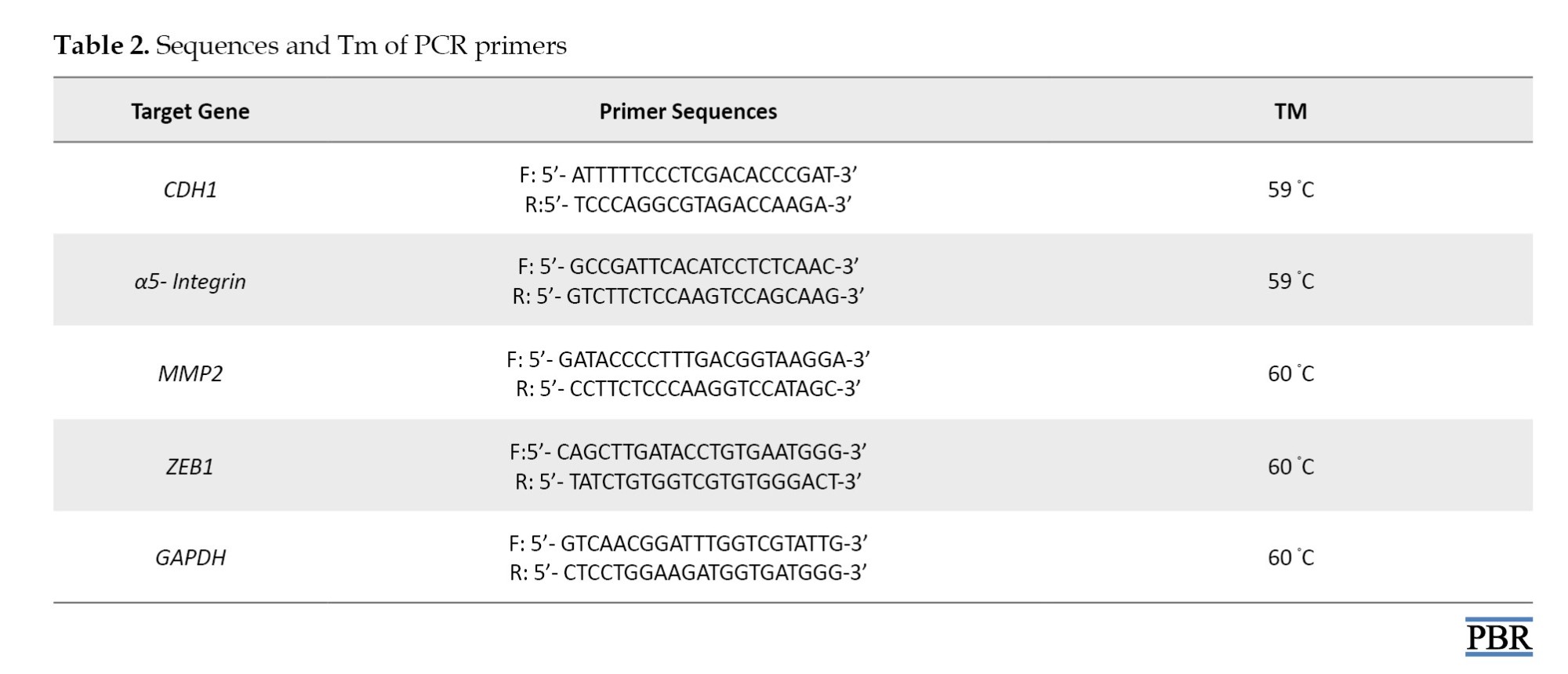
Total RNA was extracted from treated and untreated A2780 and SKOV3 cells using a HighPure RNA isolation kit (ParsTous, Iran) according to the manufacturer’s protocol. RNA concentration and purity were measured with a Nanodrop spectrophotometer, and RNA integrity was verified by assessing the A260/A280 and A260/A230 ratios. One µg of high-quality RNA was used for cDNA synthesis with the Easy cDNA Synthesis Kit (ParsTous, Iran). Quantitative real-time polymerase chain reaction (qRT-PCR) amplification was performed using RealQ Plus 2x Master Mix Green (Ampliqon, Denmark) on an Eppendorf Mastercycler EP Realplex instrument. Primer sequences used for amplification are listed in Table 2.
Cycling conditions were as follows: initial denaturation at 95 °C for 10 min, followed by 40 cycles of denaturation at 95 °C for 15s, annealing at 56 °C for 30s, and extension at 72 °C for 30s. Specificity of amplification confirmed by melting curve analysis. Relative expression levels of target genes were normalized against GAPDH as an internal control and calculated using the 2−ΔΔCt method. Primer efficiencies confirmed to fall within the acceptable range (90–110%), ensuring reliable quantification.
Induction of ovarian cancer and in vivo study design
Female Wistar rats (180-220 g, 6-8 weeks old) were obtained from the Animal Center Resources and Research of Royan Institute. Animals were housed in polypropylene cages under controlled conditions (20-22 ºC temperature, 12 hours light/dark cycle, 40-50% humidity, and no noise pollution). The animals were acclimated for 10 days and fed a standard pellet diet and water. Ethical principles regarding the treatment of laboratory animals were observed. For tumor induction, rats were anesthetized with an intraperitoneal injection of 10% ketamine and 2% xylazine. Then, a single dose of DMBA (4 mg in 0.02 mL of sesame oil) was surgically delivered to the right ovary, and the ovaries were returned intact to the abdominal cavity. The muscle and skin layers were closed using a silk suture, and a prophylactic antibiotic was administered for a week post-surgery. Then, 33 rats were randomly assigned to three groups (n=11 in each group). The complete in vivo study was described below:
Group I (control group): No DMBA injection. Group II (DMBA+saline): Received an intraperitoneal injection of normal 0.9% saline (vehicle of saponin). Group III (DMBA+QBS): Treated with QBS administered intraperitoneally (at a concentration of 16 mg/kg; based on the dose-response assay).
Treatment was initiated 10 days after DMBA administration. QBS was administered at 16 mg/kg every 12 days for three consecutive cycles, starting 10 days after DMBA administration (days 10, 22, and 34). All rats were euthanized 7 days after the final injection, and the ovaries were collected for histological analysis [27].
Histomorphological assessment
Ovarian tissue was fixed in Bouin’s solution for 24 h, dehydrated in a series of ethanol (70-100%), embedded in paraffin, and sectioned into 5 μm sesctions. After rehydration and hematoxylin and eosin staining, the changes in ovarian tissue structure were observed by an optical microscope.
Statistical analysis
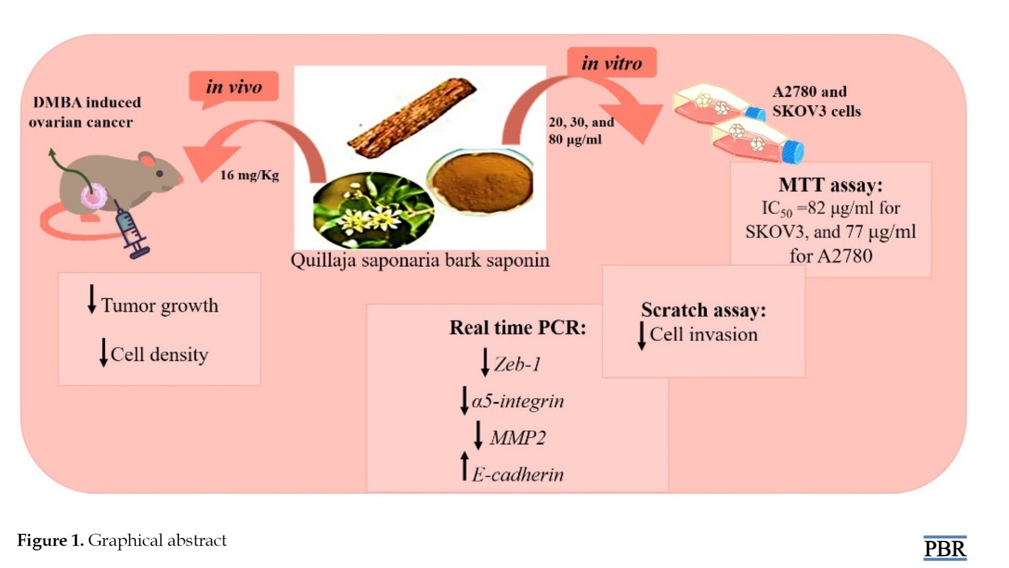
All results were obtained from three separate experiments and performed in quadruplicate (n=12). The statistical significance was evaluated using SPSS software, version 22 with one-way analysis of variance, and the MTT assay was evaluated using Duncan’s test for cell cytotoxicity, metabolic activity, and cell viability analysis. Data are presented as the Mean±SD. P<0.05 are considered significant. The experimental means were compared with those of untreated cells harvested in parallel (Figure 1).
Ovarian cancer is a fatal gynecological cancer ranking eighth among the leading causes of ca)ncer–related mortality in women worldwide. Epithelial ovarian cancer (EOC) is the most widespread subtype, including more than 95% of all ovarian cancer cases [1]. Based on surveys, ovarian cancer cells are very invasive; therefore, suppression of their invasion is considered a therapeutic approach for oncological research. In this regard, , epithelial cells must undergo epithelial–mesenchymal transition (EMT) to gain metastatic potential, a crucial step in cancer progression during which cancer cells acquire the invasive abilities necessary to complete all steps of the metastatic cascade [2, 3]. Related to EMT, metastasis of malignant epithelial cells requires lysis of the ECM by matrix metalloproteinase (MMP) enzymes. MMPs are zinc-dependent enzymes that play an essential role in degrading the basement membrane and ECM proteins. MMP2 overexpression in malignant ovarian cancer is related to the metastasis of ovarian cancer [4]. MMP2, a key member of this family, digests type IV collagen and is overexpressed in EOC, which correlates with poor survival [5-7]. Integrins are adhesion molecules in the cell surface containing α and β subunits, which have different functions in cells. Notably, tumor cells can resist anoikis by expressing ITGA5. In addition, it was reported that high expression of ITGA5 in tumor cells is associated with cell survival attenuation [8-10]. Studies showed that ITGA5 overexpression initiates transformed cells towards a more malignant phenotype and metastasis in malignant ovarian cells [11]. On the other hand, CDH1, as an epithelial marker and Ca-dependent glycoprotein, is involved in cellular connections and controls metastatic progression. Studies showed that when CDH1 is overexpressed, cancer cells are less likely to invade [12, 13]. In this regard, zinc finger E-box binding homeobox 1 (ZEB1) is a major transcription factor that plays a central role in EOC progression and stimulates EMT by downregulating epithelial genes, including CDH1, and increasing the metastatic ability of EOC [14, 15].
With the progression of ovarian cancer stage, the conventional therapeutic strategies, including surgery, chemotherapy, immunotherapy, and radiotherapy have indicated minimal success [16]. Meanwhile, natural compounds are considered promising sources of anticancer agents [17].
Saponins are triterpenoids found in many plant species, such as sunflower, chickpea, soybean, sugar beet, ginseng, and oats, and exhibit cytotoxic activity and play a protective role in plants by defending against predators. Many biological effects of saponins, such as anti-inflammatory and anti-microbial effects, cell membrane permeabilization, immune system stimulation, and cytotoxic attributes, have also been reported [18-20].
According to the literature, saponins have exhibited many pharmacological effects; however, their application is limited due to their in vitro hemolytic activity. Nevertheless, this hemolytic property is not the same in all saponins. Quillaja saponaria bark saponin (QBS), in particular, exhibits minimal hemolytic effects, so it was previously approved as a vaccine adjuvant [21]. Previous studies have demonstrated that saponin compounds can exert anti-cancer effects via modulation of EMT and invasion of ovarian and other cancer cells [19, 20]. Despite extensive evidence of the anti-proliferation and anti-invasion effects of saponins, the anti-migratory effects of QBS in ovarian cancer have not been fully elucidated. In the present study, SKOV3 and A2780 cell lines were used as ovarian cancer cell lines. A2780 cells are more sensitive than SKOV3 cells to treatment with fluorouracil, oxaliplatin, paclitaxel, etc. Furthermore, to provide more evidence supporting the potential anti-cancer properties of QBS, its effects were also tested in the Wistar rat model, which induced ovarian cancer with 7,12-Dimethylbenz[a]anthracene (DMBA) as carcinogen [19-22]. The DMBA is commonly used in animal models to induce breast cancer, skin cancer, and ovarian cancer. Studies have shown that DMBA-induced ovarian cancer resembles human ovarian cancer in terms of histology, molecular, and dissemination patterns. Thus, a DMBA-induced ovarian cancer model can be useful in human ovarian cancer studies [23, 24]. The SKOV3 cell line is a human ovarian adenocarcinoma derived from ascitic fluid, and the A2780 cell line is known as a human ovarian carcinoma isolated from tumor tissue. Besides, A2780 cells are more sensitive than SKOV3 cells to oxaliplatin, fluorouracil, gemcitabine, paclitaxel, etc. [25]. Furthermore, ITGA5, MMP2, ZEB1, and CDH1 genes were selected because of their well-documented involvement in EMT and their recognized roles in tumor progression and metastatic dissemination. In this study, we aimed to evaluate the effects of QBS on EMT-related genes in A2780 and SKOV3 human ovarian cancer cell lines.
Materials and Methods
Chemicals and reagents
QBS powder (Cat No: S4521) was purchased from Sigma-Aldrich (USA). According to the manufacturer´s specifications, the product contained 25-35% sapogenins. A2780 and SKOV3 human ovarian cancer cell lines were obtained from the Gene-Iran laboratory (Tehran, Iran), and the NIH3T3 normal fibroblast cell line was obtained from National Cell Bank of Iran (NCBI). Roswell Park Memorial Institute (RPMI)-1640 and DMEM, fetal bovine serum (FBS), penicillin/streptomycin, and trypsin/EDTA were purchased from Bioidea (Tehran, Iran). 3-(4,5-dimethylthiazol-2-yl)-2,5-diphenyltetrazolium bromide (MTT) and DMBA solutions were prepared from Merck (Germany). RNA isolation (Cat No: A101231), complementary DNA (cDNA) synthesis (Cat No: A101161), and SYBR Green master mix (Cat No: C101051, C101052) were purchased from Parstous (Tehran, Iran). The abbreviations used in this study are presented in Table 1.
Preparation of Q. saponaria bark saponin stock and working solutions
A stock solution was prepared by dissolving QBS in sterile phosphate-buffered saline. The solution was sterilized by filtration through a 0.22-µm syringe filter and stored at 4 °C until use. Immediately before each experiment, the stock solution was diluted with complete culture medium to prepare working concentrations of 20, 30, and 80 µg/mL.
Cell culture
The study selected two ovarian cancer cell lines (A2780 and SKOV3) to assess the effects of QBS. Also, NIH3T3, a normal fibroblast cell line, was also cultured to assess the selectivity of QBS against ovarian cancer cells. A2780 and SKOV3 human ovarian cancer cells were cultured in RPMI-1640 medium with 1% antibiotics and 10% FBS, and NIH3T3 cells were cultured in DMEM under standard conditions (37 °C, 5% CO2 incubator).
Cell viability assay
The MTT assay relies on the capacity of live cells to convert the yellow MTT tetrazolium salt into purple formazan crystals. MTT passes through the cell membrane and the mitochondrial inner membrane of viable cells, and it is reduced to purple formazan crystals that produce a quantifiable colorimetric signal [26]. A2780, SKOV3, and NIH3T3 cells were cultured (104 cells/well) in a 96-well plate to determine the effect of QBS on cell survival. After 24 h, NIH3T3, SKOV3, and A2780 cells were treated with QBS at 20, 30, and 80 μg/mL, or without QBS (control), for 24 hours, then 40 µL of MTT solution (5 mg/mL) was added to each well. Following 3 hours of incubation, 80 of DMSO was added to each well. Cell viability was determined. Four hours later, DMSO was added to each well to dissolve the formazan crystals, and then the absorbance of each well was recorded at 570 nm using an ELISA reader, a microplate reader that measures the absorbance of solubilized formazan, indicating cell viability. The optimal wavelength for measuring formazan is 570 nm. The MTT assay was performed in three independent biological experiments, with each experimental concentration analyzed in triplicate.
In vitro scratch wound assay
Cell migration ability was assessed using a scratch wound-healing assay. A2780 and SKOV3 cells (2×10⁵) were cultured in a 6-well plate in RPMI-1640 medium with 1% FBS; when the cell density reached 70%, a wound was created with a sterile pipette tip. Detached cells were removed, and the wound was checked after 24 h of treatment with IC50 and sub-IC50 values of 20 and 30 µg/mL. Finally, cell invasion into the wound area was assessed by inverted microscopy, and images were captured. Then, the distance of cells from the scratch area was measured using ImageJ. The average scraped width per well was measured and subtracted from the pre-treatment measurement. The change in the width for each experimental condition was compared with the control.
RNA extraction and quantitative real-time polymerase chain reaction
Total RNA was extracted from treated and untreated A2780 and SKOV3 cells using a HighPure RNA isolation kit (ParsTous, Iran) according to the manufacturer’s protocol. RNA concentration and purity were measured with a Nanodrop spectrophotometer, and RNA integrity was verified by assessing the A260/A280 and A260/A230 ratios. One µg of high-quality RNA was used for cDNA synthesis with the Easy cDNA Synthesis Kit (ParsTous, Iran). Quantitative real-time polymerase chain reaction (qRT-PCR) amplification was performed using RealQ Plus 2x Master Mix Green (Ampliqon, Denmark) on an Eppendorf Mastercycler EP Realplex instrument. Primer sequences used for amplification are listed in Table 2.
Cycling conditions were as follows: initial denaturation at 95 °C for 10 min, followed by 40 cycles of denaturation at 95 °C for 15s, annealing at 56 °C for 30s, and extension at 72 °C for 30s. Specificity of amplification confirmed by melting curve analysis. Relative expression levels of target genes were normalized against GAPDH as an internal control and calculated using the 2−ΔΔCt method. Primer efficiencies confirmed to fall within the acceptable range (90–110%), ensuring reliable quantification.
Induction of ovarian cancer and in vivo study design
Female Wistar rats (180-220 g, 6-8 weeks old) were obtained from the Animal Center Resources and Research of Royan Institute. Animals were housed in polypropylene cages under controlled conditions (20-22 ºC temperature, 12 hours light/dark cycle, 40-50% humidity, and no noise pollution). The animals were acclimated for 10 days and fed a standard pellet diet and water. Ethical principles regarding the treatment of laboratory animals were observed. For tumor induction, rats were anesthetized with an intraperitoneal injection of 10% ketamine and 2% xylazine. Then, a single dose of DMBA (4 mg in 0.02 mL of sesame oil) was surgically delivered to the right ovary, and the ovaries were returned intact to the abdominal cavity. The muscle and skin layers were closed using a silk suture, and a prophylactic antibiotic was administered for a week post-surgery. Then, 33 rats were randomly assigned to three groups (n=11 in each group). The complete in vivo study was described below:
Group I (control group): No DMBA injection. Group II (DMBA+saline): Received an intraperitoneal injection of normal 0.9% saline (vehicle of saponin). Group III (DMBA+QBS): Treated with QBS administered intraperitoneally (at a concentration of 16 mg/kg; based on the dose-response assay).
Treatment was initiated 10 days after DMBA administration. QBS was administered at 16 mg/kg every 12 days for three consecutive cycles, starting 10 days after DMBA administration (days 10, 22, and 34). All rats were euthanized 7 days after the final injection, and the ovaries were collected for histological analysis [27].
Histomorphological assessment
Ovarian tissue was fixed in Bouin’s solution for 24 h, dehydrated in a series of ethanol (70-100%), embedded in paraffin, and sectioned into 5 μm sesctions. After rehydration and hematoxylin and eosin staining, the changes in ovarian tissue structure were observed by an optical microscope.
Statistical analysis
All results were obtained from three separate experiments and performed in quadruplicate (n=12). The statistical significance was evaluated using SPSS software, version 22 with one-way analysis of variance, and the MTT assay was evaluated using Duncan’s test for cell cytotoxicity, metabolic activity, and cell viability analysis. Data are presented as the Mean±SD. P<0.05 are considered significant. The experimental means were compared with those of untreated cells harvested in parallel (Figure 1).
Results
Effects of Q. saponaria bark saponin on viability of SKOV3 and A2780 cells in a dose-dependent manner
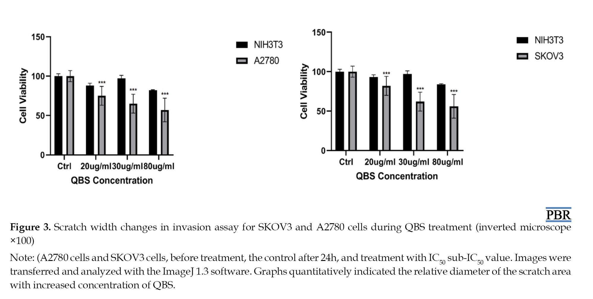
In SKOV3 cells, QBS at concentrations of 20, 30, and 80 µg/mL reduced cell survival percentage to 78.9±0.4, 61.4±0.2, and 52.6±0.1, respectively. In A2780 cells, cell viability under exposure to QBS at concentrations of 20, 30, and 80 µg/mL was 75.1±0.09, 64.4±0.06, and 45.9±0.1 (Figure 2).
The QBS also did not significantly reduce the viability of NIH3T3 fibroblast cells as compared with untreated ovarian cancer cells. According to the MTT assay, the IC50 value were 82 µg/mL for SKOV3 cells and 77 µg/mL for A2780 cells. IC50 is the most commonly used and informative measure of a drug’s efficacy. It shows how much drug is needed to reduce a biological process by half, helping measure the potency of an antagonist drug in pharmacological research. A nonlinear regression model in GraphPad Prism software, version 10.1.1 calculated IC50 values.
Q. saponaria bark saponin exerts the anti-migratory effect
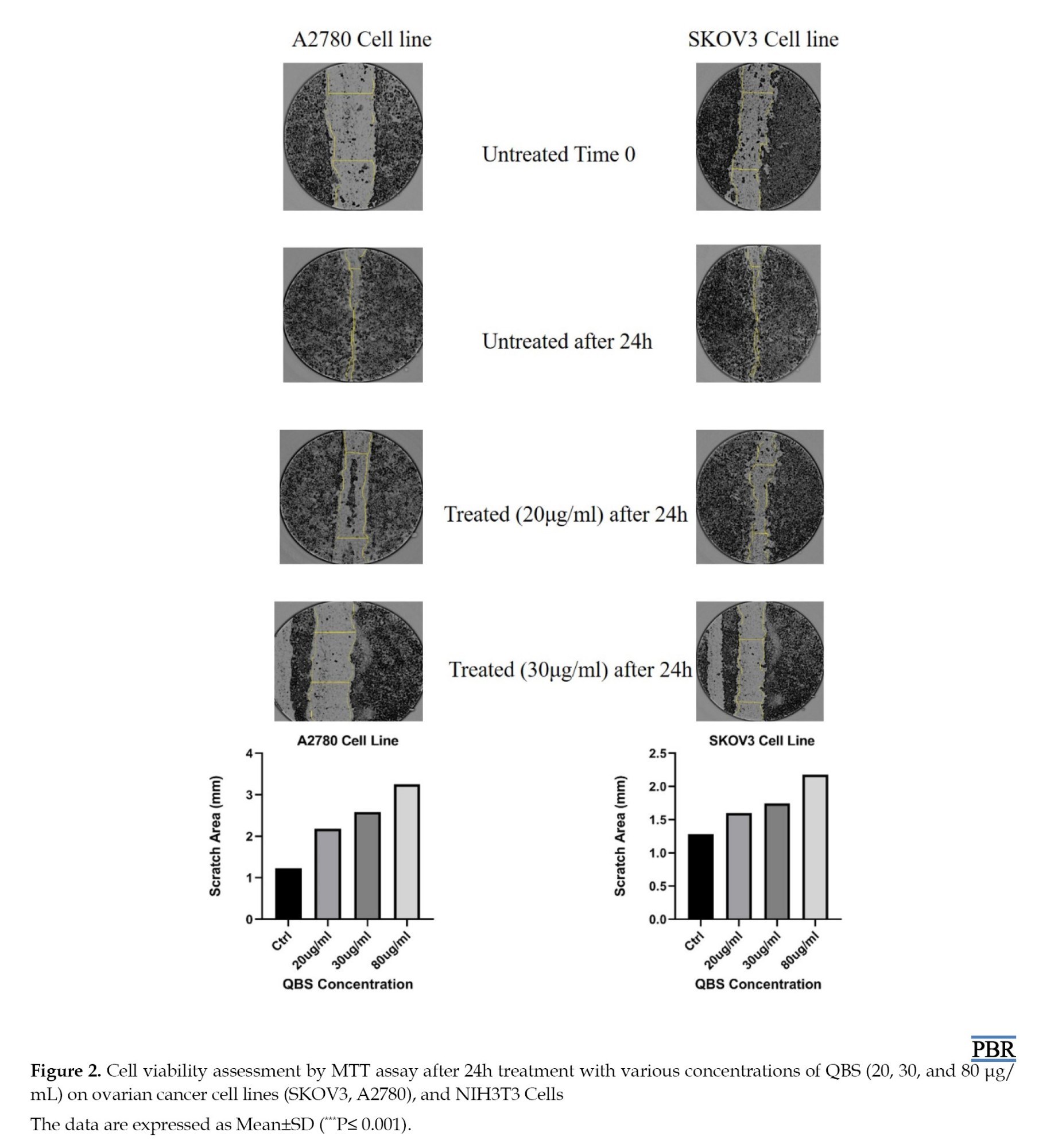
The results showed that QBS reduced cell migration in a dose-dependent manner at concentrations below IC50 (20 and 30 µg/mL). As shown in Figure 3, untreated ovarian cancer cells migrated into the scratched area and substantially closed the wound after 24 h (97%, 98%; A2780 and SKOV3 cells, respectively).
In contrast, QBS treatment reduced wound closure relative to the control, indicating decreased cell migration. This effect was more pronounced after exposure with 30 µg/mL of QBS (19%, 24%) than 20 μg/mL (33%, 42%) in both A2780 and SKOV3 cells, suggesting that QBS at 30 µg/mL may reduce the migratory potential of both ovarian cancer cell lines only after 24 h of treatment.
Gene expression analysis by quantitative real-time polymerase chain reaction
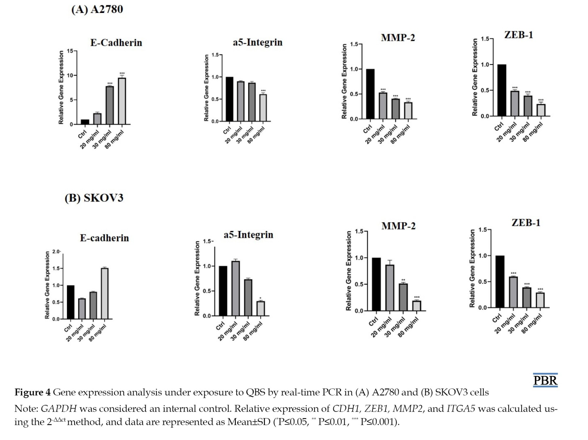
To identify the anti-EMT molecular mechanism of QBS, the messenger RNA (mRNA) expressions of factors associated with the regulation of cell adhesion were evaluated by RT-PCR (Figure 4).
Gene expression analysis by quantitative real-time polymerase chain reaction
To identify the anti-EMT molecular mechanism of QBS, the messenger RNA (mRNA) expressions of factors associated with the regulation of cell adhesion were evaluated by RT-PCR (Figure 4).
In this regard, A2780 and SKOV3 ovarian cancer cells were treated with IC50 and sub-IC50 values of QBS. Statistical analysis showed that in the SKOV3 cell line, ZEB1 expression was significantly decreased under exposure to IC50 and sub-IC50 values of QBS (***P≤0.001). The expression of MMP2 was attenuated at IC50 and sub-IC50 values of QBS (***P≤0.001 and **P≤0.01). Data analysis showed that the ITGA5 expression was significantly down-regulated at the IC50 value of QBS (*P≤0.05). CDH1 expression analysis indicated non-significant down-regulation at a sub-IC50 value of QBS, and up-regulation at the IC50 value of QBS, but was not considered statistically significant. In the A2780 ovarian cancer cell line, statistical analysis revealed that ZEB1 and MMP2 expression were significantly attenuated at the IC50 value and sub-IC50 values of QBS (***P≤0.001). ITGA5 expression was slightly decreased at the sub-IC50 value of QBS, which was not statistically significant. In addition, the expression of ITGA5 showed significant down-regulation at the IC50 value of QBS (***P≤0.001). CDH1 expression was non-significantly overexpressed at sub-IC50 values of QBS, and at the IC50 value of QBS, this overexpression was noticeable (***P≤0.001).
Histomorphological changes
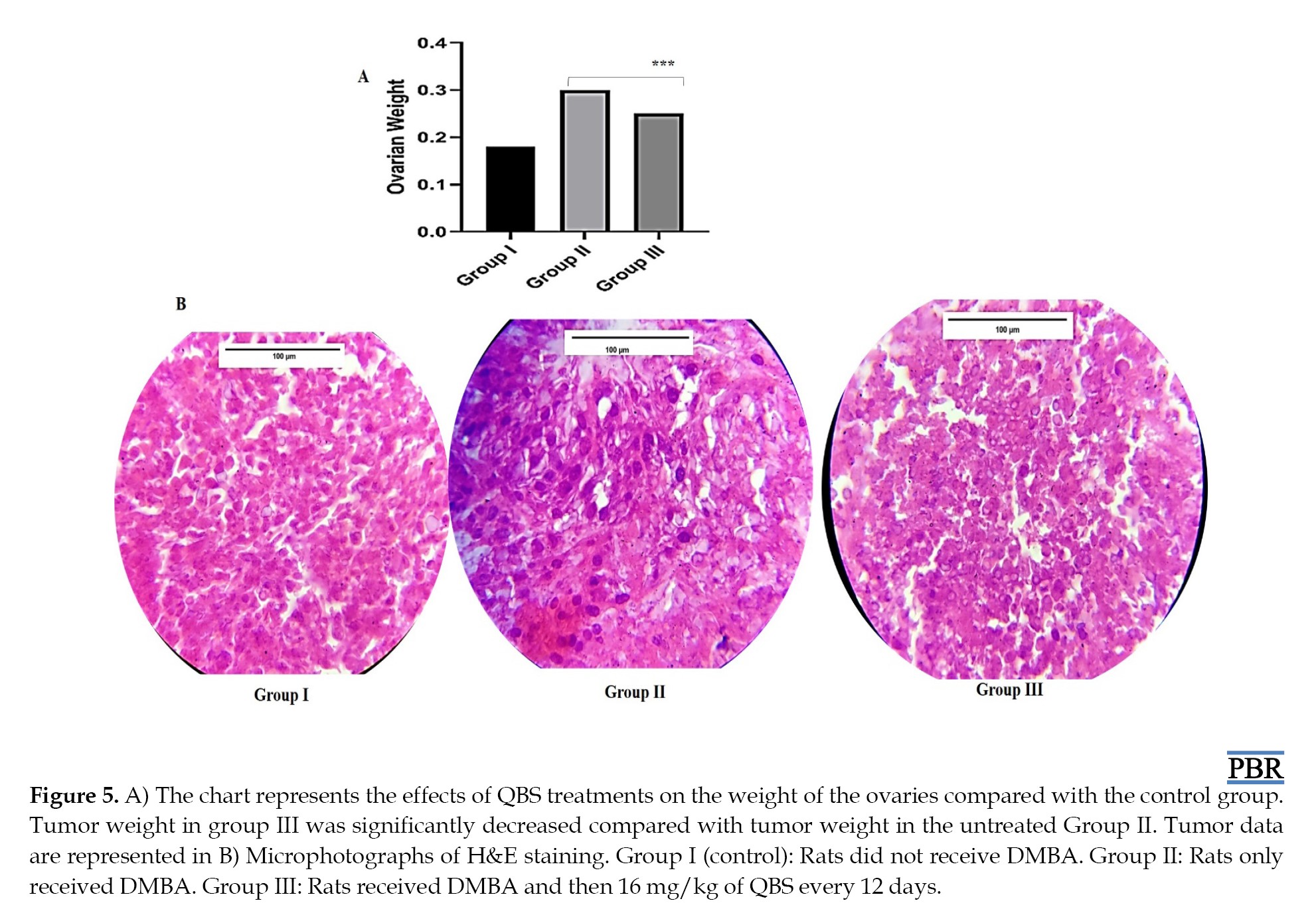
The mean ovarian weight was 0.19±0.01 in group I, 0.3±0.1 in group II, and 0.21±0.08 in group III. Ovary’ weight analysis indicated a significant increase in mean ovarian weight in the DMBA-exposed group (group II) compared to the control group (group I) (***P≤0.001), indicating successful induction of ovarian tumor formation. The results also showed that the tumor weight was significantly decreased in the QBS-administered experimental group (Group III) compared with group II (***P≤0.001), suggesting a suppressive effect of QBS on tumor growth, while there was no significant difference between the mean tumor weight in the QBS-administered group (group III) and ovarian weight in the control group (group I) (P>0.05) (Figure 5A).
As shown in Figure 5B, histopathological evaluation of ovarian tissues revealed that the control group (group I) preserved cell structure with normal stromal organization without neoplastic alterations. In the DMBA-exposed group (group II), ovarian tissues exhibited significant histopathological abnormalities characterized by dense cellular proliferation, nuclear hyperchromasia, and cellular pleomorphism, indicating tumor progression. In contrast, ovarian tissues from the QBS-administered group (group III) showed fewer histopathological changes compared to group II. QBS treatment reduced cellular density and partially improved tissue structure.
Discussion
Ovarian cancer is the most lethal gynecologic cancer, with malignant metastasis and poor prognosis. Effective components that can block metastasis and EMT in ovarian cancer may be used as a new alternative drug for the suppression of tumor invasion and metastasis [28]. Blocking or reversing EMT in tumor cells can reduce resistance to chemotherapy, radiotherapy, and immunotherapy [29].
In this study, we found that QBS has cytotoxic effects against SKOV3 and A2780 ovarian cancer cells with minimum cytotoxicity in normal fibroblast cells. Based on these findings, administration of 16 mg/kg QBS to the rat´s ovarian tumor reduced tumor cell burden and ovarian weight compared with untreated DMBA-induced ovaries, suggesting a potential inhibitory effect on DMBA-induced ovarian tumor development. In addition, the scratch wound assay indicated the inhibitory effect of QBS on ovarian cancer cell migration in the IC50 value of QBS. To confirm QBS’s inhibitory effect on cell migration, the expression of ovarian cancer EMT-related genes has been evaluated. RT-PCR analysis exhibited that QBS influenced EMT and migration in both SKOV3 and A2780 cell lines by up-regulation of CDH1 and down-regulation of ZEB1, MMP2, and ITGA5.
CDH1 is one of the most frequently expressed epithelial adhesion molecules in transformed ovarian epithelia, and decreased CDH1 expression in ovarian cancer tissue may promote disease progression and an invasive phenotype [30]. Related to CDH1, down-regulation of CDH1 causes free β-catenin to migrate the cell nucleus and promotes transcription of metastasis markers such as ZEB1 and MMPs [31]. ZEB1 is a transcriptional repressor that can inhibit the transcription of CDH1 by binding to the CDH1 promoter, and the enzymatic activity of MMP2 increases with increasing ZEB1 [32]. Further, ZEB1 overexpression inhibited cell apoptosis in EOC [33]. Therefore, suppressing ZEB1 can decrease EMT by reducing MMP2-mediated digestion and restoring CDH1 expression, making it a valuable strategy against tumor development.
α5-integrin is a focal adhesion molecule that can remodel or stabilize adhesion junctions like CDH1, and it can play a role in CDH1 endocytosis [34]. On the other hand, recent studies have shown that adhesive molecules, such as cadherins and integrins interact in many ways, including crosstalk with each other [35, 36]. It was shown that ovarian cancer cells isolated from the primary tumor started developing mesenchymal characteristics when floated in peritoneal fluid, and they could resist the anoikis by expressing factors, such as ITGA5 [10]. In addition, ITGA5 expression can induce tumor growth and migration; therefore, the connection between α5-integrin and fibronectin interaction triggers a signaling cascade to start up that induces focal adhesion molecule phosphorylation and activation via integrin-linked kinase and the integrin intracellular domain. Activation of the PI3K/AKT pathway and AKT phosphorylation inhibit apoptosis, which triggers EMT during cancer progression. On the other hand, activated AKT up-regulates ZEB1 and promotes metastasis [10, 37].
In the present study, statistical analysis showed that ZEB1 and MMP2 expression were significantly decreased at all concentrations (***P≤0.001). Expression of ITGA5 at the IC50 value significantly decreased (***P≤0.001). In addition, CDH1 at sub-IC50 and IC50 values of QBS was significantly overexpressed in A2780(***P≤0.001).
In recent years, many studies have examined the role of EMT and the molecules involved in this process, and the anti-metastatic function of naturally occurring triterpenoids, such as saponins. Zhang et al. indicated that CDH1 downregulation may play an important role in gastric cancer progression and metastasis [38]. Mou et al. showed the inhibitory effect of Paris saponin VII on A2780 cell EMT by up-regulation of CDH1 [39]. On the other hand, Chen et al. evaluated the effect of Paris saponin H on hepatocarcinoma cells in vitro and showed suppression of tumor growth and EMT under Paris saponin H treatment in vitro and in vivo [40]. Zhu et al. showed that pulsatilla saponin E suppressed A549 NSCLC cell invasion with inhibition of lipid rafts and negatively regulated the Akt/FASN pathway [41]. Kim et al. demonstrated that Panax notoginseng saponin affect induced mammary carcinogenesis in rats by decreasing tumor volume and reducing lipid peroxidation levels [42].
All of the above studies, consistent with our study, have confirmed the cytotoxic effects of saponins. Further, the special role of ZEB1, ITGA5, MMP2, and CDH1 on the EOC metastasis pathway, suppression level of MMP2, ZEB1, and ITGA5, and overexpression of CDH1 level by QBS has been considered this herbal product an essential component against some of the EMT pathways in SKOV3 and A2780 EOC cells.
Accordingly, the results of this study indicated that QBS could be an EMT suppressor agent for ovarian cancer in vitro. Nevertheless, a Western blot to detect proteins would be necessary to confirm the proposed molecular mechanisms, using another human epithelial ovarian cell line, such as human ovarian surface epithelium, and further studies on the metastasis pathways regulated by QBS seem obligatory in the rat model.
Conclusion
The current study provides data on the cytotoxicity of QBS against sensitive and resistant ovarian cancer cells and in the ovarian tumor rat model. Further, we provide valuable data associated with the involvement of EMT signaling pathway genes, such as MMP2, ZEB1, CDH1, and ITGA5, in QBS’s potential against ovarian cancer in vitro. Preclinical and clinical research are required to confirm the anti-cancer mechanism of QBS as an EMTmodulating therapeutic agent for clinical applications.
Ethical Considerations
Compliance with ethical guidelines
This study was approved by the Ethics Committee of Kharazmi University, Tehran, Iran (Code: IR.KHU.REC.1401.015).
Funding
This research did not receive any specific grant from funding agencies in the public, commercial, or not-for-profit sectors.
Authors' contributions
Conceptualization, methodology, investigation, data curation, formal analysis, writing the original draft: Raoufeh Koochaki; Supervision, study design and data interpretation: Mohammad Nabiuni; Scientific consultation, data interpretation, review & editing, critical revision: Elaheh Amini; Final approval: All authors.
Conflict of interest
The authors declared no conflicts of interest.
Acknowledgments
The authors thank the Faculty of Biological Sciences, Kharazmi University, Tehran, Iran.
References
- Arora T, Mullangi S, Vadakekut ES, Lekkala MR. Epithelial ovarian cancer. Treasure Island: StatPearls Publishing; 2024. [Link]
- Banyard J, Bielenberg DR. The role of EMT and MET in cancer dissemination. Connect Tissue Res. 2015; 56(5):403-13. [DOI:10.3109/03008207.2015.1060970] [PMID] [PMCID]
- Zhao G, Wang Q, Gu Q, Qiang W, Wei JJ, Dong P, et al. Lentiviral CRISPR/Cas9 nickase vector mediated BIRC5 editing inhibits epithelial to mesenchymal transition in ovarian cancer cells. Oncotarget. 2017; 8(55):94666. [DOI:10.18632/oncotarget.21863] [PMID] [PMCID]
- Gao Q, Li Y. Clinical significance and impact on the cell behaviors of miR-758-5p/MMP-2 axis in ovarian cancer cells. BMC Med Genomics. 2026; 19(1):31. [DOI:10.1186/s12920-025-02303-4] [PMID] [PMCID]
- Carey P, Low E, Harper E, Stack MS. Metalloproteinases in Ovarian Cancer. Int J Mol Sci. 2021; 22(7):3403. [DOI:10.3390/ijms22073403] [PMID] [PMCID]
- Medeiros NI, Gomes JAS, Fiuza JA, Sousa GR, Almeida EF, Novaes RO, et al. MMP-2 and MMP-9 plasma levels are potential biomarkers for indeterminate and cardiac clinical forms progression in chronic Chagas disease. Sci Rep. 2019; 9(1):14170. [DOI:10.1038/s41598-019-50791-z] [PMID] [PMCID]
- Nyante SJ, Wang T, Tan X, Ozdowski EF, Lawton TJ. Quantitative expression of MMPs 2, 9, 14, and collagen IV in LCIS and paired normal breast tissue. Sci Rep. 2019; 9(1):13432. [DOI:10.1038/s41598-019-48602-6] [PMID] [PMCID]
- Mitra AK. Ovarian cancer metastasis: A unique mechanism of dissemination. London: IntechOpen; 2016. [DOI:10.5772/64700]
- Moffitt L, Karimnia N, Stephens A, Bilandzic M. Therapeutic targeting of collective invasion in ovarian cancer. Int J Mol Sci. 2019; 20(6):1466. [DOI:10.3390/ijms20061466] [PMID] [PMCID]
- Wilson RB, Solass W, Archid R, Weinreich FJ, Königsrainer A, Reymond MA. Resistance to anoikis in transcoelomic shedding: The role of glycolytic enzymes. Pleura Peritoneum. 2019; 4(1):20190003. [DOI:10.1515/pp-2019-0003] [PMID] [PMCID]
- Ahmed N, Riley C, Rice GE, Quinn MA, Baker MS. Alpha(v)beta(6) integrin-A marker for the malignant potential of epithelial ovarian cancer. J Histochem Cytochem. 2002; 50(10):1371-80. [DOI:10.1177/002215540205001010] [PMID]
- Dhaliwal D, Shepherd TG. Molecular and cellular mechanisms controlling integrin-mediated cell adhesion and tumor progression in ovarian cancer metastasis: A review. Clin Exp Metastasis. 2022; 39(2):291-301. [DOI:10.1007/s10585-021-10136-5] [PMID] [PMCID]
- Tian J, Liang X, Wang D, Tian J, Liang H, Lei T, et al. TBC1D2 promotes ovarian cancer metastasis via inducing CDH1 degradation. Front Oncol. 2022; 12:766077. [DOI:10.3389/fonc.2022.766077] [PMID] [PMCID]
- Kobayashi M, Sawada K, Kimura T. Potential of integrin inhibitors for treating ovarian cancer: A literature review. Cancers. 2017; 9(7):83. [DOI:10.3390/cancers9070083] [PMID] [PMCID]
- Sakata J, Utsumi F, Suzuki S, Niimi K, Yamamoto E, Shibata K, et al. Inhibition of ZEB1 leads to inversion of metastatic characteristics and restoration of paclitaxel sensitivity of chronic chemoresistant ovarian carcinoma cells. Oncotarget. 2017; 8(59):99482-94. [DOI:10.18632/oncotarget.20107] [PMID] [PMCID]
- Afzal Ashaie M, Hoque Chowdhury E. Cadherins: The superfamily critically involved in breast cancer. Curr Pharm Des. 2016; 22(5):616-38. [DOI:10.2174/138161282205160127095338] [PMID]
- Zhang X, Zhang Z, Zhang Q, Zhang Q, Sun P, Xiang R, et al. ZEB1 confers chemotherapeutic resistance to breast cancer by activating ATM. Cell Death Dis. 2018; 9(2):57. [DOI:10.1038/s41419-017-0087-3] [PMID] [PMCID]
- Akter S, Rahman MA, Hasan MN, Akhter H, Noor P, Islam R, et al. Recent advances in ovarian cancer: Therapeutic strategies, potential biomarkers, and technological improvements. Cells. 2022; 11(4):650. [DOI:10.3390/cells11040650] [PMID] [PMCID]
- Komatsu N, Komatsu M, Ohashi R, Horii A, Hoshi K, Takato T, et al. Saponin facilitates anti‐Robo1 immunotoxin cytotoxic effects on maxillary sinus squamous cell carcinoma. J Oncol. 2020; 2020(1):9593516. [DOI:10.1155/2020/9593516] [PMID] [PMCID]
- Kozińska N, Tokarska K, Chudy M, Wojciechowski K. Cytotoxicity of Quillaja saponaria saponins towards lung cells is higher for cholesterol-rich cells. Biophysica. 2021; 1(2):126-36. [DOI:10.3390/biophysica1020010]
- Ahmed Abdel-Reheim M, Messiha BAS, Abo-Saif AA. Quillaja saponaria bark saponin protects Wistar rats against ferrous sulphate-induced oxidative and inflammatory liver damage. Pharm Biol. 2017; 55(1):1972-83. [DOI:10.1080/13880209.2017.1345950] [PMID] [PMCID]
- Reichert CL, Salminen H, Weiss J. Quillaja saponin characteristics and functional properties. Annu Rev Food Sci Technol. 2019; 10:43-73. [DOI:10.1146/annurev-food-032818-122010] [PMID]
- de Souza BR, Oliveira G, Leme G, Reis IB, Cabral FAT, de Paula JLB, et al. A novel serous ovarian carcinoma model induced by DMBA: Results from OncoTherad®(MRB-CFI-1) immunotherapy preclinical testing. Biomed Pharmacother. 2025; 182:117755. [DOI:10.1016/j.biopha.2024.117755] [PMID]
- Gonçalves ACC, Carvalho KC, Turri JAO, Simoes RdS, Carvalho JP, da Silva LFF, et al. 7, 12-Dimethylbenz (a) anthracene as a model for ovarian cancer induction in rats. Biology. 2025; 14(1):73. [DOI:10.3390/biology14010073] [PMID] [PMCID]
- Beaufort CM, Helmijr JC, Piskorz AM, Hoogstraat M, Ruigrok-Ritstier K, Besselink N, et al. Ovarian cancer cell line panel (OCCP): Clinical importance of in vitro morphological subtypes. Plos One. 2014; 9(9):e103988. [DOI:10.1371/journal.pone.0103988] [PMID] [PMCID]
- Ghasemi M, Liang S, Luu QM, Kempson I. The MTT assay: A method for error minimization and interpretation in measuring cytotoxicity and estimating cell viability. Cell viability assays: Methods and protocols. New York: Springer; 2023. [DOI:10.1007/978-1-0716-3052-5_2] [PMID]
- Keramati K, Mehranpoor M, Babakhani A, Vaezi G, Habibi F. Effect of flunixin on ovarian cancer induced by DMBA in female wistar rats. J Knowl Health Basic Med Sci. 2012; 7(1):44-9. [DOI:10.29252/jnkums.3.4.125]
- Heerboth S, Housman G, Leary M, Longacre M, Byler S, Lapinska K, et al. EMT and tumor metastasis. Clin Trans Med. 2015; 4(1):6. [DOI:10.1186/s40169-015-0048-3] [PMID] [PMCID]
- Nedeljković M, Damjanović A. Mechanisms of chemotherapy resistance in triple-negative breast cancer-how we can rise to the challenge. Cells. 2019 22; 8(9):957. [DOI:10.3390/cells8090957] [PMID] [PMCID]
- Roque R, Costa Sousa F, Figueiredo-Dias M. Epithelial-mesenchymal interconversions in ovarian cancer: The levels and functions of CDH1 in intraabdominal dissemination. Oncol Rev. 2020; 14(2):475. [DOI:10.4081/oncol.2020.475] [PMID] [PMCID]
- Mazda M, Nishi K, Naito Y, Ui-Tei K. CDH1 is transcriptionally activated via suppression of ZEB1 transcriptional repressor by small RNA-mediated gene silencing. Plos One. 2011; 6(12):e28688. [DOI:10.1371/journal.pone.0028688] [PMID] [PMCID]
- Jin M, Yang Z, Ye W, Xu H, Hua X. MicroRNA-150 predicts a favorable prognosis in patients with epithelial ovarian cancer, and inhibits cell invasion and metastasis by suppressing transcriptional repressor ZEB1. Plos One. 2014; 9(8):e103965. [DOI:10.1371/journal.pone.0103965] [PMID] [PMCID]
- Cai W, Zhang Q. The transcription factor ZEB1 mediates the progression of epithelial ovarian cancer by promoting the transcription of CircANKRD17. J Biochem Mol Toxicol. 2022; 36(8):e23086. [DOI:10.1002/jbt.23086] [PMID]
- Nam EH, Lee Y, Moon B, Lee JW, Kim S. Twist1 and AP-1 cooperatively upregulate integrin α5 expression to induce invasion and the epithelial-mesenchymal transition. Carcinogenesis. 2015; 36(3):327-37. [DOI:10.1093/carcin/bgv005] [PMID]
- Weber GF, Bjerke MA, DeSimone DW. Integrins and cadherins join forces to form adhesive networks. J Cell Sci. 2011; 124(8):1183-93. [DOI:10.1242/jcs.064618] [PMID] [PMCID]
- Barcelona-Estaje E, Dalby MJ, Cantini M, Salmeron-Sanchez M. You talking to me? Cadherin and integrin crosstalk in biomaterial design. Adv Healthc Mater. 2021; 10(6):e2002048. [DOI:10.1002/adhm.202002048] [PMID]
- Paoli P, Giannoni E, Chiarugi P. Anoikis molecular pathways and its role in cancer progression. Biochim Biophys Acta. 2013; 1833(12):3481-98. [DOI:10.1016/j.bbamcr.2013.06.026] [PMID]
- Zhang K, Yang G, Wu W, Zhang J, Xia X, Jiang T, et al. Decreased expression of caveolin-1 and CDH1 correlates with the clinicopathologic features of gastric cancer and the EMT process. Recent Pat Anticancer Drug Discov. 2016; 11(2):236-44. [DOI:10.2174/1574892811666160128151437] [PMID]
- Mou K, Wang Z, Wang X, Zhang R, Liu Y, Xu J. Paris saponin VII inhibits epithelial-to-mesenchymal transition in ovarian cancer cell migration and invasion via the GSK-3 beta/beta-catenin signaling pathway. Int J Clin Exp Med. 2017; 12(10):16771-9. [Link]
- Chen T, Lin J, Tang D, Zhang M, Wen F, Xue D, Zhang H. Paris saponin H suppresses human hepatocellular carcinoma (HCC) by inactivation of Wnt/β-catenin pathway in vitro and in vivo. Int J Clin Exp Pathol. 2019; 12(8):2875-86. [PMID]
- Zhu M, Shi W, Chen K, Hu H, Ye X, Jiang Y. Pulsatilla saponin E suppresses viability, migration, invasion and promotes apoptosis of NSCLC cells through negatively regulating Akt/FASN pathway via inhibition of flotillin-2 in lipid raft. J Recept Signal Transduct Res. 2022; 42(1):23-33. [DOI:10.1080/10799893.2020.1839764] [PMID]
- Kim TD, Thanh HN, Thuy DN, Duc LV, Thi TV, Manh HV, et al. Anticancer effects of saponin and saponin-phospholipid complex of Panax notoginseng grown in Vietnam. Asian Pac J Trop Biomed. 2016; 6(9):795-800. [DOI:10.1016/j.apjtb.2016.04.013]
Type of Study: Original Research |
Subject:
Cellular and Molecular Biology
| Rights and permissions | |
 |
This work is licensed under a Creative Commons Attribution-NonCommercial 4.0 International License. |


This is an open access article distributed under the terms of the Creative Commons Attribution License (CC-By-NC), which permits use, distribution, and reproduction in any medium, provided the original work is properly cited and is not used for commercial purposes.
Contact Information