

Volume 12, Issue 3 (2026)
Pharm Biomed Res 2026, 12(3): 187-194 |
Back to browse issues page



Download citation:
BibTeX | RIS | EndNote | Medlars | ProCite | Reference Manager | RefWorks
Send citation to:


BibTeX | RIS | EndNote | Medlars | ProCite | Reference Manager | RefWorks
Send citation to:
Arjmand M, Fazel M Y, Parvaresh A, Shamsipour F, Ghorbani R, Bagheryan A S, et al . Prolonged QT Interval in Methadone Poisoning: A 10-Year Retrospective Study of 1,000 Cases. Pharm Biomed Res 2026; 12 (3) :187-194
URL: http://pbr.mazums.ac.ir/article-1-740-en.html
URL: http://pbr.mazums.ac.ir/article-1-740-en.html
Marziyeh Arjmand1 

 , Mohammad Yousef Fazel2 , Arash Parvaresh3 , Fateme Shamsipour4 , Roshanak Ghorbani5 , Amir Sajad Bagheryan6 , Seyed Ahmadreza Ziaei7 , Niloofar Deravi6 , Mehrnoush Giahi Yazdi1 , Marjan Shariatpanahi8 , Elahe Zare9 , Hamid Owliaey *10
1- Department of Medical Science, Ya.C., Azad University, Yazd, Iran.
2- Department of Toxicology, School of Pharmacy, Shahid Beheshti University of Medical Sciences, Tehran, Iran.
3- Department of Clinical Pharmacy, Faculty of Pharmacy, Shahid Beheshti University of Medical Sciences, Tehran, Iran.
4- Student Research Committee, Department of Toxicology, School of Health, Guilan University of Medical Sciences, Rasht, Iran.
5- Universal Scientific Education and Research Network (USERN), Tehran, Iran.
6- School of Medicine, Shahid Beheshti University of Medical Sciences, Tehran, Iran.
7- Student Research Committee, Department of Medicine, School of Medicine, Fasa University of Medical Sciences, Fasa, Iran.
8- Department of Pharmacology and Toxicology, School of Pharmacy, Iran University of Medical Sciences, Tehran, Iran.
9- Clinical Research Development Center, Afshar Hospital, Shahid Sadoughi University of Medical Sciences, Yazd, Iran.
10- Department of Forensic Medicine and Clinical Toxicology, Ya.C., Islamic Azad University, Yazd, Iran.
Keywords: Methadone, Poisoning, Electrocardiography (ECG), Long QT syndrome, Cardiotoxicity, Opioid-related disorders
Full-Text [PDF 714 kb]
(53 Downloads)
| Abstract (HTML) (264 Views)
Discussion
In this 10-year retrospective observational study of 1,000 hospitalized patients with methadone poisoning, we assessed the frequency of prolonged QT intervals and their clinical correlates. Nearly half of the cohort (48%) had a QT interval ≥450 ms, and the mean QT interval of 478.6±32.1 ms was substantially above the upper normal limit reported in the general population, underscoring the high cardiotoxic potential of acute methadone toxicity through hERG channel inhibition and delayed ventricular repolarization.
The prevalence of QT prolongation observed in our study is broadly consistent with previous reports from the same geographic region. Farsi et al. reported that approximately 50.7% of patients with acute methadone overdose had a QT interval greater than 450 ms, which is comparable to our rate of 48% [15]. Other retrospective Iranian ICU studies have also documented frequent ECG abnormalities in patients with significant methadone toxicity, including STT changes and repolarization disturbances [16]. Minor differences in prevalence estimates across studies may reflect variations in sample size, case mix, and inclusion criteria; our multi-center cohort spanning a 10-year period likely captures a wider spectrum of methadone exposures and poisoning severity.
In terms of demographic correlates, we found that both younger age and female sex were associated with an increased likelihood of QT prolongation. Patients younger than 45 years had approximately 2.6 fold higher odds of prolonged QT compared with those aged ≥45 years, and female patients had about 1.4fold higher odds of QT prolongation than males, in crude analyses. These findings are consistent with prior evidence indicating that age and female sex are important noncardiologic risk factors for druginduced QT prolongation and TdP [17, 18]. Although we used a single QT cutoff of 450 ms for both sexes based on prior studies in methadonetreated populations, sexspecific thresholds based on corrected QT would better reflect underlying physiological differences and may improve risk stratification.
Our data also highlight the complex relationship between methadone dose and QT prolongation. The mean reported methadone dose in this cohort was 75.4±2.25 mg, which is within ranges where prior studies have described dosedependent effects on repolarization [19, 20]. While earlier studies have demonstrated a relatively linear association between methadone dose and QTc prolongation, particularly at higher doses, our descriptive findings suggest that clinically significant QT prolongation can occur even at comparatively lower doses in the setting of acute overdose [20]. In our dataset, a high oral dose alone appeared to be a less reliable predictor of prolonged QT than the actual ECG changes, reinforcing the notion that systemic exposure and cardiotoxicity are strongly influenced by individual variability in metabolism, comorbid conditions, and coadministered medications. Polymorphisms in CYP3A4 and CYP2B6, as well as drug–drug interactions with CYP inhibitors, may further modulate methadone levels and arrhythmic risk.
Electrolyte disturbances emerged as key modifiable contributors to QT prolongation in our cohort. The overall prevalence of hypokalemia was 32.2%, and patients with hypokalemia had more than double the odds of prolonged QT compared with those with normal potassium levels. Hypomagnesemia was also more common among patients with prolonged QT, consistent with the concept of a reduced repolarization reserve when multiple risk factors such as methadone exposure, electrolyte abnormalities, and structural heart disease coexist. Prior experimental and clinical studies have shown that hypokalemia and hypomagnesemia independently prolong ventricular repolarization, facilitate early afterdepolarizations, and increase susceptibility to TdP, particularly in patients receiving QT-prolonging drugs, including methadone [21, 22].
We also observed strong associations between QT prolongation, underlying cardiovascular comorbidity, and clinically significant arrhythmias. Comorbid cardiovascular disease was markedly more frequent in the prolonged QT group, with a crude odds ratio above 20 for QT prolongation, albeit with wide confidence intervals reflecting the low prevalence in the normal QT group. Furthermore, arrhythmias—including TdP and other malignant ventricular rhythms—were substantially more common among patients with prolonged QT, with nearly sixfold higher odds compared with patients without arrhythmias. These findings align with the Cubeddu study that demonstrated druginduced QT prolongation is closely linked to the development of TdP and sudden cardiac death, particularly in individuals with additional risk factors such as structural heart disease, electrolyte imbalances, and concurrent use of other QT-prolonging drugs [23].
Clinically, methadone poisoning in our cohort was associated with significant morbidity and mortality, with an overall inhospital fatality rate of approximately 15% and arrhythmias documented in 15.1% of patients. The combination of QT prolongation, electrolyte disturbances, and comorbid cardiovascular disease was associated with worse outcomes, mirroring prior studies in which prolonged QT or QTc predicted intensive care unit admission and death in methadoneexposed patients [19, 24]. Our results support current expert recommendations advocating early 12 lead ECG acquisition, continuous cardiac monitoring, and aggressive correction of electrolyte abnormalities—particularly potassium and magnesium—to highnormal levels in all suspected cases of methadone overdose.
Several limitations should be considered when interpreting these findings. First, the retrospective design and reliance on hospital records introduced inherent information bias and restricted our ability to control for all potential confounders. Important variables such as the precise time from ingestion to ECG recording, detailed coingestant profiles (including benzodiazepines, alcohol, and other QT-prolonging medications), serum methadone concentrations, and comprehensive measures of hepatic and renal function were not systematically documented and could not be fully accounted for. Second, QT assessment was based on the admission 12-lead ECG and automated measurements of the raw QT interval, rather than manually confirmed QTc values with sexspecific cutoffs. As a result, some misclassification of repolarization abnormalities is possible, and late QT-prolongation occurring after admission may have been missed. Third, because only aggregate data were available at the time of analysis, we could not perform multivariable regression to adjust for confounding between correlated risk factors, such as age, sex, comorbidity, and electrolyte disturbances. Crude OR therefore provide useful but unadjusted estimates of association and should be interpreted with caution. Finally, the study population consisted exclusively of hospitalized patients with sufficiently complete records, which may overrepresent more severe poisonings and limit generalizability to milder cases managed in outpatient or emergency settings.
To better delineate the causal pathways linking methadone exposure, repolarization delay, and arrhythmic outcomes, future studies should employ prospective designs with serial ECG monitoring, formal QTc measurement using standardized correction formulas, and simultaneous assessment of serum methadone levels and coadministered QT-prolonging agents. Incorporating sexspecific QTc thresholds, detailed pharmacogenetic profiling, and multivariable modeling would further clarify the independent contribution of demographic, clinical, and pharmacologic factors to QT prolongation and TdP risk in this highrisk population.
Conclusion
In this 10year retrospective study of 1,000 patients with methadone toxicity, 48% had QT intervals ≥ 450 ms, with a mean QT interval of 478.6 ms, indicating substantial cardiac toxicity and an elevated risk of TdP and ventricular fibrillation. Prolonged QT was more frequent in younger patients and in women and was strongly associated with electrolyte disturbances,particularly hypokalemia, and preexisting cardiovascular disease, all of which were linked to higher rates of malignant arrhythmias and an inhospital mortality rate of 15%. These findings reinforce the need for routine 12lead ECG screening, serial monitoring, and prompt correction of electrolyte abnormalities in all patients with suspected methadone overdose, along with careful evaluation of concomitant QT-prolonging medications and underlying cardiac risk factors.
Ethical Considerations
Compliance with ethical guidelines
The Ethics Committee of the Islamic Azad University, Yazd, Iran (Code: IR.IAU.YAZD.REC.1403.105), in accordance with the principles of the Declaration of Helsinki for research involving human subjects.
Funding
This research did not receive any grant from funding agencies in the public, commercial, or non-profit sectors.
Authors' contributions
Data interpretation: Marziyeh Arjmand, Hamid Owliaey and Marjan Shariatpanahi; Data collection, and drafting of the initial manuscript: Marziyeh Arjmand; Data collection and statistical analysis: Niloofar Deravi; Study design and critical revision of the manuscript: Mehrnoush Giahi Yazdi; Literature review: Marjan Shariatpanahi; Data collection and manuscript editing: Elahe Zare; Study design, supervision, and critically revised: Hamid Owliaey; Final approval: All authors.
Conflict of interest
The authors declared no conflicts of interest.
Full-Text: (27 Views)
Introduction
The global opioid epidemic is one of the top public health issues of the 21st century. According to the United Nations Office on Drugs and Crime, there are an estimated 60 million opioid users worldwide [1]. The rise in overdose deaths due to synthetic opioids has been staggering [1]. The Middle East is a major area with an increase in opioid abuse; for example, in Iran, the drug pattern is dominated by traditional opium along with an increasing amount of methadone that is usually diverted for recreation [2, 3]. Methadone has been the gold standard for treating people suffering from opioid dependence since the 1960s, and it has been shown to be effective in reducing withdrawal symptoms, curbing illicit drug use, and decreasing the transmission of blood-borne pathogens [4-8]. However, it has disadvantages, such as high lipophilicity and a long, unpredictable half-life ranging from 8 to 60 hours.
Methadone is most notably linked to prolonged QT intervals, reflecting the duration of ventricular repolarization on electrocardiography (ECG). A long QT interval increases the risk of untreated (irregularities in heart rhythm) and may occur as a result of two conditions: 1) After depolarization, ventricular recovery resulting in electric shock; and 2) when a premature ventricular contraction (PVC) occurs on top of the prolonged QT. The PVC can result in a potentially fatal arrhythmia called torsades de pointes (TdP) – a form of polymorphic ventricular tachycardia – which can lead to ventricular fibrillation and sudden cardiac death [9]. Unlike many other opioids, methadone acts as a potent inhibitor of the IKr potassium current, which is mediated by the human ether-à-go-go-related gene (hERG) [10]. By blocking these channels, methadone delays potassium efflux during phase 3 of the cardiac action potential, thereby lengthening repolarization time.
The effect of QT interval prolongation by methadone is primarily dose-dependent but can also be affected by many intrinsic patient-specific factors. Examples of factors or conditions that decrease the repolarization reserve include advanced age, being female, underlying structural heart disease, and a genetic predisposition to congenital QT syndrome [11]. Methadone is also metabolized via the cytochrome P450 (CYP450) liver enzyme system. The main CYP450 enzymes involved are CYP3A4, CYP2B6, and CYP2C19. As a result, methadone combined with any CYP450 enzyme inhibitor (for example, erythromycin, a common antibiotic, or some antiretroviral agents) may increase methadone levels in the bloodstream, even when dosages seem reasonable [12].
Systemic physiological disturbances compound acute poisoning or overdose. Opioid toxicity produces a significant decrease in respiratory drive, leading to hypoxia and severe acidosis. These changes affect the electrical stability of the myocardial membranes. Electrolytes are often not retained by chronic users of certain substances because of inadequate nutrition or other medical conditions. Electrolyte abnormalities are critical because hypokalemia and hypomagnesemia can independently increase QT interval length and lower the threshold for TdP [13]. Previous studies have shown that a QT interval exceeding 500 ms is a critical threshold for heightened arrhythmic risk, but significant prolongation can occur even at therapeutic dosages [14].
According to the article, there is a lack of information regarding the frequency of dangerous QT prolongation resulting from methadone overdose in Yazd, Iran. To fill this gap, the researchers will examine 1,000 cases of poisoning from two referral hospitals from 2014-2023 by measuring the QT interval (QT≥450 ms) on the ECG and determining what factors (age, dose, comorbidity, and electrolytes) predict a person’s risk of overdose so that Iran can implement better protocols for overdoses, such as routine electrocardiogram (ECG) monitoring, and timely correction of electrolyte abnormalities to reduce the incidence of fatal cardiac complications.
Materials and Methods
Study design and setting
This retrospective observational (cross-sectional) study was designed to determine the frequency and clinical correlates of QT interval prolongation among patients with methadone poisoning. The study was conducted at two major referral centers for toxicological emergencies in Yazd Province, Iran (Shah Vali Hospital in Yazd City and Shahid Beheshti Hospital in Taft City).
Participants
The study population consisted of 1,000 patients admitted with a confirmed diagnosis of methadone poisoning between January 2014 and December 2023. A consecutive sampling method was used to identify eligible cases from the hospital archives.
Patients were included if they had a clinical diagnosis of methadone poisoning established by the treating physician, were hospitalized (beyond an emergency room visit only) in one of the participating centers during the study period, and had sufficiently complete medical records, including electrocardiographic and laboratory data, to allow extraction of the study variables. Patients with missing core clinical or ECG information or those managed solely as outpatients or discharged from the emergency department without admission were excluded.
Instruments and measures
Data were extracted using a standardized checklist adapted from previous toxicological and cardiology studies. The following domains were recorded:
Demographic and clinical characteristics: Age (years), gender, and the reported dose of methadone consumed (milligrams). Comorbidities and complications: Presence of underlying diseases (e.g. cardiovascular disease, diabetes, hypertension), and in-hospital complications such as apnea, aspiration pneumonia, acute respiratory distress syndrome, and rhabdomyolysis.
Laboratory data: Serum electrolyte levels, specifically potassium, magnesium, calcium, and sodium, were recorded to identify abnormalities such as hypokalemia or hypomagnesemia. Clinical outcomes: The patient’s outcome was categorized as discharge (survival) or death.
Electrocardiographic analysis
The primary outcome measure was the QT interval, obtained from the initial 12lead ECG performed at admission. QT intervals were taken from the automated ECG report generated by the hospital’s standard ECG machines and recorded in milliseconds. For descriptive purposes, the QT interval was analyzed as both a continuous variable and a categorical variable.
Based on prior literature in methadone-treated populations, QT intervals were categorized a priori into two groups:
Group 1 (Normal/Borderline): <450 ms; group 2 (Prolonged): ≥450 ms
This cut-off (≥450 ms) has been suggested as a threshold requiring closer clinical observation in methadone users.
Data collection
Data were collected using both physical medical records and electronic hospital information systems. The researchers obtained necessary administrative approvals from hospital administration and the University Research Council prior to data extraction. Trained research personnel reviewed the archives and extracted relevant data into a standardized data collection form. To protect patient confidentiality, all identifying information was removed, and each case was assigned a unique study ID code.
Statistical analysis
Statistical analyses were conducted using IBM SPSS software, version 26. A two sided P<0.05 was considered statistically significant. Categorical variables were summarized as frequencies and percentages, and continuous variables as Mean±SD with minimum and maximum values.
The normality of continuous variables was evaluated using the Kolmogorov–Smirnov test, which indicated that all continuous variables were approximately normally distributed (P>0.05), supporting the use of parametric tests. The independent samples t-test was used to compare the mean QT interval between patients with normal/borderline QT and those with prolonged QT.
Associations between prolonged QT (≥450 ms) and categorical predictors (age group <45 vs ≥45 years, sex, comorbid status, electrolyte imbalance, and clinical outcome) were initially assessed using Pearson’s chisquare test. For key 2×2 comparisons, crude odds ratios (ORs) with 95% confidence intervals (CIs) were calculated from contingency tables to quantify the strength of association between clinical factors and the presence of QT prolongation. No multivariable regression models were performed because the dataset is retrospective and lacks individuallevel data beyond aggregated counts.
Results
A total of 1,000 patients with methadone overdose were included in the analysis. The mean age was 32.8±9.6 years (range: 18–65 years), with 42.0% (n=420) younger than 45 years and 58.0% (n=580) aged 45 years or older (Table 1).
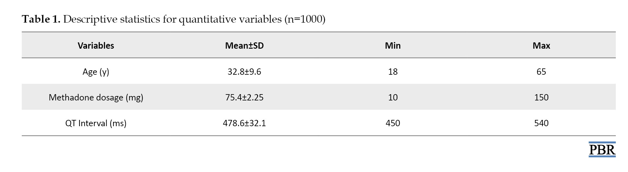
The mean reported methadone dose was 75.4±2.25 mg (range: 10-150 mg). The mean QT interval for the entire cohort was 478.6±32.1 ms (range: 450-540 ms), and the median QT interval was 466.0 ms (Table 1).
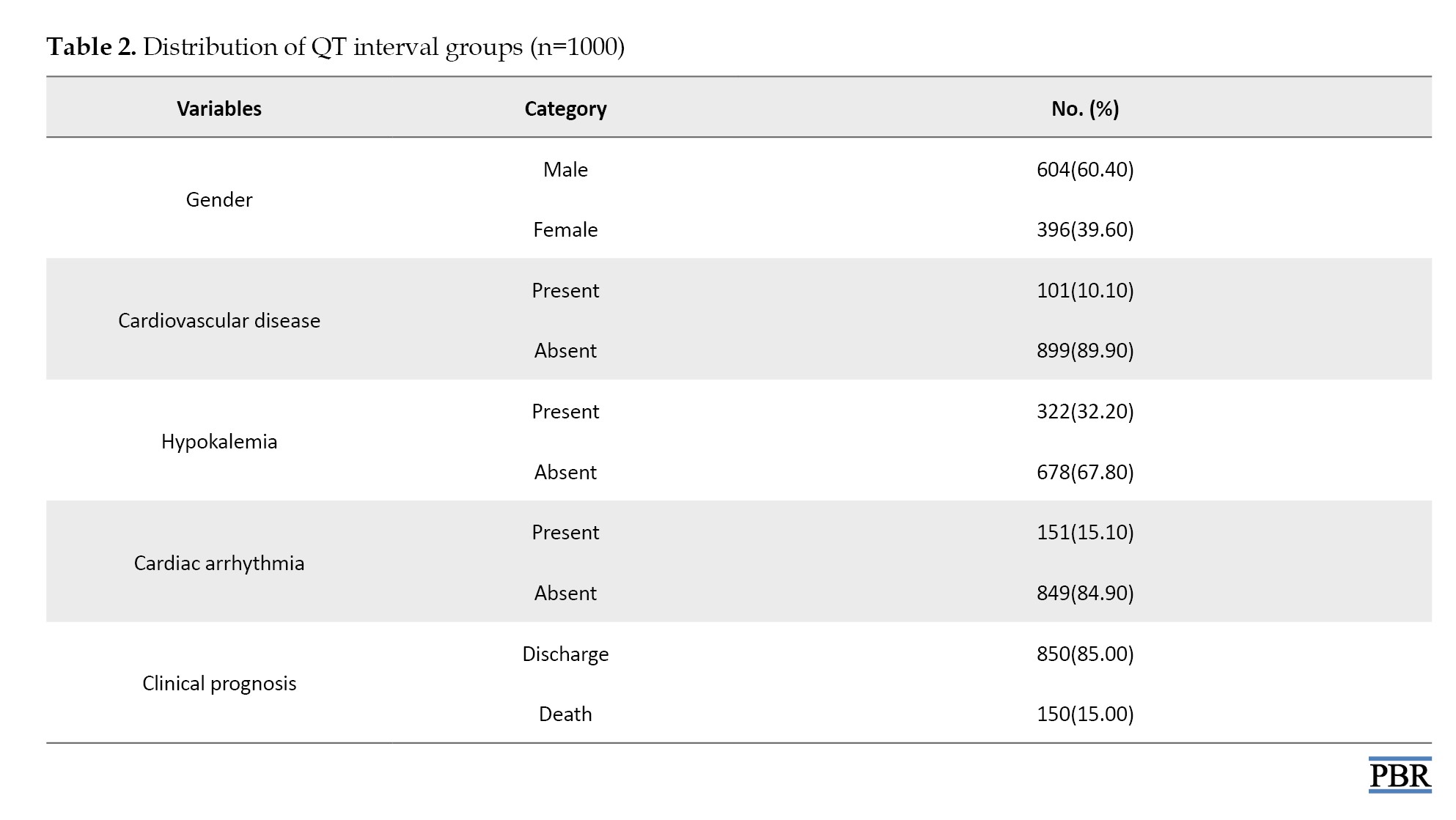
QT intervals were classified a priori into two categories: Normal/borderline (<450 ms) and prolonged (≥450 ms). Overall, 52.0% (n=520) of patients had normal/borderline QT intervals, whereas 48.0% (n=480) exhibited QT prolongation (≥450 ms) (Table 2).
As expected, the mean QT interval was substantially higher in the prolonged group compared with the normal/borderline group (540±70 ms vs 420±50 ms, respectively).
Regarding demographic correlates, a statistically significant association was observed between age group and QT status (P=0.02, Table 3).
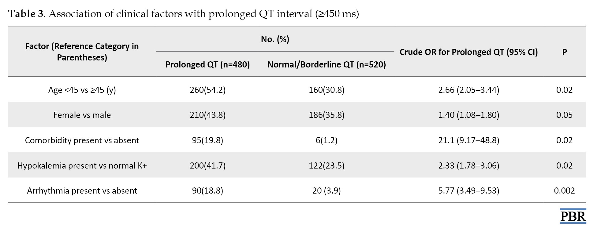
Among patients with prolonged QT intervals, 54.16% (n=260) were <45 years and 45.83% (n=220) were ≥45 years, compared with 30.76% (n=160) and 69.23% (n=360) in the normal/borderline QT group, respectively. This corresponded to a crude OR of approximately 2.66 (95% CI, 2.05%, 3.44%) for QT prolongation in patients younger than 45 years compared with those aged ≥45 years.
Gender was also significantly associated with QT status (P=0.05, Table 3). In the prolonged QT group, 56.25% (n=270) were male, and 43.75% (n=210) were female, whereas in the normal/borderline QT group, 64.23% (n=334) were male and 35.77% (n=186) were female. Using female sex as the exposure, the crude OR for prolonged QT was approximately 1.40 (95% CI, 1.08%, 1.80%) compared with male patients.
Comorbid cardiovascular disease was more frequent among patients with prolonged QT intervals. Comorbid conditions were present in 19.8% (n=95) of patients in the prolonged QT group, compared with 1.2% (n=6) in the normal/borderline group (P=0.02, Table 3). This represented a markedly elevated crude OR of approximately 21.1 (95% CI, 9.17%, 48.75%) for QT prolongation in patients with documented comorbid cardiovascular disease relative to those without comorbidity.
Electrolyte abnormalities were also associated with QT prolongation (P=0.02, Table 3). Hypokalemia was observed in 41.6% (n = 200) of patients with prolonged QT intervals and 23.46% (n=122) of those with normal/borderline QT intervals. The corresponding crude OR for QT prolongation in the presence of hypokalemia was approximately 2.33 (95% CI, 1.78%, 3.06%). Hypomagnesemia was present in 31.25% (n=150) of patients in the prolonged QT group and was notably less common in the normal/borderline group, further supporting the role of electrolyte disturbances in repolarization abnormalities (Tables 2 and 3).
Cardiac arrhythmias, including TdP, were significantly more frequent among patients with prolonged QT. Overall, 15.1% (n=151) of the cohort experienced clinically documented arrhythmias, and TdP was reported in 81.8% (n=90) of patients within the prolonged QT group. Compared with patients without arrhythmia, those with arrhythmias had higher odds of belonging to the prolonged QT group (crude OR: 5.77; 95% CI, 3.49%, 9.53%; P=0.002, Table 3).
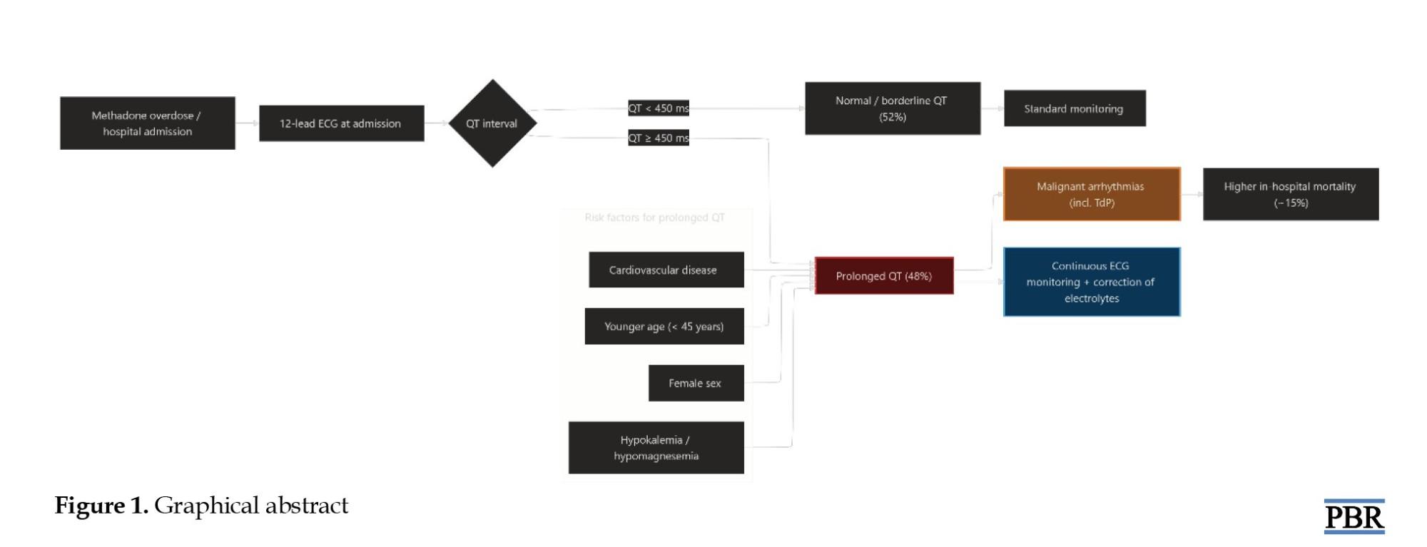
Clinical outcomes also differed according to QT status. The overall in-hospital mortality rate was 15.0% (n=150), with the majority of patients discharged alive (85.0%, n=850) (Table 2). Patients with prolonged QT intervals, particularly those with concomitant electrolyte imbalances and comorbid cardiovascular disease, showed a worse prognosis, consistent with the higher burden of malignant arrhythmias observed in this group. A summary of these results is presented in Figure 1.
The global opioid epidemic is one of the top public health issues of the 21st century. According to the United Nations Office on Drugs and Crime, there are an estimated 60 million opioid users worldwide [1]. The rise in overdose deaths due to synthetic opioids has been staggering [1]. The Middle East is a major area with an increase in opioid abuse; for example, in Iran, the drug pattern is dominated by traditional opium along with an increasing amount of methadone that is usually diverted for recreation [2, 3]. Methadone has been the gold standard for treating people suffering from opioid dependence since the 1960s, and it has been shown to be effective in reducing withdrawal symptoms, curbing illicit drug use, and decreasing the transmission of blood-borne pathogens [4-8]. However, it has disadvantages, such as high lipophilicity and a long, unpredictable half-life ranging from 8 to 60 hours.
Methadone is most notably linked to prolonged QT intervals, reflecting the duration of ventricular repolarization on electrocardiography (ECG). A long QT interval increases the risk of untreated (irregularities in heart rhythm) and may occur as a result of two conditions: 1) After depolarization, ventricular recovery resulting in electric shock; and 2) when a premature ventricular contraction (PVC) occurs on top of the prolonged QT. The PVC can result in a potentially fatal arrhythmia called torsades de pointes (TdP) – a form of polymorphic ventricular tachycardia – which can lead to ventricular fibrillation and sudden cardiac death [9]. Unlike many other opioids, methadone acts as a potent inhibitor of the IKr potassium current, which is mediated by the human ether-à-go-go-related gene (hERG) [10]. By blocking these channels, methadone delays potassium efflux during phase 3 of the cardiac action potential, thereby lengthening repolarization time.
The effect of QT interval prolongation by methadone is primarily dose-dependent but can also be affected by many intrinsic patient-specific factors. Examples of factors or conditions that decrease the repolarization reserve include advanced age, being female, underlying structural heart disease, and a genetic predisposition to congenital QT syndrome [11]. Methadone is also metabolized via the cytochrome P450 (CYP450) liver enzyme system. The main CYP450 enzymes involved are CYP3A4, CYP2B6, and CYP2C19. As a result, methadone combined with any CYP450 enzyme inhibitor (for example, erythromycin, a common antibiotic, or some antiretroviral agents) may increase methadone levels in the bloodstream, even when dosages seem reasonable [12].
Systemic physiological disturbances compound acute poisoning or overdose. Opioid toxicity produces a significant decrease in respiratory drive, leading to hypoxia and severe acidosis. These changes affect the electrical stability of the myocardial membranes. Electrolytes are often not retained by chronic users of certain substances because of inadequate nutrition or other medical conditions. Electrolyte abnormalities are critical because hypokalemia and hypomagnesemia can independently increase QT interval length and lower the threshold for TdP [13]. Previous studies have shown that a QT interval exceeding 500 ms is a critical threshold for heightened arrhythmic risk, but significant prolongation can occur even at therapeutic dosages [14].
According to the article, there is a lack of information regarding the frequency of dangerous QT prolongation resulting from methadone overdose in Yazd, Iran. To fill this gap, the researchers will examine 1,000 cases of poisoning from two referral hospitals from 2014-2023 by measuring the QT interval (QT≥450 ms) on the ECG and determining what factors (age, dose, comorbidity, and electrolytes) predict a person’s risk of overdose so that Iran can implement better protocols for overdoses, such as routine electrocardiogram (ECG) monitoring, and timely correction of electrolyte abnormalities to reduce the incidence of fatal cardiac complications.
Materials and Methods
Study design and setting
This retrospective observational (cross-sectional) study was designed to determine the frequency and clinical correlates of QT interval prolongation among patients with methadone poisoning. The study was conducted at two major referral centers for toxicological emergencies in Yazd Province, Iran (Shah Vali Hospital in Yazd City and Shahid Beheshti Hospital in Taft City).
Participants
The study population consisted of 1,000 patients admitted with a confirmed diagnosis of methadone poisoning between January 2014 and December 2023. A consecutive sampling method was used to identify eligible cases from the hospital archives.
Patients were included if they had a clinical diagnosis of methadone poisoning established by the treating physician, were hospitalized (beyond an emergency room visit only) in one of the participating centers during the study period, and had sufficiently complete medical records, including electrocardiographic and laboratory data, to allow extraction of the study variables. Patients with missing core clinical or ECG information or those managed solely as outpatients or discharged from the emergency department without admission were excluded.
Instruments and measures
Data were extracted using a standardized checklist adapted from previous toxicological and cardiology studies. The following domains were recorded:
Demographic and clinical characteristics: Age (years), gender, and the reported dose of methadone consumed (milligrams). Comorbidities and complications: Presence of underlying diseases (e.g. cardiovascular disease, diabetes, hypertension), and in-hospital complications such as apnea, aspiration pneumonia, acute respiratory distress syndrome, and rhabdomyolysis.
Laboratory data: Serum electrolyte levels, specifically potassium, magnesium, calcium, and sodium, were recorded to identify abnormalities such as hypokalemia or hypomagnesemia. Clinical outcomes: The patient’s outcome was categorized as discharge (survival) or death.
Electrocardiographic analysis
The primary outcome measure was the QT interval, obtained from the initial 12lead ECG performed at admission. QT intervals were taken from the automated ECG report generated by the hospital’s standard ECG machines and recorded in milliseconds. For descriptive purposes, the QT interval was analyzed as both a continuous variable and a categorical variable.
Based on prior literature in methadone-treated populations, QT intervals were categorized a priori into two groups:
Group 1 (Normal/Borderline): <450 ms; group 2 (Prolonged): ≥450 ms
This cut-off (≥450 ms) has been suggested as a threshold requiring closer clinical observation in methadone users.
Data collection
Data were collected using both physical medical records and electronic hospital information systems. The researchers obtained necessary administrative approvals from hospital administration and the University Research Council prior to data extraction. Trained research personnel reviewed the archives and extracted relevant data into a standardized data collection form. To protect patient confidentiality, all identifying information was removed, and each case was assigned a unique study ID code.
Statistical analysis
Statistical analyses were conducted using IBM SPSS software, version 26. A two sided P<0.05 was considered statistically significant. Categorical variables were summarized as frequencies and percentages, and continuous variables as Mean±SD with minimum and maximum values.
The normality of continuous variables was evaluated using the Kolmogorov–Smirnov test, which indicated that all continuous variables were approximately normally distributed (P>0.05), supporting the use of parametric tests. The independent samples t-test was used to compare the mean QT interval between patients with normal/borderline QT and those with prolonged QT.
Associations between prolonged QT (≥450 ms) and categorical predictors (age group <45 vs ≥45 years, sex, comorbid status, electrolyte imbalance, and clinical outcome) were initially assessed using Pearson’s chisquare test. For key 2×2 comparisons, crude odds ratios (ORs) with 95% confidence intervals (CIs) were calculated from contingency tables to quantify the strength of association between clinical factors and the presence of QT prolongation. No multivariable regression models were performed because the dataset is retrospective and lacks individuallevel data beyond aggregated counts.
Results
A total of 1,000 patients with methadone overdose were included in the analysis. The mean age was 32.8±9.6 years (range: 18–65 years), with 42.0% (n=420) younger than 45 years and 58.0% (n=580) aged 45 years or older (Table 1).
The mean reported methadone dose was 75.4±2.25 mg (range: 10-150 mg). The mean QT interval for the entire cohort was 478.6±32.1 ms (range: 450-540 ms), and the median QT interval was 466.0 ms (Table 1).
QT intervals were classified a priori into two categories: Normal/borderline (<450 ms) and prolonged (≥450 ms). Overall, 52.0% (n=520) of patients had normal/borderline QT intervals, whereas 48.0% (n=480) exhibited QT prolongation (≥450 ms) (Table 2).
As expected, the mean QT interval was substantially higher in the prolonged group compared with the normal/borderline group (540±70 ms vs 420±50 ms, respectively).
Regarding demographic correlates, a statistically significant association was observed between age group and QT status (P=0.02, Table 3).
Among patients with prolonged QT intervals, 54.16% (n=260) were <45 years and 45.83% (n=220) were ≥45 years, compared with 30.76% (n=160) and 69.23% (n=360) in the normal/borderline QT group, respectively. This corresponded to a crude OR of approximately 2.66 (95% CI, 2.05%, 3.44%) for QT prolongation in patients younger than 45 years compared with those aged ≥45 years.
Gender was also significantly associated with QT status (P=0.05, Table 3). In the prolonged QT group, 56.25% (n=270) were male, and 43.75% (n=210) were female, whereas in the normal/borderline QT group, 64.23% (n=334) were male and 35.77% (n=186) were female. Using female sex as the exposure, the crude OR for prolonged QT was approximately 1.40 (95% CI, 1.08%, 1.80%) compared with male patients.
Comorbid cardiovascular disease was more frequent among patients with prolonged QT intervals. Comorbid conditions were present in 19.8% (n=95) of patients in the prolonged QT group, compared with 1.2% (n=6) in the normal/borderline group (P=0.02, Table 3). This represented a markedly elevated crude OR of approximately 21.1 (95% CI, 9.17%, 48.75%) for QT prolongation in patients with documented comorbid cardiovascular disease relative to those without comorbidity.
Electrolyte abnormalities were also associated with QT prolongation (P=0.02, Table 3). Hypokalemia was observed in 41.6% (n = 200) of patients with prolonged QT intervals and 23.46% (n=122) of those with normal/borderline QT intervals. The corresponding crude OR for QT prolongation in the presence of hypokalemia was approximately 2.33 (95% CI, 1.78%, 3.06%). Hypomagnesemia was present in 31.25% (n=150) of patients in the prolonged QT group and was notably less common in the normal/borderline group, further supporting the role of electrolyte disturbances in repolarization abnormalities (Tables 2 and 3).
Cardiac arrhythmias, including TdP, were significantly more frequent among patients with prolonged QT. Overall, 15.1% (n=151) of the cohort experienced clinically documented arrhythmias, and TdP was reported in 81.8% (n=90) of patients within the prolonged QT group. Compared with patients without arrhythmia, those with arrhythmias had higher odds of belonging to the prolonged QT group (crude OR: 5.77; 95% CI, 3.49%, 9.53%; P=0.002, Table 3).
Clinical outcomes also differed according to QT status. The overall in-hospital mortality rate was 15.0% (n=150), with the majority of patients discharged alive (85.0%, n=850) (Table 2). Patients with prolonged QT intervals, particularly those with concomitant electrolyte imbalances and comorbid cardiovascular disease, showed a worse prognosis, consistent with the higher burden of malignant arrhythmias observed in this group. A summary of these results is presented in Figure 1.
Discussion
In this 10-year retrospective observational study of 1,000 hospitalized patients with methadone poisoning, we assessed the frequency of prolonged QT intervals and their clinical correlates. Nearly half of the cohort (48%) had a QT interval ≥450 ms, and the mean QT interval of 478.6±32.1 ms was substantially above the upper normal limit reported in the general population, underscoring the high cardiotoxic potential of acute methadone toxicity through hERG channel inhibition and delayed ventricular repolarization.
The prevalence of QT prolongation observed in our study is broadly consistent with previous reports from the same geographic region. Farsi et al. reported that approximately 50.7% of patients with acute methadone overdose had a QT interval greater than 450 ms, which is comparable to our rate of 48% [15]. Other retrospective Iranian ICU studies have also documented frequent ECG abnormalities in patients with significant methadone toxicity, including STT changes and repolarization disturbances [16]. Minor differences in prevalence estimates across studies may reflect variations in sample size, case mix, and inclusion criteria; our multi-center cohort spanning a 10-year period likely captures a wider spectrum of methadone exposures and poisoning severity.
In terms of demographic correlates, we found that both younger age and female sex were associated with an increased likelihood of QT prolongation. Patients younger than 45 years had approximately 2.6 fold higher odds of prolonged QT compared with those aged ≥45 years, and female patients had about 1.4fold higher odds of QT prolongation than males, in crude analyses. These findings are consistent with prior evidence indicating that age and female sex are important noncardiologic risk factors for druginduced QT prolongation and TdP [17, 18]. Although we used a single QT cutoff of 450 ms for both sexes based on prior studies in methadonetreated populations, sexspecific thresholds based on corrected QT would better reflect underlying physiological differences and may improve risk stratification.
Our data also highlight the complex relationship between methadone dose and QT prolongation. The mean reported methadone dose in this cohort was 75.4±2.25 mg, which is within ranges where prior studies have described dosedependent effects on repolarization [19, 20]. While earlier studies have demonstrated a relatively linear association between methadone dose and QTc prolongation, particularly at higher doses, our descriptive findings suggest that clinically significant QT prolongation can occur even at comparatively lower doses in the setting of acute overdose [20]. In our dataset, a high oral dose alone appeared to be a less reliable predictor of prolonged QT than the actual ECG changes, reinforcing the notion that systemic exposure and cardiotoxicity are strongly influenced by individual variability in metabolism, comorbid conditions, and coadministered medications. Polymorphisms in CYP3A4 and CYP2B6, as well as drug–drug interactions with CYP inhibitors, may further modulate methadone levels and arrhythmic risk.
Electrolyte disturbances emerged as key modifiable contributors to QT prolongation in our cohort. The overall prevalence of hypokalemia was 32.2%, and patients with hypokalemia had more than double the odds of prolonged QT compared with those with normal potassium levels. Hypomagnesemia was also more common among patients with prolonged QT, consistent with the concept of a reduced repolarization reserve when multiple risk factors such as methadone exposure, electrolyte abnormalities, and structural heart disease coexist. Prior experimental and clinical studies have shown that hypokalemia and hypomagnesemia independently prolong ventricular repolarization, facilitate early afterdepolarizations, and increase susceptibility to TdP, particularly in patients receiving QT-prolonging drugs, including methadone [21, 22].
We also observed strong associations between QT prolongation, underlying cardiovascular comorbidity, and clinically significant arrhythmias. Comorbid cardiovascular disease was markedly more frequent in the prolonged QT group, with a crude odds ratio above 20 for QT prolongation, albeit with wide confidence intervals reflecting the low prevalence in the normal QT group. Furthermore, arrhythmias—including TdP and other malignant ventricular rhythms—were substantially more common among patients with prolonged QT, with nearly sixfold higher odds compared with patients without arrhythmias. These findings align with the Cubeddu study that demonstrated druginduced QT prolongation is closely linked to the development of TdP and sudden cardiac death, particularly in individuals with additional risk factors such as structural heart disease, electrolyte imbalances, and concurrent use of other QT-prolonging drugs [23].
Clinically, methadone poisoning in our cohort was associated with significant morbidity and mortality, with an overall inhospital fatality rate of approximately 15% and arrhythmias documented in 15.1% of patients. The combination of QT prolongation, electrolyte disturbances, and comorbid cardiovascular disease was associated with worse outcomes, mirroring prior studies in which prolonged QT or QTc predicted intensive care unit admission and death in methadoneexposed patients [19, 24]. Our results support current expert recommendations advocating early 12 lead ECG acquisition, continuous cardiac monitoring, and aggressive correction of electrolyte abnormalities—particularly potassium and magnesium—to highnormal levels in all suspected cases of methadone overdose.
Several limitations should be considered when interpreting these findings. First, the retrospective design and reliance on hospital records introduced inherent information bias and restricted our ability to control for all potential confounders. Important variables such as the precise time from ingestion to ECG recording, detailed coingestant profiles (including benzodiazepines, alcohol, and other QT-prolonging medications), serum methadone concentrations, and comprehensive measures of hepatic and renal function were not systematically documented and could not be fully accounted for. Second, QT assessment was based on the admission 12-lead ECG and automated measurements of the raw QT interval, rather than manually confirmed QTc values with sexspecific cutoffs. As a result, some misclassification of repolarization abnormalities is possible, and late QT-prolongation occurring after admission may have been missed. Third, because only aggregate data were available at the time of analysis, we could not perform multivariable regression to adjust for confounding between correlated risk factors, such as age, sex, comorbidity, and electrolyte disturbances. Crude OR therefore provide useful but unadjusted estimates of association and should be interpreted with caution. Finally, the study population consisted exclusively of hospitalized patients with sufficiently complete records, which may overrepresent more severe poisonings and limit generalizability to milder cases managed in outpatient or emergency settings.
To better delineate the causal pathways linking methadone exposure, repolarization delay, and arrhythmic outcomes, future studies should employ prospective designs with serial ECG monitoring, formal QTc measurement using standardized correction formulas, and simultaneous assessment of serum methadone levels and coadministered QT-prolonging agents. Incorporating sexspecific QTc thresholds, detailed pharmacogenetic profiling, and multivariable modeling would further clarify the independent contribution of demographic, clinical, and pharmacologic factors to QT prolongation and TdP risk in this highrisk population.
Conclusion
In this 10year retrospective study of 1,000 patients with methadone toxicity, 48% had QT intervals ≥ 450 ms, with a mean QT interval of 478.6 ms, indicating substantial cardiac toxicity and an elevated risk of TdP and ventricular fibrillation. Prolonged QT was more frequent in younger patients and in women and was strongly associated with electrolyte disturbances,particularly hypokalemia, and preexisting cardiovascular disease, all of which were linked to higher rates of malignant arrhythmias and an inhospital mortality rate of 15%. These findings reinforce the need for routine 12lead ECG screening, serial monitoring, and prompt correction of electrolyte abnormalities in all patients with suspected methadone overdose, along with careful evaluation of concomitant QT-prolonging medications and underlying cardiac risk factors.
Ethical Considerations
Compliance with ethical guidelines
The Ethics Committee of the Islamic Azad University, Yazd, Iran (Code: IR.IAU.YAZD.REC.1403.105), in accordance with the principles of the Declaration of Helsinki for research involving human subjects.
Funding
This research did not receive any grant from funding agencies in the public, commercial, or non-profit sectors.
Authors' contributions
Data interpretation: Marziyeh Arjmand, Hamid Owliaey and Marjan Shariatpanahi; Data collection, and drafting of the initial manuscript: Marziyeh Arjmand; Data collection and statistical analysis: Niloofar Deravi; Study design and critical revision of the manuscript: Mehrnoush Giahi Yazdi; Literature review: Marjan Shariatpanahi; Data collection and manuscript editing: Elahe Zare; Study design, supervision, and critically revised: Hamid Owliaey; Final approval: All authors.
Conflict of interest
The authors declared no conflicts of interest.
References
- United Nations. World drug report 2025 [Internet]. 2025 [Updated 2026 September 1]. Available from: [Link]
- Momtazi S, Noroozi A, Rawson RA. An overview of iran drug treatment and harm reduction programs. In: el-Guebaly N, Carrà G, Galanter M, editors. Textbook of addiction treatment: International perspectives. Milano: Springer Milan; 2015. [DOI:10.1007/978-88-470-5322-9_25]
- Soltaninejad K, Hassanian-Moghaddam H, Shadnia S. Methadone Related Poisoning on the Rise in Tehran, Iran. Asia Pac J Med Toxicol. 2014; 3(3):104-9. [Link]
- Bruce RD. Methadone as HIV prevention: high volume methadone sites to decrease HIV incidence rates in resource limited settings. Int J Drug Policy. 2010; 21(2):122-4. [DOI:10.1016/j.drugpo.2009.10.004] [PMID] [PMCID]
- Fullerton CA, Kim M, Thomas CP, Lyman DR, Montejano LB, Dougherty RH, et al. Medication-assisted treatment with methadone: assessing the evidence. Psychiatr Serv. 2014; 65(2):146-57. [DOI:10.1176/appi.ps.201300235] [PMID]
- Gowing L, Farrell MF, Bornemann R, Sullivan LE, Ali R. Oral substitution treatment of injecting opioid users for prevention of HIV infection. Cochrane Database Syst Rev. 2011(8):Cd004145. [DOI:10.1002/14651858.CD004145.pub4] [PMID]
- MacArthur GJ, Minozzi S, Martin N, Vickerman P, Deren S, Bruneau J, et al. Opiate substitution treatment and HIV transmission in people who inject drugs: Systematic review and meta-analysis. BMJ. 2012; 345:e5945. [DOI:10.1136/bmj.e5945] [PMID] [PMCID]
- Mattick RP, Breen C, Kimber J, Davoli M. Methadone maintenance therapy versus no opioid replacement therapy for opioid dependence. Cochrane Database Syst Rev. 2009; 2009(3):Cd002209. [DOI:10.1002/14651858.CD002209.pub2] [PMID] [PMCID]
- Krantz Mori J, Palmer Robert B, Haigney Mark CP. Cardiovascular complications of opioid use. JACC. 2021; 77(2):205-23. [DOI:10.1016/j.jacc.2020.11.002] [PMID]
- Katchman AN, McGroary KA, Kilborn MJ, Kornick CA, Manfredi PL, Woosley RL, et al. Influence of opioid agonists on cardiac human ether-a-go-go-related gene K+ currents. The Journal of Pharmacology and Experimental Therapeutics. 2002; 303(2):688-94. [DOI:10.1124/jpet.102.038240] [PMID]
- Amin AS, Pinto YM, Wilde AA. Long QT syndrome: beyond the causal mutation. J Physiol. 2013; 591(17):4125-39. [DOI:10.1113/jphysiol.2013.254920] [PMID] [PMCID]
- Greenblatt DJ. Drug interactions with methadone: Time to revise the product label. Clin Pharmacol Drug Dev. 2014; 3(4):249-51. [DOI:10.1002/cpdd.137] [PMID]
- Cabahug MC, Vempati A. Torsade de pointes due to hypokalemia and hypomagnesemia. J Educ Teach Emerg Med. 2022; 7(4):S27-51. [DOI:10.5070/M57459219] [PMID] [PMCID]
- Khan SA, Emmanuel S, Shantha Kumar V, Nerella R, Shaman Ameen B, Patel D, et al. Long QT syndrome with drugs used in the management of arrhythmias: A systematic review. Cureus. 2024; 16(7):e65857. [DOI:10.7759/cureus.65857]
- Farsi D, Mirafzal A, Hassanian-Moghaddam H, Azizi Z, Jamshidnejad N, Zehtabchi S. The correlation between prolonged corrected QT interval with the frequency of respiratory arrest, endotracheal intubation, and mortality in acute methadone overdose. Cardiovasc Toxicol. 2014; 14(4):358-67. [DOI:10.1007/s12012-014-9259-x] [PMID]
- Soroosh D, Neamatshahi M, Zarmehri B, Nakhaee S, Mehrpour O. Drug-induced prolonged corrected QT interval in patients with methadone and opium overdose. Subst Abuse Treat Prev Policy. 2019; 14(1):8. [DOI:10.1186/s13011-019-0196-3] [PMID] [PMCID]
- Trinkley KE, Page RL, 2nd, Lien H, Yamanouye K, Tisdale JE. QT interval prolongation and the risk of torsades de pointes: essentials for clinicians. Curr Med Res Opin. 2013; 29(12):1719-26. [DOI:10.1185/03007995.2013.840568] [PMID]
- Khatib R, Sabir FRN, Omari C, Pepper C, Tayebjee MH. Managing drug-induced QT prolongation in clinical practice. Postgrad Med J. 2021; 97(1149):452-8. [DOI:10.1136/postgradmedj-2020-138661] [PMID] [PMCID]
- Juba KM, Khadem TM, Hutchinson DJ, Brown JE. Methadone and corrected QT prolongation in pain and palliative care patients: A case-control study. J Palliat Med. 2017; 20(7):722-8. [DOI:10.1089/jpm.2016.0411] [PMID]
- Huh B, Park CH. Retrospective analysis of low-dose methadone and QTc prolongation in chronic pain patients. Korean J Anesthesiol. 2010; 58(4):338-43. [DOI:10.4097/kjae.2010.58.4.338] [PMID] [PMCID]
- Kumar V, Khosla S, Stancu M. Torsade de pointes induced by hypokalemia from imipenem and piperacillin. Case Rep Cardiol. 2017; 2017(1):4565182. [DOI:10.1155/2017/4565182] [PMID] [PMCID]
- Marill KA, Miller ES. Hypokalemia in women and methadone therapy are the strongest non-cardiologic factors associated with QT prolongation in an emergency department setting. J Electrocardiol. 2017; 50(4):416-23. [DOI:10.1016/j.jelectrocard.2017.02.001] [PMID] [PMCID]
- Cubeddu LX. QT prolongation and fatal arrhythmias: A review of clinical implications and effects of drugs. Am J Ther. 2003; 10(6):452-7. [DOI:10.1097/00045391-200311000-00013] [PMID]
- Martin JA, Campbell A, Killip T, Kotz M, Krantz MJ, Kreek MJ, et al. QT interval screening in methadone maintenance treatment: report of a SAMHSA expert panel. J Addict Dis. 2011; 30(4):283-306. [DOI:10.1080/10550887.2011.610710] [PMID] [PMCID]
Type of Study: Original Research |
Subject:
General
| Rights and permissions | |
 |
This work is licensed under a Creative Commons Attribution-NonCommercial 4.0 International License. |


This is an open access article distributed under the terms of the Creative Commons Attribution License (CC-By-NC), which permits use, distribution, and reproduction in any medium, provided the original work is properly cited and is not used for commercial purposes.
Contact Information