

Volume 11, Issue 4 (2025)
Pharm Biomed Res 2025, 11(4): 277-298 |
Back to browse issues page


Download citation:
BibTeX | RIS | EndNote | Medlars | ProCite | Reference Manager | RefWorks
Send citation to:


BibTeX | RIS | EndNote | Medlars | ProCite | Reference Manager | RefWorks
Send citation to:
Banalia A, Puri D. Green Synthesis of Metallic Nanoparticles and Their Multifaceted Applications: A Review Study. Pharm Biomed Res 2025; 11 (4) :277-298
URL: http://pbr.mazums.ac.ir/article-1-700-en.html
URL: http://pbr.mazums.ac.ir/article-1-700-en.html


1- Department of Pharmaceutics, School of Pharmacy, Graphic Era Hill University, Dehradun, India.
Full-Text [PDF 1486 kb]
(168 Downloads)
| Abstract (HTML) (457 Views)
Full-Text: (97 Views)
Introduction
Over the past 20 years, nanotechnology has greatly ranked among the most important technologies studied and rapidly expanding sciences because of its applicability in several human welfare domains [1].
The term “Nano” was obtained from Greek word “nanos,” which means diminutive. The units of measurement for nanoparticles (NPs) are nanometers. They are popular because of their sub-microscopic particle-size and large surface area [2].
There are four main categories of NPs depending on their chemical makeup: Carbon-based (carbon nanotubes and nanofibers, etc.), bioorganic-based (liposomes, micelles, etc.), metal- and metal oxide-based (Ag, Cu, etc.), and composite based [3]. NPs have extraordinary physical, thermal, optical, magnetic electrical, and chemical, properties compared to their non-nano material [4]. They’ve wide scope in Pharmaceuticals, as well as catalysis, householding products, environmental sensors, automotive industries, and nanobiotechnology. Additionally, nanotechnology helps the early diagnosis of serious illnesses like cancer. Examples of NPs include dendrimers, metal NPs, liposomes, fullerenes, and nanodroplets [5].
Metallic NPs are an important and most-studied class of materials with various applications. Numerous studies are being conducted on the production of metal ion NPs from microorganisms and botanical extracts [6]. Silver NPs, AgNPs, are the most often used of all the produced NPs and found in over 25% of consumer goods. Research shows that gold NPs (AuNPs) has biological uses as muscle relaxant, antibacterial, and enzyme control. AgNPs prevent gram-positive and gram-negative bacteria from growing and acting. Copper NPs (CuNPs) possess great promise as drug delivery vehicles, anti-cancer drugs, and enhancers of photodynamic treatment. Palladium NPs (PdNPs) are used as catalysts, dye degradation, and anti-microbial. Finally, iron NPs (FeNPs) inhibit bacterial growth [7]. NPs can be synthesized by three different techniques: physical, chemical, biological (Figure 1) [5].
.PNG)
This review explores how biology and physical sciences collaborate to produce metal NPs in a “green” manner for different applications. According to the research, bacteria and plant extracts are novel sources of producing NPs. To achieve this goal, it is essential to use natural resources (such organic means) and the best solvent systems.
Advantages of green synthesis over synthetic method [8–10]
Green synthesis has the following advantages
1) By using fewer dangerous components and toxic organic solvents, green chemistry provides substantial economic and environmental benefits over conventional synthetic processes. 2) They improve the sustainability in drug synthesis by using renewable resources while preserving the efficacy and quality of medications. 3) Green synthesis reduces the use of resources and improves the atom economy. 4) This method lowers toxicity and eliminates using dangerous procedures. 5) It uses cutting-edge techniques, including water as a solvent and microwave synthesis. 6) Real time monitoring can takes place. 7) During green synthesis, catalysts are utilized sparingly and have a high rate of small-scale reaction.
Green synthesis
Although the traditional methods have long been used for decades, research has shown that green methods are more outstanding in producing NPs because they are easier to outline, less expensive, and less likely to fail [10]. Numerous resources, including plants and their extracts, algae, fungi, yeast, bacteria, and viruses, can be used to carry out the green synthesis of NPs [11]. Proteins (amino acids), phytochemicals (alkaloids, flavonoids, reducing sugars, polyphenols), and other substances are present in the biomaterials and serve as capping and reducing agents throughout the synthesis process for generating metal NPs from their precursor metal salts [12]. The reduction of the metal precursor to its subsequent NPs may be initially confirmed by observing the color shift of the colloidal solution [9, 10]. Many biotechnological uses, such as bioremediation and bioleaching, have been created since bacteria can interact with, extract, and collect metals from their surroundings [3].
Because of lipid based amphipathic membrane, they can interact with their environment, promote variety of oxidation-reduction reactions, and allow biochemical transformations. Utilizing plants as opposed to other environmentally beneficial biological systems, such as bacteria and fungus, such doing away with costly and time-consuming isolation and processing techniques [13]. Use of plants and their extracts is more safer and efficacious for the production of NPs than other biological systems of producing NPs [14]. Summary of concepts and principles behind the green chemistry are shown in Figures 2 and 3.
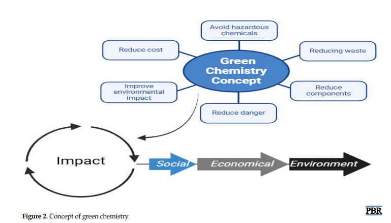
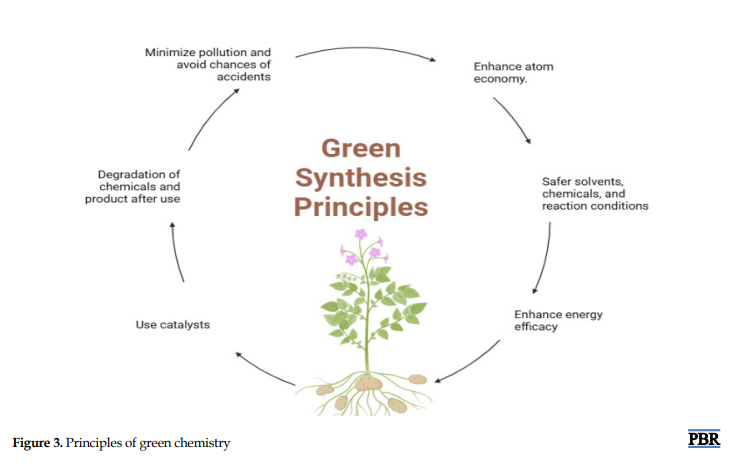
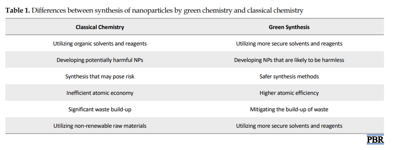
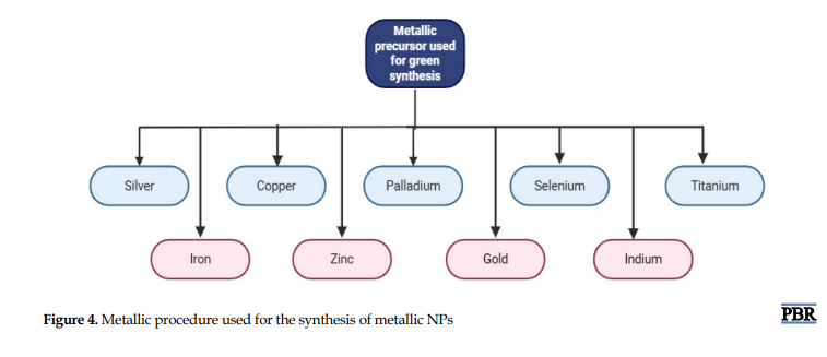
Different metallic precursors used for the synthesis are given in Figure 4. Finally, Table 1 presents the differences between synthesis of NPs by green chemistry and classical chemistry. The major advantage of green chemistry over classical chemistry is that green chemistry is more eco-friendly
Chemical transformations during green synthesis
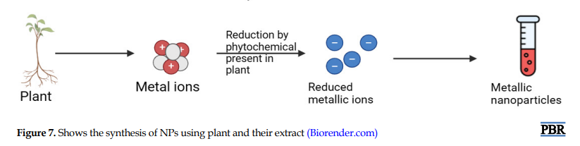
The primary ingredients of AgNPs green synthesis are silver metal ion solution (0.1-10 mM) and a reducing biological agent. This eco-friendly method avoids the use of toxic chemicals [15]. Reaction should be completed at room temperature to mild heating and pH 7–10 is optimal for many systems. The green synthesis of AgNPs included the reduction of silver ions (Ag⁺) to elemental silver (Ag⁰) using plant extracts, microbes, or other biological agents, which is followed by agglomeration into clusters. These clusters eventually form metallic colloidal silver particles [16]. In most cases, the reducing agents or other constituents present in the cells act as stabilizing and capping agents. This process includes the following steps [17].
a. Silver salt dissociation:
In the first step, silver nitrate is dissociated into silver ion.
AgNO3→Ag++NO3−
b. Reduction of Ag⁺ to Ag⁰ by phytochemicals:
In the presence of reducing agent like plant extract, microbes, or biomolecules, Ag+ is reduced to Ag0.
Ag++[Reducing agent from plant/microbe]→Ag0↓+ [Oxidized by-products]
Example is using a polyphenol like catechol.
2Ag++C6H4(OH)2→2Ag0+C6H4O2+2H+
c. Nucleation and growth:
In this step, reduced Ag0 atoms are nucleated to form small clusters and then these clusters grow into NPs.
d. Stabilization (capping):
At the end, biomolecules (like proteins, terpenoids, etc.) cap the NPs to prevent aggregation.
Synthesis of metallic NPs using bacteria
Numerous bacteria have demonstrated the capacity to synthesize metallic NPs; each has specific pros and cons. Critical metals need to enter the cytoplasm through the cell wall (extracellular and intracellular) [18]. Because of their capacity to reduce metal ions, bacteria are excellent options for creating NPs. Prokaryotic and actinomycetes bacteria have been used extensively in the synthesis of metal/metal oxide NPs [19].
Bacteria can participate actively in creating NPs, serve as a bioscaffold for mineralization, or function as a biocatalyst for the synthesis of inorganic materials. During the incubation period, bacteria in broth medium can produce extracellular or intracellular nanomaterials [10]. Bacterial species with different morphologies and the internal and exterior environment of a cell frequently affect the crystalline and non-crystalline phases of particle creation [20, 21].
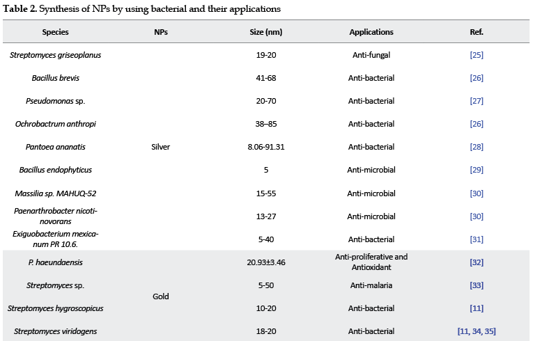
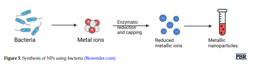
Shivaji et al. synthesized AgNPs using bacterial culture that remain stable for 8 months in dark. Bacteria used in this experiment were Bacillus indicus, B. cecembensis, Arthrobacter kerguelensis, A. gangotriensis, P. antarctica, P. proteolytica, and P. meridiana. Created NPs were bactericidal [22]. Sharma et al. synthesized gold NPs using Marinobacter pelagius [23]. Tiwari et al. manufactured copper NPs using copper-resistant B. cereus. Synthesized NPs shows antimicrobial effects [24]. Hasan et al. synthesized iron nanoparticle using B. proteolyticus UPMC1508. Created NPs were bactericidal and anti-cancer [25]. Liu et al. synthesized selenium NPs using B. paramycoides. Created NPs shows anti-bacterial and anti-oxidant properties [26] (Table 2). Figure 5 shows the process of NPs synthesis using bacteria.Silver salt dissociation:
.PNG)
Synthesis of NPs using fungi
Fungi are considered good candidates because they can produce monodisperse NPs with highly defined dimension, various chemical compositions and sizes. Also, fungi release greater quantity of proteins that lead to a higher level of nanoparticle production [66]. Fungi can produce various compounds with different applications. Over 6400 bioactive chemicals are produced by ascomycetes, imperfect fungi, and other microscopic filamentous fungi [67]. Fungi are at the forefront of research for the production of biological metal NPs because of their tolerance and capacity for metal biomagnification [68-73]. The ability to synthesize enormous amounts of proteins and enzymes, some of which may be utilized for the quick and sustainable production of NPs, gives fungi an edge over other microbes [67].
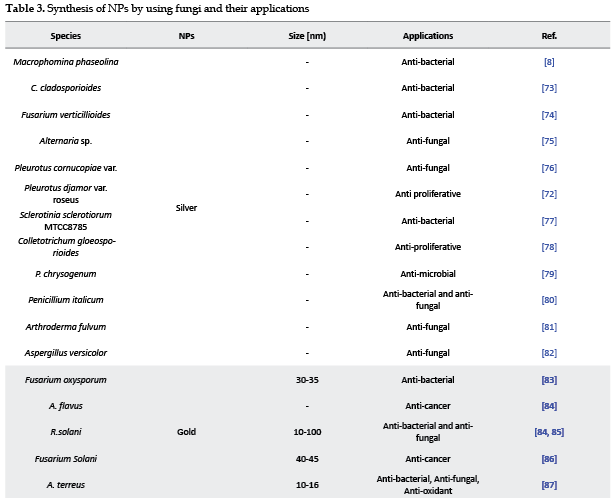
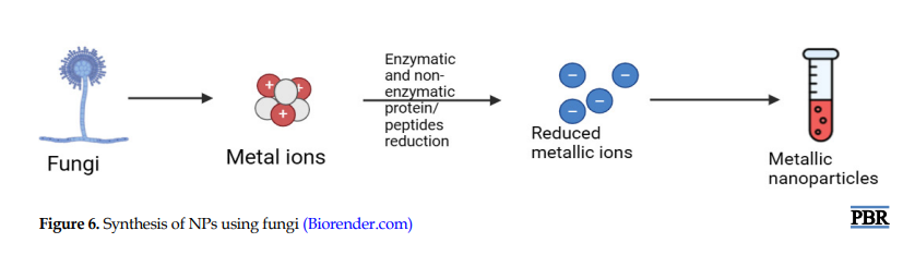
Raut et al. synthesized silver NPs using fungi, such as Cladosporium cladosporioides, Penicillium chrysogenum, and Purpureocillium lilacinum. Created NPs shows anti-microbial NPs [72]. Iranmanesh et al. synthesized gold NPs using 12 fungi; out of 12 fungi, 8 could successfully synthesize gold NPs. Further out of 8 fungi, 3 are investigated: Fusarium oxysporum, Aspergillus flavus, Rhizoctonia solani, and Verticillium dahliae [72]. Kamal et al. synthesized iron NPs using fungi, ie, Daedalea mushroom. It is first time that iron NPs are synthesized by using mushroom. Created NPs were analyzed against the pathogenic fungus Aspergillus niger [74]. Fatima and Wahid synthesized copper NPs using fungi Schizophyllum commune. Created NPs were analyzed against multidrug-resistant organisms (MDROs) like Escherichia coli, Salmonella abony, Staphylococcus aureus, and Klebsiella pneumoniae [75]. Hussein et al. synthesized selenium NPs by using 4 different fungi: Aspergillus quadrilineatus, Aspergillus ochraceus, A. terreus, Fusarium equiseti. Synthesized NPs were anti-bacterial and anti-fungal [76] (Table 3). Figure 6 shows the process of synthesized of NPs using fungi.
.PNG)
.PNG)
Synthesis of NPs using plant and their extracts
Although the capacity of plant extracts to reduce metals has been known since the early 1900s, little is known about the specifics of the reducing chemicals at play. Compared to whole plant tissue, using plant extracts to make NPs is simpler. Plant extract-mediated synthesis is a growing area of interest [25]. These days, plant extracts serve as capping and reducing agents throughout the nanoparticle manufacturing process, which is way better than microbial, chemical synthesis [109-114].
Zahir et al. synthesized silver NPs using aqueous plant extract of Euphorbia prostrata. Created NPs were analyzed with pesticidal activity against Sitophilus oryzae L [115]. Hazarika et al. synthesized palladium NPs using leaf extract of Garcinia pedunculata Roxb. Created NPs were analyzed with anti-microbial activity Cronobacter sakazakii strain AMD04 [116]. Majumdar et al. synthesized gold NPs by using leaf extract of Acacia nilotica (Babool). Created NPs were analyzed with catalyst property which is used to reduce 4-nitrophenol to 4-aminophenol [117]. Vasantharaj et al. synthesized iron NPs using Ruellia tuberosa (RT) leaf extract. Created NPs were analyzed with potent anti-bacterial activity against K. pneumoniae, E. coli and average anti-bacterial activity against S. aureus [118]. Alao et al. synthesized copper NPs using ethanolic extract of Kigelia africana fruit. Created NPs were analyzed with anti-microbial activity against E. coli, Shigella sp., S. aureus, Pseudomonas aeruginosa, and Salmonella typhi [119] (Table 4). Figure 7 shows the synthesis of nanoparticles using plant and their extract.
Characterization of metallic NPs [164-178]
Characterization of NPs can be done using ultraviolet (UV) (UV–Vis spectroscopy), Fourier transforms infrared spectroscopy (FT-IR), scanning electronic microscopy (SEM), transmission electron microscopy (TEM), dynamic light scattering (DLS), and X-ray diffraction (XRD). Figure 8 shows a summary of techniques used for the characterization of metallic NPs.
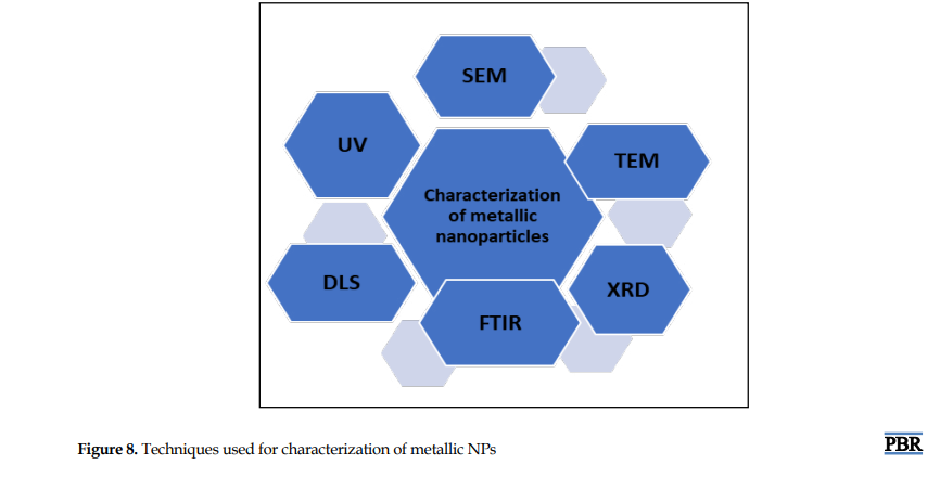
UV-Visible: Examining the optical properties of NPs requires the use of UV-visible absorption spectroscopy. This method makes it easier to analyze the size of NPs and enables the quantitative evaluation of their creation. In essence, it entails examining a sample's reaction to electromagnetic waves having wavelengths between 190 and 700 nm.
FT-IR: FT-IR spectroscopy is used to get information about various functional groups based on the peak positions observed in the spectrum. Additionally, this analysis may provide information about the stability and capping of NPs.
SEM: It is used to investigate NPs. The synthesised NPs' size, shape, morphology, and distribution are all ascertained using this analytical method. The morphological structural alterations before and after the therapy are assessed using the SEM analysis.
TEM: When compared to SEM, TEM has two important advantages: It delivers higher resolution and permits more thorough analytical examinations. The necessity of a high vacuum environment, the need for a very tiny sample size, and the time-consuming nature of sample preparation—all of which are critical for TEM—are disadvantages, though.
XRD: Materials' atomic structures can be examined using XRD. This method is useful for figuring out a substance's qualitative and quantitative properties. The size and structure of crystalline NPs are identified and confirmed using XRD analysis. To determine the particle size of nanomaterials, XRD data are subjected to the Debye–Scherrer formula, which links the width of the Bragg reflection to the subsequent equation: Kλ/β cos θ = d. The Scherrer constant, K, the X-ray wavelength, β, the full width at half maximum, diffraction angle (half of the Bragg angle) connected to the lattice plane, and particle size, d, which is measured in nanometers (nm), are all represented in this equation.
DLS: Small particle size and distribution may be examined with DLS on a scale ranging from submicron levels to 1 nm. This method depends on how light and NPs interact. Narrow size distributions, particularly those between 2 and 500 nm, may be measured using it.
Biodistribution of NPs
Biodistribution is a technique to determine whether the compound of interest is distributed throughout the body of animal or human and how long it stays in tissue or the body.
Determining the biodistribution of the NPs after in vivo treatment in humans and animals is a crucial step in the translational evaluation of nanomedicines. Several methods are available to assess the biodistribution of NPs. Existing methods of evaluating biodistribution of NPs with their pros and cons are discussed below [149–152].
Histology
Pros
1) A comparatively economical approach; 2) typically regarded as a qualitative method for assessing biodistribution; 3) facilitates the examination of extensive tissue samples; 4) enables the investigation of the precise cellular interactions of NPs within tissues, and 5) does not necessitate the use of contrast materials.
Cons
1) Low-resolution imaging of NPs inside tissue slices is possible with “light and fluorescence microscopy; 2) A small number of tissue slices are typically analyzed to determine the biodistribution of NPs over an entire organ, 3) this method is time-consuming and labor-intensive; 4) tissue structure and resolution may be harmed by the freezing procedure used for cryostat sectioning, especially when light microscopy is being employed,; 5) human mistake is a possible while preparing and analyzing slides; 6) It might be difficult to distinguish between particular cell types and NPs in tissue slices,; 7) labelling NPs with fluorescent dyes for fluorescence imaging of histological sections may change the NPs' ‘physicochemical’ characteristics and impact how they behave in vivo, and 8) when fluorescently tagged, NPs are exposed to light during the in vivo injection procedure, photobleaching may happen, which might cause problems with tissue harvesting and processing.
Electron microscopy
Pros
1) Able to provide thorough information at very high magnification about the biodistribution of NPs; 2) facilitates the observation of nanoparticle accumulation within cells and their specific localization in cellular organelles; 3) typically regarded as a semi-quantitative approach, and 4) this approach is mostly used to evaluate the cellular interaction of NPs in vitro; just a few research studies use it to examine the biodistribution of NPs following in vivo delivery.
Cons
1) A more costly method compared to conventional histology; 2) inability to assess large tissue samples; 3) this technique is labor-intensive; 4) typically, a small number of extremely thin tissue slices are analyzed to determine the distribution of NPs throughout an entire organ; 5) a relatively large quantity of NPs must be consumed; 6) to accurately detect the nanomaterial within tissues and cells, another identification technique are needed; 7) the application of high-voltage electron beams may have an impact on the characterisation of soft materials; 8) images may have burn-in regions, which might result in artifacts, and 9) not all nanoparticle kinds may be suitable for the sample preparation method.
Liquid scintillation counting (LSC)
Pros
1) A technique that is sensitive, specific, and quantitative, and 2) LSC can assess the biodistribution of NPs at the level of organs or tissues.
Cons
1) This approach can be quite demanding, particularly due to the requirement to process and dissolve the collected tissues before conducting LSC analysis; 2) it may not accurately represent the biodistribution of the entire organ if only a small segment is sampled for LSC; 3) LSC provides little information about the precise cellular interactions or the buildup of NPs in the tissues, and 4) the cocktail used, together with variables like sample composition, volume, temperature, and the counting device employed, all affect the data's quality and consistency.
Measurement of drug concentration in tissues
Pros
1) A numerical evaluation of biodistribution that may be used to analyze whole or partial tissue samples; 2) it can serve as a supplementary quantitative tool to reinforce the biodistribution findings obtained through qualitative methods, and 3) using contrast agents to enhance imaging outcomes, integrating imaging molecules into NPs, or being exposed to ionizing radiation are not necessary for this method.
Cons
1) Assessing the payload's biodistribution is the main objective of this indirect method; 2) if the drug separates from the NPs too quickly after in vivo delivery, it might, however, provide conflicting findings; 3) the quality of tissue preparation and the extraction procedure, which may be both time-consuming and tedious, have a major impact on the accuracy of drug concentration readings, and 4) additionally, this method does not offer insights into the real-time biodistribution at various time intervals in animal subjects.
In vivo optical imaging
Pros
1) A straightforward and non-invasive method that is easy to implement; 2) quick image capture durations; 3) eliminates the need for exposure to ionizing radiation; 4) allows for real-time imaging and can be conducted at various time intervals; 5) it is possible to assess the biodistribution of NPs at the tissue or organ level; 6) the resulting pictures typically exhibit high snsitivity along with improved spatial and temporal resolution, and 7) This approach is typically thought of as a qualitative evaluation of biodistribution.
Cons
1) Less than 1 cm of tissue can be penetrated, and as tissue depth grows, it may become less effective; 2) Comparatively lacks the spatial resolution of CT and MRI imaging methods; 3) fluorophores can be used to mark NPs, changing their physicochemical properties and in vivo behavior; 4) numerous fluorophores are prone to photobleaching during imaging procedures, which can diminish their sensitivity; 5) because tissue autofluorescence is a significant obstacle that might impede the interpretation of results, fluorophores should have greater signal-to-background ratios; 6) does not yield information about the specific cellular association or accumulation of NPs within tissues, and 7) is unable to visualize individual NPs, focusing instead on measuring overall fluorescence intensity.
Computed tomography (CT)
Pros
1) Produces reliable, high-quality images that may be used to assess the NPs' biodiversity; 2) it has no restrictions on the invasion of tissue and provides comparatively quick picture capture times; 3) generally considered a qualitative evaluation of biodistribution; 4) identifying nanoparticle’s biodistribution at both the tissue and organ levels, and 5) it is possible to track the distribution of NPs throughout the body both in real time and at different intervals of time.
Cons
1) Includes being exposed to ionized radiation; 2) Lacks details on the certain cells interactions of NPs; 3) to increase clarity and distinguish between different tissue types, contrast-enhancing imaging substances are frequently required; 4) there may be potential complications when NPs tagged with contrast agents are utilized alongside other contrast imaging agents to enhance anatomical and tissue imaging; 5) the sensitivity of nanoparticle contrast agents is lower than that of other imaging modalities, such as nuclear imaging, and 6) when contrast agents are added to NPs, their physicochemical properties and behavior within a biological organism can be altered.
Magnetic resonance imaging (MRI)
Pros
1) Non-invasive and straightforward method; 2) eliminates the risk of exposure to ionizing radiation,; 3) generates images with superior spatial resolution in comparison to techniques like optical; 4) improves the ability to distinguish between fat, muscle, water, and soft tissue by providing more contrast for soft tissues compared to CT; 5) unrestricted by the depth of the tissue, penetration is unbounded; 6) capable of assessing NPs distribution at the tissue level, and 7) enables real-time evaluation of NP biodistribution across various time intervals.
Cons
1) A comparatively expensive method; 2) exhibits slow image capture and extended post-processing durations; 3) typically necessitates a significantly larger volume of contrast agents due to its potential low sensitivity; 4) not suitable for individuals with metallic implants or devices, and 5) the integration of contrast agents into NPs may modify their physicochemical characteristics and in-vivo performance.
Nuclear medicine imaging (PET and SPECT)
Pros
1) Capable of quantitative evaluation of biodistribution; 2) it is possible to track the biodistribution of NPs in real time; 3) this method allows for the imaging of biochemical processes; 4) it is not limited by the constraints of tissue penetration; 5) because this extremely sensitive method uses less radiolabels, it has less of an effect on adjacent tissues and cellular activity; 6) PET offers greater sensitivity compared to SPECT and delivers more precise localization of radiation events; 7) by substituting positron-emitting isotopes with naturally existing atoms, PET can better see molecular processes; 8) SPECT is capable of simultaneously imaging many radionuclide probes and is easier to get, and 9) SPECT examination are considerably more cost-effective than PET scans, largely due to the simpler preparation of its radionuclides, easier availability, and generally longer half-lives compared to those used in PET.
Cons
1) A comparatively expensive method; 2) consists of ionizing radiation exposure; 3) shows sluggish rates of picture capture; 4) because radiolabels deteriorate with time, they are not appropriate for longitudinal investigations; 5) provides a poor spatial resolution and insufficient anatomical details, which often necessitates its combination with other imaging techniques like MRI or CT; 6) because certain NPs may exhibit differing compatibility and imaging performance across various approaches, the choice of radionuclide and radiolabelling methodology needs to be carefully considered; 7) compared to PET, SPECT has a reduced photon detection efficiency and resolution, and 8) PET generally necessitates the use of a cyclotron or generator.
Conclusion
Since the traditional techniques for creating NPs are expensive and provide highly hazardous products, it is urgent to lower the risk of environmental toxicity from the various substances that are employed in both chemical and physical processes. Green synthesis is one of the alternate techniques that has been found to discover to create NPs. In this review, green synthesis of Au, Ag, Fe, Cu, and Pd NPs with their applications has been discussed. Synthesis of metallic NPs by using green method, i.e, plant and their extract, bacteria, fungi has shown enormous promise in a number of fields, including industry and medicine.
Ethical Considerations
Compliance with ethical guidelines
This article is a review paper with no human or animal sample.
Funding
This review received no specific grant from funding agencies in the public, commercial, or not-for-profit sectors.
Authors' contributions
Conceptualization, study design, data collection, and draft manuscript: Ankush Banalia; Data analysis and data interpretation: Dinesh Puri.
Conflict of interest
The authors declared no conflict of interest.
Acknowledgments
The authors are thankful to the School of Pharmacy, Graphic Era Hill University for offering guidance and technical.
References
Over the past 20 years, nanotechnology has greatly ranked among the most important technologies studied and rapidly expanding sciences because of its applicability in several human welfare domains [1].
The term “Nano” was obtained from Greek word “nanos,” which means diminutive. The units of measurement for nanoparticles (NPs) are nanometers. They are popular because of their sub-microscopic particle-size and large surface area [2].
There are four main categories of NPs depending on their chemical makeup: Carbon-based (carbon nanotubes and nanofibers, etc.), bioorganic-based (liposomes, micelles, etc.), metal- and metal oxide-based (Ag, Cu, etc.), and composite based [3]. NPs have extraordinary physical, thermal, optical, magnetic electrical, and chemical, properties compared to their non-nano material [4]. They’ve wide scope in Pharmaceuticals, as well as catalysis, householding products, environmental sensors, automotive industries, and nanobiotechnology. Additionally, nanotechnology helps the early diagnosis of serious illnesses like cancer. Examples of NPs include dendrimers, metal NPs, liposomes, fullerenes, and nanodroplets [5].
Metallic NPs are an important and most-studied class of materials with various applications. Numerous studies are being conducted on the production of metal ion NPs from microorganisms and botanical extracts [6]. Silver NPs, AgNPs, are the most often used of all the produced NPs and found in over 25% of consumer goods. Research shows that gold NPs (AuNPs) has biological uses as muscle relaxant, antibacterial, and enzyme control. AgNPs prevent gram-positive and gram-negative bacteria from growing and acting. Copper NPs (CuNPs) possess great promise as drug delivery vehicles, anti-cancer drugs, and enhancers of photodynamic treatment. Palladium NPs (PdNPs) are used as catalysts, dye degradation, and anti-microbial. Finally, iron NPs (FeNPs) inhibit bacterial growth [7]. NPs can be synthesized by three different techniques: physical, chemical, biological (Figure 1) [5].
This review explores how biology and physical sciences collaborate to produce metal NPs in a “green” manner for different applications. According to the research, bacteria and plant extracts are novel sources of producing NPs. To achieve this goal, it is essential to use natural resources (such organic means) and the best solvent systems.
Advantages of green synthesis over synthetic method [8–10]
Green synthesis has the following advantages
1) By using fewer dangerous components and toxic organic solvents, green chemistry provides substantial economic and environmental benefits over conventional synthetic processes. 2) They improve the sustainability in drug synthesis by using renewable resources while preserving the efficacy and quality of medications. 3) Green synthesis reduces the use of resources and improves the atom economy. 4) This method lowers toxicity and eliminates using dangerous procedures. 5) It uses cutting-edge techniques, including water as a solvent and microwave synthesis. 6) Real time monitoring can takes place. 7) During green synthesis, catalysts are utilized sparingly and have a high rate of small-scale reaction.
Green synthesis
Although the traditional methods have long been used for decades, research has shown that green methods are more outstanding in producing NPs because they are easier to outline, less expensive, and less likely to fail [10]. Numerous resources, including plants and their extracts, algae, fungi, yeast, bacteria, and viruses, can be used to carry out the green synthesis of NPs [11]. Proteins (amino acids), phytochemicals (alkaloids, flavonoids, reducing sugars, polyphenols), and other substances are present in the biomaterials and serve as capping and reducing agents throughout the synthesis process for generating metal NPs from their precursor metal salts [12]. The reduction of the metal precursor to its subsequent NPs may be initially confirmed by observing the color shift of the colloidal solution [9, 10]. Many biotechnological uses, such as bioremediation and bioleaching, have been created since bacteria can interact with, extract, and collect metals from their surroundings [3].
Because of lipid based amphipathic membrane, they can interact with their environment, promote variety of oxidation-reduction reactions, and allow biochemical transformations. Utilizing plants as opposed to other environmentally beneficial biological systems, such as bacteria and fungus, such doing away with costly and time-consuming isolation and processing techniques [13]. Use of plants and their extracts is more safer and efficacious for the production of NPs than other biological systems of producing NPs [14]. Summary of concepts and principles behind the green chemistry are shown in Figures 2 and 3.
Different metallic precursors used for the synthesis are given in Figure 4. Finally, Table 1 presents the differences between synthesis of NPs by green chemistry and classical chemistry. The major advantage of green chemistry over classical chemistry is that green chemistry is more eco-friendly
Chemical transformations during green synthesis
The primary ingredients of AgNPs green synthesis are silver metal ion solution (0.1-10 mM) and a reducing biological agent. This eco-friendly method avoids the use of toxic chemicals [15]. Reaction should be completed at room temperature to mild heating and pH 7–10 is optimal for many systems. The green synthesis of AgNPs included the reduction of silver ions (Ag⁺) to elemental silver (Ag⁰) using plant extracts, microbes, or other biological agents, which is followed by agglomeration into clusters. These clusters eventually form metallic colloidal silver particles [16]. In most cases, the reducing agents or other constituents present in the cells act as stabilizing and capping agents. This process includes the following steps [17].
a. Silver salt dissociation:
In the first step, silver nitrate is dissociated into silver ion.
AgNO3→Ag++NO3−
b. Reduction of Ag⁺ to Ag⁰ by phytochemicals:
In the presence of reducing agent like plant extract, microbes, or biomolecules, Ag+ is reduced to Ag0.
Ag++[Reducing agent from plant/microbe]→Ag0↓+ [Oxidized by-products]
Example is using a polyphenol like catechol.
2Ag++C6H4(OH)2→2Ag0+C6H4O2+2H+
c. Nucleation and growth:
In this step, reduced Ag0 atoms are nucleated to form small clusters and then these clusters grow into NPs.
d. Stabilization (capping):
At the end, biomolecules (like proteins, terpenoids, etc.) cap the NPs to prevent aggregation.
Synthesis of metallic NPs using bacteria
Numerous bacteria have demonstrated the capacity to synthesize metallic NPs; each has specific pros and cons. Critical metals need to enter the cytoplasm through the cell wall (extracellular and intracellular) [18]. Because of their capacity to reduce metal ions, bacteria are excellent options for creating NPs. Prokaryotic and actinomycetes bacteria have been used extensively in the synthesis of metal/metal oxide NPs [19].
Bacteria can participate actively in creating NPs, serve as a bioscaffold for mineralization, or function as a biocatalyst for the synthesis of inorganic materials. During the incubation period, bacteria in broth medium can produce extracellular or intracellular nanomaterials [10]. Bacterial species with different morphologies and the internal and exterior environment of a cell frequently affect the crystalline and non-crystalline phases of particle creation [20, 21].
Shivaji et al. synthesized AgNPs using bacterial culture that remain stable for 8 months in dark. Bacteria used in this experiment were Bacillus indicus, B. cecembensis, Arthrobacter kerguelensis, A. gangotriensis, P. antarctica, P. proteolytica, and P. meridiana. Created NPs were bactericidal [22]. Sharma et al. synthesized gold NPs using Marinobacter pelagius [23]. Tiwari et al. manufactured copper NPs using copper-resistant B. cereus. Synthesized NPs shows antimicrobial effects [24]. Hasan et al. synthesized iron nanoparticle using B. proteolyticus UPMC1508. Created NPs were bactericidal and anti-cancer [25]. Liu et al. synthesized selenium NPs using B. paramycoides. Created NPs shows anti-bacterial and anti-oxidant properties [26] (Table 2). Figure 5 shows the process of NPs synthesis using bacteria.Silver salt dissociation:
Synthesis of NPs using fungi
Fungi are considered good candidates because they can produce monodisperse NPs with highly defined dimension, various chemical compositions and sizes. Also, fungi release greater quantity of proteins that lead to a higher level of nanoparticle production [66]. Fungi can produce various compounds with different applications. Over 6400 bioactive chemicals are produced by ascomycetes, imperfect fungi, and other microscopic filamentous fungi [67]. Fungi are at the forefront of research for the production of biological metal NPs because of their tolerance and capacity for metal biomagnification [68-73]. The ability to synthesize enormous amounts of proteins and enzymes, some of which may be utilized for the quick and sustainable production of NPs, gives fungi an edge over other microbes [67].
Raut et al. synthesized silver NPs using fungi, such as Cladosporium cladosporioides, Penicillium chrysogenum, and Purpureocillium lilacinum. Created NPs shows anti-microbial NPs [72]. Iranmanesh et al. synthesized gold NPs using 12 fungi; out of 12 fungi, 8 could successfully synthesize gold NPs. Further out of 8 fungi, 3 are investigated: Fusarium oxysporum, Aspergillus flavus, Rhizoctonia solani, and Verticillium dahliae [72]. Kamal et al. synthesized iron NPs using fungi, ie, Daedalea mushroom. It is first time that iron NPs are synthesized by using mushroom. Created NPs were analyzed against the pathogenic fungus Aspergillus niger [74]. Fatima and Wahid synthesized copper NPs using fungi Schizophyllum commune. Created NPs were analyzed against multidrug-resistant organisms (MDROs) like Escherichia coli, Salmonella abony, Staphylococcus aureus, and Klebsiella pneumoniae [75]. Hussein et al. synthesized selenium NPs by using 4 different fungi: Aspergillus quadrilineatus, Aspergillus ochraceus, A. terreus, Fusarium equiseti. Synthesized NPs were anti-bacterial and anti-fungal [76] (Table 3). Figure 6 shows the process of synthesized of NPs using fungi.
Synthesis of NPs using plant and their extracts
Although the capacity of plant extracts to reduce metals has been known since the early 1900s, little is known about the specifics of the reducing chemicals at play. Compared to whole plant tissue, using plant extracts to make NPs is simpler. Plant extract-mediated synthesis is a growing area of interest [25]. These days, plant extracts serve as capping and reducing agents throughout the nanoparticle manufacturing process, which is way better than microbial, chemical synthesis [109-114].
Zahir et al. synthesized silver NPs using aqueous plant extract of Euphorbia prostrata. Created NPs were analyzed with pesticidal activity against Sitophilus oryzae L [115]. Hazarika et al. synthesized palladium NPs using leaf extract of Garcinia pedunculata Roxb. Created NPs were analyzed with anti-microbial activity Cronobacter sakazakii strain AMD04 [116]. Majumdar et al. synthesized gold NPs by using leaf extract of Acacia nilotica (Babool). Created NPs were analyzed with catalyst property which is used to reduce 4-nitrophenol to 4-aminophenol [117]. Vasantharaj et al. synthesized iron NPs using Ruellia tuberosa (RT) leaf extract. Created NPs were analyzed with potent anti-bacterial activity against K. pneumoniae, E. coli and average anti-bacterial activity against S. aureus [118]. Alao et al. synthesized copper NPs using ethanolic extract of Kigelia africana fruit. Created NPs were analyzed with anti-microbial activity against E. coli, Shigella sp., S. aureus, Pseudomonas aeruginosa, and Salmonella typhi [119] (Table 4). Figure 7 shows the synthesis of nanoparticles using plant and their extract.
Characterization of metallic NPs [164-178]
Characterization of NPs can be done using ultraviolet (UV) (UV–Vis spectroscopy), Fourier transforms infrared spectroscopy (FT-IR), scanning electronic microscopy (SEM), transmission electron microscopy (TEM), dynamic light scattering (DLS), and X-ray diffraction (XRD). Figure 8 shows a summary of techniques used for the characterization of metallic NPs.
UV-Visible: Examining the optical properties of NPs requires the use of UV-visible absorption spectroscopy. This method makes it easier to analyze the size of NPs and enables the quantitative evaluation of their creation. In essence, it entails examining a sample's reaction to electromagnetic waves having wavelengths between 190 and 700 nm.
FT-IR: FT-IR spectroscopy is used to get information about various functional groups based on the peak positions observed in the spectrum. Additionally, this analysis may provide information about the stability and capping of NPs.
SEM: It is used to investigate NPs. The synthesised NPs' size, shape, morphology, and distribution are all ascertained using this analytical method. The morphological structural alterations before and after the therapy are assessed using the SEM analysis.
TEM: When compared to SEM, TEM has two important advantages: It delivers higher resolution and permits more thorough analytical examinations. The necessity of a high vacuum environment, the need for a very tiny sample size, and the time-consuming nature of sample preparation—all of which are critical for TEM—are disadvantages, though.
XRD: Materials' atomic structures can be examined using XRD. This method is useful for figuring out a substance's qualitative and quantitative properties. The size and structure of crystalline NPs are identified and confirmed using XRD analysis. To determine the particle size of nanomaterials, XRD data are subjected to the Debye–Scherrer formula, which links the width of the Bragg reflection to the subsequent equation: Kλ/β cos θ = d. The Scherrer constant, K, the X-ray wavelength, β, the full width at half maximum, diffraction angle (half of the Bragg angle) connected to the lattice plane, and particle size, d, which is measured in nanometers (nm), are all represented in this equation.
DLS: Small particle size and distribution may be examined with DLS on a scale ranging from submicron levels to 1 nm. This method depends on how light and NPs interact. Narrow size distributions, particularly those between 2 and 500 nm, may be measured using it.
Biodistribution of NPs
Biodistribution is a technique to determine whether the compound of interest is distributed throughout the body of animal or human and how long it stays in tissue or the body.
Determining the biodistribution of the NPs after in vivo treatment in humans and animals is a crucial step in the translational evaluation of nanomedicines. Several methods are available to assess the biodistribution of NPs. Existing methods of evaluating biodistribution of NPs with their pros and cons are discussed below [149–152].
Histology
Pros
1) A comparatively economical approach; 2) typically regarded as a qualitative method for assessing biodistribution; 3) facilitates the examination of extensive tissue samples; 4) enables the investigation of the precise cellular interactions of NPs within tissues, and 5) does not necessitate the use of contrast materials.
Cons
1) Low-resolution imaging of NPs inside tissue slices is possible with “light and fluorescence microscopy; 2) A small number of tissue slices are typically analyzed to determine the biodistribution of NPs over an entire organ, 3) this method is time-consuming and labor-intensive; 4) tissue structure and resolution may be harmed by the freezing procedure used for cryostat sectioning, especially when light microscopy is being employed,; 5) human mistake is a possible while preparing and analyzing slides; 6) It might be difficult to distinguish between particular cell types and NPs in tissue slices,; 7) labelling NPs with fluorescent dyes for fluorescence imaging of histological sections may change the NPs' ‘physicochemical’ characteristics and impact how they behave in vivo, and 8) when fluorescently tagged, NPs are exposed to light during the in vivo injection procedure, photobleaching may happen, which might cause problems with tissue harvesting and processing.
Electron microscopy
Pros
1) Able to provide thorough information at very high magnification about the biodistribution of NPs; 2) facilitates the observation of nanoparticle accumulation within cells and their specific localization in cellular organelles; 3) typically regarded as a semi-quantitative approach, and 4) this approach is mostly used to evaluate the cellular interaction of NPs in vitro; just a few research studies use it to examine the biodistribution of NPs following in vivo delivery.
Cons
1) A more costly method compared to conventional histology; 2) inability to assess large tissue samples; 3) this technique is labor-intensive; 4) typically, a small number of extremely thin tissue slices are analyzed to determine the distribution of NPs throughout an entire organ; 5) a relatively large quantity of NPs must be consumed; 6) to accurately detect the nanomaterial within tissues and cells, another identification technique are needed; 7) the application of high-voltage electron beams may have an impact on the characterisation of soft materials; 8) images may have burn-in regions, which might result in artifacts, and 9) not all nanoparticle kinds may be suitable for the sample preparation method.
Liquid scintillation counting (LSC)
Pros
1) A technique that is sensitive, specific, and quantitative, and 2) LSC can assess the biodistribution of NPs at the level of organs or tissues.
Cons
1) This approach can be quite demanding, particularly due to the requirement to process and dissolve the collected tissues before conducting LSC analysis; 2) it may not accurately represent the biodistribution of the entire organ if only a small segment is sampled for LSC; 3) LSC provides little information about the precise cellular interactions or the buildup of NPs in the tissues, and 4) the cocktail used, together with variables like sample composition, volume, temperature, and the counting device employed, all affect the data's quality and consistency.
Measurement of drug concentration in tissues
Pros
1) A numerical evaluation of biodistribution that may be used to analyze whole or partial tissue samples; 2) it can serve as a supplementary quantitative tool to reinforce the biodistribution findings obtained through qualitative methods, and 3) using contrast agents to enhance imaging outcomes, integrating imaging molecules into NPs, or being exposed to ionizing radiation are not necessary for this method.
Cons
1) Assessing the payload's biodistribution is the main objective of this indirect method; 2) if the drug separates from the NPs too quickly after in vivo delivery, it might, however, provide conflicting findings; 3) the quality of tissue preparation and the extraction procedure, which may be both time-consuming and tedious, have a major impact on the accuracy of drug concentration readings, and 4) additionally, this method does not offer insights into the real-time biodistribution at various time intervals in animal subjects.
In vivo optical imaging
Pros
1) A straightforward and non-invasive method that is easy to implement; 2) quick image capture durations; 3) eliminates the need for exposure to ionizing radiation; 4) allows for real-time imaging and can be conducted at various time intervals; 5) it is possible to assess the biodistribution of NPs at the tissue or organ level; 6) the resulting pictures typically exhibit high snsitivity along with improved spatial and temporal resolution, and 7) This approach is typically thought of as a qualitative evaluation of biodistribution.
Cons
1) Less than 1 cm of tissue can be penetrated, and as tissue depth grows, it may become less effective; 2) Comparatively lacks the spatial resolution of CT and MRI imaging methods; 3) fluorophores can be used to mark NPs, changing their physicochemical properties and in vivo behavior; 4) numerous fluorophores are prone to photobleaching during imaging procedures, which can diminish their sensitivity; 5) because tissue autofluorescence is a significant obstacle that might impede the interpretation of results, fluorophores should have greater signal-to-background ratios; 6) does not yield information about the specific cellular association or accumulation of NPs within tissues, and 7) is unable to visualize individual NPs, focusing instead on measuring overall fluorescence intensity.
Computed tomography (CT)
Pros
1) Produces reliable, high-quality images that may be used to assess the NPs' biodiversity; 2) it has no restrictions on the invasion of tissue and provides comparatively quick picture capture times; 3) generally considered a qualitative evaluation of biodistribution; 4) identifying nanoparticle’s biodistribution at both the tissue and organ levels, and 5) it is possible to track the distribution of NPs throughout the body both in real time and at different intervals of time.
Cons
1) Includes being exposed to ionized radiation; 2) Lacks details on the certain cells interactions of NPs; 3) to increase clarity and distinguish between different tissue types, contrast-enhancing imaging substances are frequently required; 4) there may be potential complications when NPs tagged with contrast agents are utilized alongside other contrast imaging agents to enhance anatomical and tissue imaging; 5) the sensitivity of nanoparticle contrast agents is lower than that of other imaging modalities, such as nuclear imaging, and 6) when contrast agents are added to NPs, their physicochemical properties and behavior within a biological organism can be altered.
Magnetic resonance imaging (MRI)
Pros
1) Non-invasive and straightforward method; 2) eliminates the risk of exposure to ionizing radiation,; 3) generates images with superior spatial resolution in comparison to techniques like optical; 4) improves the ability to distinguish between fat, muscle, water, and soft tissue by providing more contrast for soft tissues compared to CT; 5) unrestricted by the depth of the tissue, penetration is unbounded; 6) capable of assessing NPs distribution at the tissue level, and 7) enables real-time evaluation of NP biodistribution across various time intervals.
Cons
1) A comparatively expensive method; 2) exhibits slow image capture and extended post-processing durations; 3) typically necessitates a significantly larger volume of contrast agents due to its potential low sensitivity; 4) not suitable for individuals with metallic implants or devices, and 5) the integration of contrast agents into NPs may modify their physicochemical characteristics and in-vivo performance.
Nuclear medicine imaging (PET and SPECT)
Pros
1) Capable of quantitative evaluation of biodistribution; 2) it is possible to track the biodistribution of NPs in real time; 3) this method allows for the imaging of biochemical processes; 4) it is not limited by the constraints of tissue penetration; 5) because this extremely sensitive method uses less radiolabels, it has less of an effect on adjacent tissues and cellular activity; 6) PET offers greater sensitivity compared to SPECT and delivers more precise localization of radiation events; 7) by substituting positron-emitting isotopes with naturally existing atoms, PET can better see molecular processes; 8) SPECT is capable of simultaneously imaging many radionuclide probes and is easier to get, and 9) SPECT examination are considerably more cost-effective than PET scans, largely due to the simpler preparation of its radionuclides, easier availability, and generally longer half-lives compared to those used in PET.
Cons
1) A comparatively expensive method; 2) consists of ionizing radiation exposure; 3) shows sluggish rates of picture capture; 4) because radiolabels deteriorate with time, they are not appropriate for longitudinal investigations; 5) provides a poor spatial resolution and insufficient anatomical details, which often necessitates its combination with other imaging techniques like MRI or CT; 6) because certain NPs may exhibit differing compatibility and imaging performance across various approaches, the choice of radionuclide and radiolabelling methodology needs to be carefully considered; 7) compared to PET, SPECT has a reduced photon detection efficiency and resolution, and 8) PET generally necessitates the use of a cyclotron or generator.
Conclusion
Since the traditional techniques for creating NPs are expensive and provide highly hazardous products, it is urgent to lower the risk of environmental toxicity from the various substances that are employed in both chemical and physical processes. Green synthesis is one of the alternate techniques that has been found to discover to create NPs. In this review, green synthesis of Au, Ag, Fe, Cu, and Pd NPs with their applications has been discussed. Synthesis of metallic NPs by using green method, i.e, plant and their extract, bacteria, fungi has shown enormous promise in a number of fields, including industry and medicine.
Ethical Considerations
Compliance with ethical guidelines
This article is a review paper with no human or animal sample.
Funding
This review received no specific grant from funding agencies in the public, commercial, or not-for-profit sectors.
Authors' contributions
Conceptualization, study design, data collection, and draft manuscript: Ankush Banalia; Data analysis and data interpretation: Dinesh Puri.
Conflict of interest
The authors declared no conflict of interest.
Acknowledgments
The authors are thankful to the School of Pharmacy, Graphic Era Hill University for offering guidance and technical.
References
- Tsekhmistrenko SI, Bityutskyy VS, Tsekhmistrenko OS, Horalskyi LP, Tymoshok NO, Spivak MY. Bacterial synthesis of nanoparticles: A green approach. Biosyst Divers. 2020; 28(1):9-17. [Link]
- Bordiwala RV. Green synthesis and Applications of Metal Nanoparticles.- A Review Article. Results Chem. 2023; 5:100832. [DOI:10.1016/j.rechem.2023.100832]
- Dikshit P, Kumar J, Das A, Sadhu S, Sharma S, Singh S, et al. Green Synthesis of Metallic Nanoparticles: Applications and Limitations. Catalysts. 2021; 11(8):902. [DOI:10.3390/catal11080902]
- Pantidos N. Biological Synthesis of Metallic Nanoparticles by Bacteria, Fungi and Plants. J Nanomedicine Nanotechnol. 2014; 05(05). [DOI:10.4172/2157-7439.1000233]
- Vijayaram S, Razafindralambo H, Sun YZ, Vasantharaj S, Ghafarifarsani H, Hoseinifar SH, et al. Applications of Green Synthesized Metal Nanoparticles - a Review. Biol Trace Elem Res. 2024; 202(1):360-86. [DOI:10.1007/s12011-023-03645-9] [PMID]
- Vijayakumar S. Eco-friendly synthesis of gold nanoparticles using fruit extracts and in vitro anticancer studies. J Saudi Chem Soc. 2019; 23(6):753–61. [DOI:10.1016/j.jscs.2018.12.002]
- Ying S, Guan Z, Ofoegbu PC, Clubb P, Rico C, He F, et al. Green synthesis of nanoparticles: Current developments and limitations. Environ Technol Innov. 2022; 26:102336. [DOI:10.1016/j.eti.2022.102336]
- Sharma A, Arora SK. A review study on green synthesis of schiff bases. Indian J Appl Res. 2023; 69-72. [DOI:10.36106/ijar/9529205]
- Laddha PR, Sitaphale GR, Charhate KB, Tathe PR. Green Chemistry in Pharmaceutical Synthesis: Sustainable Strategies for Drug Production. Int J Adv Res Sci Commun Technol. 2024; 4(2):323-30. [DOI:10.48175/IJARSCT-19854]
- Tauro SJ, Gawad JB. Green Chemistry: A Boon to Pharmaceutical Synthesis. Int J Sci Res. 2012; 2(7):67-9. [DOI:10.15373/22778179/JULY2013/22]
- Rafique M, Sadaf I, Rafique MS, Tahir MB. A review on green synthesis of silver nanoparticles and their applications. Artif Cells Nanomed Biotechnol. 2017; 45(7):1272-91.[DOI:10.1080/21691401.2016.1241792] [PMID]
- Gour A, Jain NK. Advances in green synthesis of nanoparticles. Artif Cells Nanomedicine Biotechnol. 2019; 47(1):844-51. [DOI:10.1080/21691401.2019.1577878] [PMID]
- Gu Z, Guo Y, Mao X, Qi M, Ge B. Green Synthesis of Nanomaterials Mediated by Herbal Medicine: Mechanism, Characterization, and Application Prospects. Nat Prod Commun. 2025; 20(8). [DOI:10.1177/1934578X251365007]
- Huq A, Khan AA, Alshehri JM, Rahman Sh, Balusamy SR, Akter S. Bacterial mediated green synthesis of silver nanoparticles and their antibacterial and antifungal activities against drug-resistant pathogens. R Soc Open Sci. 2023; 10(10):230796. [DOI:10.1098/rsos.230796]
- Singh P, Kim YJ, Zhang D, Yang DC. Biological Synthesis of Nanoparticles from Plants and Microorganisms. Trends Biotechnol. 2016; 34(7):588-99. [DOI:10.1016/j.tibtech.2016.02.006] [PMID]
- Singh J, Dutta T, Kim KH, Rawat M, Samddar P, Kumar P. ‘Green’ synthesis of metals and their oxide nanoparticles: Applications for environmental remediation. J Nanobiotechnol. 2018; 16(1):84. [DOI:10.1186/s12951-018-0408-4]
- Chopra H, Bibi S, Singh I, Hasan MM, Khan MS, Yousafi Q, et al. Green Metallic Nanoparticles: Biosynthesis to Applications. Front Bioeng Biotechnol. 2022; 10:874742. [DOI:10.3389/fbioe.2022.874742] [PMID]
- Hebeish A, El-Rafie MHM, El-Sheikh MA, El-Naggar ME. Nanostructural features of silver nanoparticles synthesized in starch–polyacrylic acid graft copolymer matrix. Carbohydr Polym. 2013; 92(1):407–13.
- Asif M, Yasmin R, Asif R, Ambreen A, Mustafa M, Umbreen S. Green Synthesis of Silver Nanoparticles (AgNPs), Structural Characterization, and their Antibacterial Potential. Dose-Response. 2022; 20(2):15593258221088709. [DOI:10.1177/15593258221088709] [PMID]
- Nabila MI, Kannabiran K. Biosynthesis, characterization and antibacterial activity of copper oxide nanoparticles (CuO NPs) from actinomycetes. Biocatal Agric Biotechnol. 2018; 15:56-62. [DOI:10.1016/j.bcab.2018.05.011]
- Omajali JB, Mikheenko IP, Merroun ML, Wood J, Macaskie LE. Characterization of intracellular palladium nanoparticles synthesized by Desulfovibrio desulfuricans and Bacillus benzeovorans. J Nanoparticle Res. 2015; 17:264. [DOI:10.1007/s11051-015-3067-5] [PMID]
- Shivaji S, Madhu S, Singh S. Extracellular synthesis of antibacterial silver nanoparticles using psychrophilic bacteria. Process Biochem. 2011; 46(9):1800-7. [DOI:10.1016/j.procbio.2011.06.008]
- Sharma N, Pinnaka AK, Raje M, Fnu A, Bhattacharyya MS, Choudhury AR. Exploitation of marine bacteria for production of gold nanoparticles. Microb Cell Fact. 2012; 11:86. [DOI:10.1186/1475-2859-11-86] [PMID]
- Tiwari M, Jain P, Chandrashekhar Hariharapura R, Narayanan K, Bhat K. U, Udupa N, et al. Biosynthesis of copper nanoparticles using copper-resistant Bacillus cereus, a soil isolate. Process Biochem. 2016; 51(10):1348-56. [DOI:10.1016/j.procbio.2016.08.008]
- Hasan YR, Wong FW, Ashari SE, Halim M, Mohamad R. Bacillus proteolyticus UPMC1508: A novel bacterial strain capable of biologically synthesize iron oxide nanoparticles. Biologia. 2025; 80(3):697-714. [Link]
- Liu P, Long H, Cheng H, Liang M, Liu Z, Han Z, et al. Highly-efficient synthesis of biogenic selenium nanoparticles by Bacillus paramycoides and their antibacterial and antioxidant activities. Front Bioeng Biotechnol. 2023; 11:1227619. [DOI:10.3389/fbioe.2023.1227619] [PMID]
- Vijayabharathi R, Sathya A, Gopalakrishnan S. Extracellular biosynthesis of silver nanoparticles using Streptomyces griseoplanus SAI-25 and its antifungal activity against Macrophomina phaseolina , the charcoal rot pathogen of sorghum. Biocatal Agric Biotechnol. 2018; 14:166-71. [DOI:10.1016/j.bcab.2018.03.006]
- Salem SS, Fouda A. Green Synthesis of Metallic Nanoparticles and Their Prospective Biotechnological Applications: an Overview. Biol Trace Elem Res. 2021; 199(1):344-70. [DOI:10.1007/s12011-020-02138-3] [PMID]
- Paul D, Sinha SN. Extracellular Synthesis of Silver Nanoparticles Using Pseudomonas Aeruginosa KUPSB12 and Its Antibacterial Activity. Jordan J Biol Sci. 2014; 7(4):245-50. [Link]
- Monowar T, Rahman MS, Bhore SJ, Raju G, Sathasivam KV. Silver Nanoparticles Synthesized by Using the Endophytic Bacterium Pantoea ananatis are Promising Antimicrobial Agents against Multidrug Resistant Bacteria. Molecules. 2018; 23(12):3220. [DOI:10.3390/molecules23123220] [PMID]
- Gan L, Zhang S, Zhang Y, He S, Tian Y. Biosynthesis, characterization and antimicrobial activity of silver nanoparticles by a halotolerant Bacillus endophyticus SCU-L. Prep Biochem Biotechnol. 2018; 48(7):582-8. [DOI:10.1080/10826068.2018.1476880] [PMID]
- Huq MdA, Akter S. Biosynthesis, Characterization and Antibacterial Application of Novel Silver Nanoparticles against Drug Resistant Pathogenic Klebsiella pneumoniae and Salmonella Enteritidis. Molecules. 2021; 26(19):5996. [DOI:10.3390/molecules26195996] [PMID]
- Huq MA, Akter S. Bacterial Mediated Rapid and Facile Synthesis of Silver Nanoparticles and Their Antimicrobial Efficacy against Pathogenic Microorganisms. Materials (Basel). 2021; 14(10):2615. [DOI:10.3390/ma14102615] [PMID]
- Patil MP, Kang MJ, Niyonizigiye I, Singh A, Kim JO, Seo YB, et al. Extracellular synthesis of gold nanoparticles using the marine bacterium Paracoccus haeundaensis BC74171T and evaluation of their antioxidant activity and antiproliferative effect on normal and cancer cell lines. Colloids Surf B Biointerfaces. 2019; 183:110455. [DOI:10.1016/j.colsurfb.2019.110455] [PMID]
- Karthik L, Kumar G, Keswani T, Bhattacharyya A, Reddy BP, Rao KV. Marine actinobacterial mediated gold nanoparticles synthesis and their antimalarial activity. Nanomedicine. 2013; 9(7):951-60. [DOI:10.1016/j.nano.2013.02.002] [PMID]
- Sadhasivam S, Shanmugam P, Veerapandian M, Subbiah R, Yun K. Biogenic synthesis of multidimensional gold nanoparticles assisted by Streptomyces hygroscopicus and its electrochemical and antibacterial properties. Biometals. 2012; 25(2):351-60. [DOI:10.1007/s10534-011-9506-6] [PMID]
- Sidhu AK, Agrawal SB, Verma N, Kaushal P, Sharma M. Fungal-mediated synthesis of multimetallic nanoparticles: mechanisms, unique properties, and potential applications. Front Nanotechnol. 2025; 7:1549713. [Link]
- Patra JK, Fraceto LF, Das G, Campos EVR. Green Nanoparticles: Synthesis and Biomedical Applications. Cham: Springer International Publishing; 2020 [DOI:10.1007/978-3-030-39246-]
- Ebrahiminezhad A, Zare M, Kiyanpour S, Berenjian A, Niknezhad SV, Ghasemi Y. Biosynthesis of xanthangum-coated INPs by using Xanthomonas campestris. IET Nanobiotechnol. 2018; 12(3):254-8. [DOI:10.1049/iet-nbt.2017.0199]
- Jacob PJ, Masarudin MJ, Hussein MZ, Rahim RA. Facile aerobic construction of iron based ferromagnetic nanostructures by a novel microbial nanofactory isolated from tropical freshwater wetlands. Microb Cell Fact. 2017; 16(1):175.[DOI:10.1186/s12934-017-0789-3] [PMID]
- Crespo KA, Baronetti JL, Quinteros MA, Páez PL, Paraje MG. Intra- and Extracellular Biosynthesis and Characterization of Iron Nanoparticles from Prokaryotic Microorganisms with Anticoagulant Activity. Pharm Res. 2017; 34(3):591-8. [DOI:10.1007/s11095-016-2084-0] [PMID]
- Fatemi M, Mollania N, Momeni-Moghaddam M, Sadeghifar F. Extracellular biosynthesis of magnetic iron oxide nanoparticles by Bacillus cereus strain HMH1: Characterization and in vitro cytotoxicity analysis on MCF-7 and 3T3 cell lines. J Biotechnol. 2018; 270:1-11. [DOI:10.1016/j.jbiotec.2018.01.021] [PMID]
- Kianpour S, Ebrahiminezhad A, Negahdaripour M, Mohkam M, Mohammadi F, Niknezhad SV, et al. Characterization of biogenic Fe (III) - binding exopolysaccharide nanoparticles produced by Ralstonia sp. SK03. Biotechnol Prog. 2018; 34(5):1167-76. [DOI:10.1002/btpr.2660] [PMID]
- Attea SA, Ghareeb MA, Kelany AK, Elhakim HKA, Allemailem KS, Bukhari SI, et al. Biosynthesis of Iron Oxide Nanoparticles by Marine Streptomyces sp. SMGL39 with Antibiofilm Activity: In Vitro and In Silico Study. Molecules. 2024; 29(19):4784. [DOI:10.3390/molecules29194784] [PMID]
- Santhoshkumar J, Agarwal H, Menon S, Rajeshkumar S, Venkat Kumar S. A biological synthesis of copper nanoparticles and its potential applications. In: Kumar Shukla A, Iravani S, editors. Green Synthesis, Characterization and Applications of Nanoparticles. Amsterdam: Elsevier; 2019. [Link]
- Venil CK, Dufossé L, Devi PR, Velmurugan P. Bacterial-mediated synthesis of copper nanoparticles and their biological applications. Appl Microbiol Biotechnol. 2020; 104(13):5355–68.
- Hasan SS, Singh S, Parikh RY, Dharne MS, Patole MS, Prasad BL, et al. Bacterial synthesis of copper/copper oxide nanoparticles. J Nanosci Nanotechnol. 2008; 8(6):3191-6. [DOI:10.1166/jnn.2008.095] [PMID]
- Venil CK, Venkatachalam R. 135 Publications 2,315 CItations See Profile. 2010.
- Mohamed SH, Othman BA, Abd-Elhalim BT, Seada MNA. Copper nanoparticles biosynthesis by Priestia megaterium and its application as antibacterial and antitumor agents. Sci Rep. 2024; 14(1):23615. [DOI:10.1038/s41598-024-72598-3] [PMID]
- Torres SK, Campos VL, León CG, Rodríguez-Llamazares SM, Rojas SM, González M, et al. Biosynthesis of selenium nanoparticles by Pantoea agglomerans and their antioxidant activity. J Nanoparticle Res. 2012; 14(11):1236. [DOI:10.1007/s11051-012-1236-3]
- Kora AJ, Rastogi L. Bacteriogenic synthesis of selenium nanoparticles by Escherichia coli ATCC 35218 and its structural characterisation. IET Nanobiotechnol. 2017; 11(2):179-84. [DOI:10.1049/iet-nbt.2016.0011] [PMID]
- Ullah A, Yin X, Wang F, Xu B, Mirani ZA, Xu B, et al. Biosynthesis of Selenium Nanoparticles (via Bacillus subtilis BSN313), and Their Isolation, Characterization, and Bioactivities. Molecules. 2021; 26(18):5559. [DOI:10.3390/molecules26185559]
- Pouri S, Motamedi H, Honary S, Kazeminezhad I. Biological synthesis of selenium nanoparticles and evaluation of their bioavailability. Braz Arch Biol Technol. 2018; 60(0). [DOI:10.1590/1678-4324-2017160452]
- Shoeibi S, Mashreghi M. Biosynthesis of selenium nanoparticles using Enterococcus faecalis and evaluation of their antibacterial activities. J Trace Elem Med Biol. 2017; 39:135-9. [DOI:10.1016/j.jtemb.2016.09.003] [PMID]
- Bunge M, Søbjerg LS, Rotaru AE, Gauthier D, Lindhardt AT, Hause G, et al. Formation of palladium(0) nanoparticles at microbial surfaces. Biotechnol Bioeng. 2010; 107(2):206-15. [DOI:10.1002/bit.22801] [PMID]
- Deplanche K, Caldelari I, Mikheenko IP, Sargent F, Macaskie LE. Involvement of hydrogenases in the formation of highly catalytic Pd(0) nanoparticles by bioreduction of Pd(II) using Escherichia coli mutant strains. Microbiology. 2010; 156(9):2630-40. [DOI:10.1099/mic.0.036681-0] [PMID]
- Hennebel T, Van Nevel S, Verschuere S, De Corte S, De Gusseme B, Cuvelier C, et al. Palladium nanoparticles produced by fermentatively cultivated bacteria as catalyst for diatrizoate removal with biogenic hydrogen. Appl Microbiol Biotechnol. 2011; 91(5):1435-45. [DOI:10.1007/s00253-011-3329-9] [PMID]
- Cui J, Zhu N, Kang N, Ha C, Shi C, Wu P. Biorecovery mechanism of palladium as nanoparticles by Enterococcus faecalis: From biosorption to bioreduction. Chem Eng J. 2017; 328:1051-7. [DOI:10.1016/j.cej.2017.07.124]
- Ghosh S. Copper and palladium nanostructures: A bacteriogenic approach. Appl Microbiol Biotechnol. 2018; 102(18):7693-701. [DOI:10.1007/s00253-018-9180-5] [PMID]
- Wu R, Tian X, Xiao Y, Ulstrup J, Christensen HE, Zhao F, et al. Selective electrocatalysis of biofuel molecular oxidation using palladium nanoparticles generated on Shewanella oneidensis MR-1. J Mater Chem. 2018; 6(23):10655-62. [DOI:10.1039/C8TA01318G]
- Ogi T, Honda R, Tamaoki K, Saito N, Konishi Y. Biopreparation of highly dispersed pd nanoparticles on bacterial cell and their catalytic activity for polymer electrolyte fuel cell. MRS Proceedings. 2010; 1272:1272-PP06. [DOI:10.1557/PROC-1272-PP06-03]
- Jayaseelan C, Rahuman AA, Kirthi AV, Marimuthu S, Santhoshkumar T, Bagavan A, et al. Novel microbial route to synthesize ZnO nanoparticles using Aeromonas hydrophila and their activity against pathogenic bacteria and fungi. Spectrochim Acta A Mol Biomol Spectrosc. 2012; 90:78-84. [DOI:10.1016/j.saa.2012.01.006] [PMID]
- Tripathi RM, Bhadwal AS, Gupta RK, Singh P, Shrivastav A, et al. ZnO nanoflowers: Novel biogenic synthesis and enhanced photocatalytic activity. J Photochem Photobiol B. 2014; 141:288-95. [DOI:10.1016/j.jphotobiol.2014.10.001] [PMID]
- Prasad K, Jha AK. ZnO Nanoparticles: Synthesis and Adsorption Study. Nat Sci. 2009; 01(02):129-35. [DOI:10.4236/ns.2009.12016]
- Kundu D, Hazra C, Chatterjee A, Chaudhari A, Mishra S. Extracellular biosynthesis of zinc oxide nanoparticles using Rhodococcus pyridinivorans NT2: Multifunctional textile finishing, biosafety evaluation and in vitro drug delivery in colon carcinoma. J Photochem Photobiol B. 2014; 140:194-204. [DOI:10.1016/j.jphotobiol.2014.08.001] [PMID]
- Jayaseelan C, Rahuman AA, Roopan SM, Kirthi AV, Venkatesan J, Kim SK, et al. Biological approach to synthesize TiO2 nanoparticles using Aeromonas hydrophila and its antibacterial activity. Spectrochim Acta A Mol Biomol Spectrosc. 2013; 107:82-9. [DOI:10.1016/j.saa.2012.12.083] [PMID]
- Órdenes-Aenishanslins NA, Saona LA, Durán-Toro VM, Monrás JP, Bravo DM, Pérez-Donoso JM. Use of titanium dioxide nanoparticles biosynthesized by Bacillus mycoides in quantum dot sensitized solar cells. Microb Cell Factories. 2014; 13(1):90. [DOI:10.1186/s12934-014-0090-7] [PMID]
- Jha AK, Prasad K, Kulkarni AR. Synthesis of TiO2 nanoparticles using microorganisms. Colloids Surf B Biointerfaces. 2009 ; 71(2):226-9. [DOI:10.1016/j.colsurfb.2009.02.007] [PMID]
- Hulkoti NI, Taranath TC. Biosynthesis of nanoparticles using microbes-A review. Colloids Surf B Biointerfaces. 2014; 121:474-83. [DOI:10.1016/j.colsurfb.2014.05.027] [PMID]
- Guilger-Casagrande M, de Lima R. Synthesis of Silver Nanoparticles Mediated by Fungi: A Review. Front Bioeng Biotechnol. 2019; 7:287. [DOI:10.3389/fbioe.2019.00287] [PMID]
- Dhoble SM, Kulkarni NS. Biosynthesis and characterization of different metal nanoparticles by using fungi. Sch Acad J Biosci. 2016; 4(11):1022-31. [Link]
- Răut I, Constantin M, Șuică-Bunghez R, Firincă C, Alexandrescu E, Gîfu IC, et al. Extracellular Biosynthesis, Characterization and Antimicrobial Activity of Silver Nanoparticles Synthesized by Filamentous Fungi. J Fungi. 2024; 10(11):798. [DOI:10.3390/jof10110798] [PMID]
- Iranmanesh S, Shahidi Bonjar GH, Baghizadeh A. Study of the biosynthesis of gold nanoparticles by using several saprophytic fungi. SN Appl Sci. 2020; 2(11):1851. [Link]
- Kamal A, Saba M, Kamal A, Batool M, Asif M, Al-Mohaimeed AM, et al. Bioinspired Green Synthesis of Bimetallic Iron and Zinc Oxide Nanoparticles Using Mushroom Extract and Use against Aspergillus niger; The Most Devastating Fungi of the Green World. Catalysts. 2023; 13(2):400. [DOI:10.3390/catal13020400]
- Fatima F, Wahid I. Eco-friendly synthesis of silver and copper nanoparticles by Shizophyllum commune fungus and its biomedical applications. Int J Environ Sci Technol. 2022; 19(8):7915-26. [Link]
- Hussein HG, El-Sayed ER, Younis NA, Hamdy AEHA, Easa SM. Harnessing endophytic fungi for biosynthesis of selenium nanoparticles and exploring their bioactivities. AMB Express. 2022; 12(1):68. [DOI:10.1186/s13568-022-01408-8] [PMID]
- Mekkawy AI, El-Mokhtar MA, Nafady NA, Yousef N, Hamad MA, El-Shanawany SM, et al. In vitro and in vivo evaluation of biologically synthesized silver nanoparticles for topical applications: effect of surface coating and loading into hydrogels. Int J Nanomedicine. 2017; 12:759-77. [DOI:10.2147/IJN.S124294] [PMID]
- Win TT, Khan S, Fu P. Fungus- (Alternaria sp.) Mediated Silver Nanoparticles Synthesis, Characterization, and Screening of Antifungal Activity against Some Phytopathogens. Rossi M, editor. J Nanotechnol. 2020; 2020:1-9. [DOI:10.1155/2020/8828878]
- Owaid MN, Raman J, Lakshmanan H, Al-Saeedi SS, Sabaratnam V, Abed IA. Mycosynthesis of silver nanoparticles by Pleurotus cornucopiae var. citrinopileatus and its inhibitory effects against Candida sp. Mater Lett. 2015; 153:186-90. [DOI:10.1016/j.matlet.2015.04.023]
- Raman J, Reddy GR, Lakshmanan H, Selvaraj V, Gajendran B, Nanjian R, et al. Mycosynthesis and characterization of silver nanoparticles from Pleurotus djamor var. roseus and their in vitro cytotoxicity effect on PC3 cells. Process Biochem. 2015; 50(1):140-7. [DOI:10.1016/j.procbio.2014.11.003]
- Saxena J, Sharma PK, Sharma MM, Singh A. Process optimization for green synthesis of silver nanoparticles by Sclerotinia sclerotiorum MTCC 8785 and evaluation of its antibacterial properties. SpringerPlus. 2016; 5(1):861. [DOI:10.1186/s40064-016-2558-x] [PMID]
- Gupta P, Singh S, Rai N, Verma A, Tiwari H, Kamble SC, et al. Unveiling the cytotoxic and anti-proliferative potential of green-synthesized silver nanoparticles mediated by Colletotrichum gloeosporioides. RSC Adv. 2024; 14(6):4074-88. [DOI:10.1039/D3RA06145K] [PMID]
- Abd El Aty AA, Mohamed AA, Zohair MM, Soliman AAF. Statistically controlled biogenesis of silver nano-size by Penicillium chrysogenum MF318506 for biomedical application. Biocatal Agric Biotechnol. 2020; 25:101592. [DOI:10.1016/j.bcab.2020.101592]
- Taha ZK, Hawar SN, Sulaiman GM. Extracellular biosynthesis of silver nanoparticles from Penicillium italicum and its antioxidant, antimicrobial and cytotoxicity activities. Biotechnol Lett. 2019; 41(8-9):899-914. [DOI:10.1007/s10529-019-02699-x] [PMID]
- Xue B, He D, Gao S, Wang D, Yokoyama K, Wang L. Biosynthesis of silver nanoparticles by the fungus Arthroderma fulvum and its antifungal activity against genera of Candida, Aspergillus and Fusarium. Int J Nanomedicine. 2016; 11:1899-906. [DOI:10.2147/IJN.S98339] [PMID]
- Elgorban AM, Aref SM, Seham SM, Elhindi KM, Bahkali AH, Sayed SR, et al. Extracellular synthesis of silver nanoparticles using Aspergillus versicolor and evaluation of their activity on plant pathogenic fungi. Mycosphere. 2016; 7(6):844-52. [Link]
- Naimi-Shamel N, Pourali P, Dolatabadi S. Green synthesis of gold nanoparticles using Fusarium oxysporum and antibacterial activity of its tetracycline conjugant. J Mycol Médicale. J Mycol Med. 2019; 29(1):7-13. [DOI:10.1016/j.mycmed.2019.01.005] [PMID]
- Abdel-Fatah SS, El-Sherbiny GM, Khalaf M, Baz AFE, El-Sayed ASA, El-Batal AI. Boosting the Anticancer Activity of Aspergillus flavus “endophyte of Jojoba” Taxol via Conjugation with Gold Nanoparticles Mediated by γ-Irradiation. Appl Biochem Biotechnol. 2022; 194(8):3558-81. [DOI:10.1007/s12010-022-03906-8] [PMID]
- Soltani Nejad M, Samandari Najafabadi N, Aghighi S, Pakina E, Zargar M. Evaluation of Phoma sp. Biomass as an Endophytic Fungus for Synthesis of Extracellular Gold Nanoparticles with Antibacterial and Antifungal Properties. Molecules. 2022; 27(4):1181. [DOI:10.3390/molecules27041181] [PMID]
- Clarance P, Luvankar B, Sales J, Khusro A, Agastian P, Tack JC, et al. Green synthesis and characterization of gold nanoparticles using endophytic fungi Fusarium solani and its in-vitro anticancer and biomedical applications. Saudi J Biol Sci. 2020; 27(2):706-12. [DOI:10.1016/j.sjbs.2019.12.026] [PMID]
- Mishra RC, Kalra R, Dilawari R, Goel M, Barrow CJ. Bio-Synthesis of Aspergillus terreus Mediated Gold Nanoparticle: Antimicrobial, Antioxidant, Antifungal and In Vitro Cytotoxicity Studies. Materials. 2022; 15(11):3877. [DOI:10.3390/ma15113877] [PMID]
- Mohamed YM, Azzam AM, Amin BH, Safwat NA. Mycosynthesis of iron nanoparticles by Alternaria alternata and its antibacterial activity. Afr J Biotechnol. 2015; 14(14):1234-41. [DOI:10.5897/AJB2014.14286]
- Sidkey N. Biosynthesis, characterization and antimicrobial activity of iron oxide nanoparticles synthesized by fungi. Al-Azhar J Pharm Sci. 2020; 62(2):164-79. [DOI:10.21608/ajps.2020.118382]
- Parveen S, Wani AH, Shah MA, Devi HS, Bhat MY, Koka JA. Preparation, characterization and antifungal activity of iron oxide nanoparticles. Microb Pathog. 2018; 115:287-292. [PMID] [DOI:10.1016/j.micpath.2017.12.068] [PMID]
- Ghareib M, Abdallah W, Tahon MA, Tallima A. Biosynthesis of copper oxide nanoparticles using the preformed biomass of aspergillus fumigatus and their antibacterial and photocatalytic activities. Digest Nanomater Biostruct. 2019; 14(2):291-303.
- Noor S, Shah Z, Javed A, Ali A, Hussain SB, Zafar S, et al. A fungal based synthesis method for copper nanoparticles with the determination of anticancer, antidiabetic and antibacterial activities. J Microbiol Methods. 2020; 174:105966. [DOI:10.1016/j.mimet.2020.105966] [PMID]
- Waris A, Din M, Ali A, Ali M, Afridi S, Baset A, et al. A comprehensive review of green synthesis of copper oxide nanoparticles and their diverse biomedical applications. Inorg Chem Commun. 2021; 123:108369. [DOI:10.1016/j.inoche.2020.108369]
- Kovačec E, Regvar M, Van Elteren JT, Arčon I, Papp T, Makovec D, et al. Biotransformation of copper oxide nanoparticles by the pathogenic fungus Botrytis cinerea. Chemosphere. 2017; 180:178-85. [DOI:10.1016/j.chemosphere.2017.04.022] [PMID]
- Majumder DR. Bioremediation: Copper Nanoparticles from Electronic-waste. Int J Eng Sci Technol. 2012; 4(10).
- Kasana RC, Panwar NR, Kaul RK, Kumar P. Biosynthesis and effects of copper nanoparticles on plants. Environ Chem Lett. 2017; 15(2):233-40. [DOI:10.1007/s10311-017-0615-5]
- Li Q, Gadd GM. Biosynthesis of copper carbonate nanoparticles by ureolytic fungi. Appl Microbiol Biotechnol. 2017; 101(19):7397-407. [DOI:10.1007/s00253-017-8451-x] [PMID]
- Mohammed EJ, Abdelaziz AEM, Mekky AE, Mahmoud NN, Sharaf M, Al-Habibi MM, et al. Biomedical Promise of Aspergillus Flavus-Biosynthesized Selenium Nanoparticles: A Green Synthesis Approach to Antiviral, Anticancer, Anti-Biofilm, and Antibacterial Applications. Pharmaceuticals. 2024; 17(7):915. [DOI:10.3390/ph17070915] [PMID]
- Gharieb MM, Soliman AM, Omara MS. Biosynthesis of selenium nanoparticles by potential endophytic fungi Penicillium citrinum and Rhizopus arrhizus: Characterization and maximization. Biomass Convers Biorefinery. 2025; 15(2):2319-28. [DOI:10.1007/s13399-023-05084-x]
- Bafghi MH, Darroudi M, Zargar M, Zarrinfar H, Nazari R. Biosynthesis of selenium nanoparticles by Aspergillus flavus and Candida albicans for antifungal applications. Micro Nano Lett. 2021; 16(14):656-69. [DOI:10.1049/mna2.12096]
- Mosallam FM, El-Sayyad GS, Fathy RM, El-Batal AI. Biomolecules-mediated synthesis of selenium nanoparticles using Aspergillus oryzae fermented Lupin extract and gamma radiation for hindering the growth of some multidrug-resistant bacteria and pathogenic fungi. Microb Pathog. 2018; 122:108-16. [DOI:10.1016/j.micpath.2018.06.013] [PMID]
- Nassar AA, Eid AM, Atta HM, El Naghy WS, Fouda A. Exploring the antimicrobial, antioxidant, anticancer, biocompatibility, and larvicidal activities of selenium nanoparticles fabricated by endophytic fungal strain Penicillium verhagenii. Sci Rep. 2023; 13(1):9054. [DOI:10.1038/s41598-023-35360-9] [PMID]
- Mohana S, Sumathi S. Multi-functional biological effects of palladium nanoparticles synthesized using agaricus bisporus. J Clust Sci. 2020; 31(2):391-400. [DOI:10.1007/s10876-019-01652-2]
- Chandrasekaran R, Gnanasekar S, Seetharaman P, Keppanan R, Arockiaswamy W, Sivaperumal S. Formulation of Carica papaya latex-functionalized silver nanoparticles for its improved antibacterial and anticancer applications. J Mol Liq. 2016; 219:232-8. [DOI:10.1016/j.molliq.2016.03.038]
- Agarwal H, Venkat Kumar S, Rajeshkumar S. A review on green synthesis of zinc oxide nanoparticles - An eco-friendly approach. Resour-Effic Technol. 2017; 3(4):406-13. [DOI:10.1016/j.reffit.2017.03.002]
- Survase AA, Kanase SS. Green synthesis of TiO2 nanospheres from isolated Aspergillus eucalypticola SLF1 and its multifunctionality in nanobioremediation of C. I. Reactive Blue 194 with antimicrobial and antioxidant activity. Ceram Int. 2023; 49(10):14964-80. [DOI:10.1016/j.ceramint.2023.01.079]
- Abd El Hamid DK, Desouky EM, AbdEllatif S, Abed N, Mahfouz AY. Green Synthesis and Characterization of Titanium Dioxide Nanoparticles by Aspergillus niger DS22 and Its Potential Application in Medical Fields. Egypt J Bot. 2024; 64(2):629-53. [Link]
- Rajakumar G, Rahuman AA, Roopan SM, Khanna VG, Elango G, Kamaraj C, et al. Fungus-mediated biosynthesis and characterization of TiO2 nanoparticles and their activity against pathogenic bacteria. Spectrochim Acta A Mol Biomol Spectrosc. 2012; 91:23-9. [DOI:10.1016/j.saa.2012.01.011] [PMID]
- Lingaraju K, Basavaraj RB, Jayanna K, S.Bhavana, Devaraja S, Kumar Swamy HM, et al. Biocompatible fabrication of TiO2 nanoparticles: Antimicrobial, anticoagulant, antiplatelet, direct hemolytic and cytotoxicity properties. Inorg Chem Commun. 2021; 127:108505. [DOI:10.1016/j.inoche.2021.108505]
- Sorbiun M, Shayegan Mehr E, Ramazani A, Mashhadi Malekzadeh A. Biosynthesis of metallic nanoparticles using plant extracts and evaluation of their antibacterial properties. Nanochemistry Res. 2018; 3(1):1-16. [DOI:10.22036/ncr.2018.01.001]
- Zahir A, Bagavan A, Kamaraj C, Elango G, Abdul Rahuman A. Efficacy of plant-mediated synthesized silver nanoparticles against sitophilus oryzae. J Biopestic. 2012; 5(0):95-102. [DOI:10.57182/jbiopestic.5.0.95-102]
- Hazarika M, Borah D, Bora P, Silva AR, Das P. Biogenic synthesis of palladium nanoparticles and their applications as catalyst and antimicrobial agent. Plos One. 2017; 12(9):e0184936. [DOI:10.1371/journal.pone.0184936] [PMID]
- Majumdar R, Bag BG, Maity N. Biosynthesis of iron oxide nanoparticles using leaf extract of Ruellia tuberosa: Antimicrobial properties and their applications in photocatalytic degradation. Int Nano Lett. 2013; 3(1):53. [DOI:10.1186/2228-5326-3-53]
- Vasantharaj S, Sathiyavimal S, Senthilkumar P, LewisOscar F, Pugazhendhi A. Biosynthesis of iron oxide nanoparticles using leaf extract of Ruellia tuberosa: Antimicrobial properties and their applications in photocatalytic degradation. J Photochem Photobiol B. 2019; 192:74-82. [DOI:10.1016/j.jphotobiol.2018.12.025] [PMID]
- Alao I, Oyekunle I, Iwuozor K, Emenike E. Green synthesis of Copper Nanoparticles and Investigation of its Antimicrobial Properties. Adv J Chem-Sect B Nat Prod Med Chem.2022; 4(1):39-52. [DOI:10.22034/ajcb.2022.323779.1106]
- Rasheed T, Bilal M, Iqbal HMN, Li C. Green biosynthesis of silver nanoparticles using leaves extract of Artemisia vulgaris and their potential biomedical applications. Colloids Surf B Biointerfaces. 2017; 158:408-15. [DOI:10.1016/j.colsurfb.2017.07.020] [PMID]
- Azahra S, Parisa N, Fatmawati F, Amalia E, Larasati V. Antibacterial efficacy of Aloe vera sap against Staphylococcus aureus and Escherichia coli. Bioscientia Med J Biomed Transl Res. 2019; 3(2):29-37. [DOI:10.32539/bsm.v3i2.87]
- Sheela SM, Vimala JR, Bharathy MS, Agila A, Theeshma J, Santhiya J. Green Biosynthesis of Silver Nanoparticles using Aqueous Extract of Pseuderanthemum reticulatum Leaves and their Antioxidant and Anti-Aging Activities. Asian J Chem. 2024; 36(12):2765-71. [DOI:10.14233/ajchem.2024.32576]
- Almasoud N, Alomar TS, Aldehaish HA, Awad MA, Alwahibi MS, Alsalem KA, et al. Green biogenic synthesis of silver nanoparticles: a thoroughly exploration of characterization and biological efficacy. Dig J Nanomater Biostructures. 2024; 19(4):1791-806. [DOI:10.15251/DJNB.2024.194.1791]
- Jagtap UB, Bapat VA. Green synthesis of silver nanoparticles using Artocarpus heterophyllus Lam. seed extract and its antibacterial activity. Ind Crops Prod. 2013; 46:132-7. [DOI:10.1016/j.indcrop.2013.01.019]
- Singhal G, Bhavesh R, Kasariya K, Sharma AR, Singh RP. Biosynthesis of silver nanoparticles using Ocimum sanctum (Tulsi) leaf extract and screening its antimicrobial activity. J Nanopart Res. 2011; 13(7):2981-8. [Link]
- Vijayakumar M, Priya K, Nancy FT, Noorlidah A, Ahmed ABA. Biosynthesis, characterisation and anti-bacterial effect of plant-mediated silver nanoparticles using Artemisia nilagirica. Ind Crops Prod. 2013; 41:235-40. [DOI:10.1016/j.indcrop.2012.04.017]
- Vijay Kumar PPN, Pammi SVN, Kollu P, Satyanarayana KVV, Shameem U. Green synthesis and characterization of silver nanoparticles using Boerhaavia diffusa plant extract and their anti bacterial activity. Ind Crops Prod. 2014; 52:562-6. [DOI:10.1016/j.indcrop.2013.10.050]
- MubarakAli D, Thajuddin N, Jeganathan K, Gunasekaran M. Plant extract mediated synthesis of silver and gold nanoparticles and its antibacterial activity against clinically isolated pathogens. Colloids Surf B Biointerfaces. 2011; 85(2):360-5. [DOI:10.1016/j.colsurfb.2011.03.009] [PMID]
- A, Joshi B, Kumar AR, Zinjarde S. Banana peel extract mediated synthesis of gold nanoparticles. Colloids Surf B Biointerfaces. 2010; 80(1):45-50. [DOI:10.1016/j.colsurfb.2010.05.029] [PMID]
- Bindhu MR, Vijaya Rekha P, Umamaheswari T, Umadevi M. Antibacterial activities of Hibiscus cannabinus stem-assisted silver and gold nanoparticles. Mater Lett. 2014; 131:194-7. [DOI:10.1016/j.matlet.2014.05.172]
- Muniyappan N, Nagarajan NS. Green synthesis of gold nanoparticles using Curcuma pseudomontana essential oil, its biological activity and cytotoxicity against human ductal breast carcinoma cells T47D. J Environ Chem Eng. 2014; 2(4):2037-44. [DOI:10.1016/j.jece.2014.03.004]
- Muthuvel A, Adavallan K, Balamurugan K, Krishnakumar N. Biosynthesis of gold nanoparticles using Solanum nigrum leaf extract and screening their free radical scavenging and antibacterial properties. Biomed Prev Nutr. 2014; 4(2):325-32. [DOI:10.1016/j.bionut.2014.03.004]
- Tahir K, Nazir S, Li B, Khan AU, Khan ZUH, Gong PY, et al. Nerium oleander leaves extract mediated synthesis of gold nanoparticles and its antioxidant activity. Mater Lett. 2015; 156:198-201. [DOI:10.1016/j.matlet.2015.05.062]
- Ahmed S, Annu, Ikram S, Yudha S S. Biosynthesis of gold nanoparticles: A green approach. J Photochem Photobiol B. 2016; 161:141-53. [DOI:10.1016/j.jphotobiol.2016.04.034] [PMID]
- Wang Z. Iron Complex Nanoparticles Synthesized by Eucalyptus Leaves. ACS Sustain Chem Eng. 2013; 1(12):1551-4. [DOI:10.1021/sc400174a]
- Vitta Y, Figueroa M, Calderon M, Ciangherotti C. Synthesis of iron nanoparticles from aqueous extract of Eucalyptus robusta Sm and evaluation of antioxidant and antimicrobial activity. Mater Sci Energy Technol. 2020; 3:97-103. [DOI:10.1016/j.mset.2019.10.014]
- Saif S, Tahir A, Chen Y. Green Synthesis of Iron Nanoparticles and Their Environmental Applications and Implications. Nanomaterials. 2016; 6(11):209. [DOI:10.3390/nano6110209] [PMID]
- Naseem T, Farrukh MA. Antibacterial activity of green synthesis of iron nanoparticles using lawsonia inermis and gardenia jasminoides leaves extract. J Chem. 2015; 2015:1-7. [DOI:10.1155/2015/912342]
- Eftekhari K, Pasha KM, Tarigopula SP, Sura M, Rayalu J. Biosynthesis and characterization of silver and iron nanoparticles from spinacia oleracea and their antimicrobial studies Int J Plant Anim Environ Sci. 2015; 5(1):116-22. [Link]
- Elkhateeb O, Atta MB, Mahmoud E. Biosynthesis of iron oxide nanoparticles using plant extracts and evaluation of their antibacterial activity. AMB Express. 2024; 14(1):92. [DOI:10.1186/s13568-024-01746-9] [PMID]
- Patel BH, Channiwala MZ, Chaudhari SB, Mandot AA. Biosynthesis of copper nanoparticles; its characterization and efficacy against human pathogenic bacterium. J Environ Chem Eng. 2016; 4(2):2163-9. [DOI:10.1016/j.jece.2016.03.046]
- Adamu S, Pindiga NY, Nuhu AH, Ibrahim A, Yakubu MS. Green synthesis of copper and iron nanoparticles from extracts of eucalyptus with their antimicrobial activities. Sci World J. 2024; 19(1):279-83. [Link]
- Shaikh RR, Mirza SS, Sawant MR, Dare SB. Biosynthesis of Copper Nanoparticles using Vitis vinifera Leaf Extract and Its Antimicrobial Activity. Der Pharm Let. 2016, 8 (4):265-72. [Link]
- Essa HL, Abdelfattah MS, Marzouk AS, Shedeed Z, Guirguis HA, El-Sayed MMH. Biogenic copper nanoparticles from Avicennia marina leaves: Impact on seed germination, detoxification enzymes, chlorophyll content and uptake by wheat seedlings. Plos One . 2021; 16(4):e0249764. [DOI:10.1371/journal.pone.0249764]
- Ghidan AY, Al-Antary TM, Awwad AM. Green synthesis of copper oxide nanoparticles using Punica granatum peels extract: Effect on green peach Aphid. Environ Nanotechnol Monit Manag. 2016; 6:95-8. [DOI:10.1016/j.enmm.2016.08.002]
- Rehana D, Mahendiran D, Kumar RS, Rahiman AK. Evaluation of antioxidant and anticancer activity of copper oxide nanoparticles synthesized using medicinally important plant extracts. Biomed Pharmacother. 2017; 89:1067-77. [DOI:10.1016/j.biopha.2017.02.101] [PMID]
- Sundaram CS, Sivakumar J, Kumar SS, Ramesh P, Zin T, Rao UM. Antibacterial and anticancer potential of Brassica oleracea var acephala using biosynthesised copper nanoparticles. Med J Malaysia. 2020; 75(6):677-84. [Link]
- Chandrasekaran R, Yadav SA, Sivaperumal S. Phytosynthesis and Characterization of Copper Oxide Nanoparticles using the Aqueous Extract of Beta vulgaris L and Evaluation of their Antibacterial and Anticancer Activities. J Clust Sci. 2020; 31(1):221-30. [Link]
- Mandava K, Kadimcharla K, Keesara NR, Fatima SN, Bommena P, Batchu UR. Green Synthesis of Stable Copper Nanoparticles and Synergistic Activity with Antibiotics. Indian J Pharm Sci. 2017 ; 79(5):695-700. [Link]
- Cui D, Liang T, Sun L, Meng L, Yang C, Wang L, et al. Green synthesis of selenium nanoparticles with extract of hawthorn fruit induced HepG2 cells apoptosis. Pharm Biol. 2018; 56(1):528-34. [DOI:10.1080/13880209.2018.1510974] [PMID]
- Alagesan V, Venugopal S. Green synthesis of selenium nanoparticle using leaves extract of withania somnifera and its biological applications and photocatalytic activities. Bionanoscience. 2019; 9(1):105-16. [DOI:10.1007/s12668-018-0566-8]
- Vyas J, Rana S. Antioxidant activity and biogenic synthesis of selenium nanoparticles using the leaf extract of aloe vera. Int J Curr Pharm Res. 2017; 9(4):147-52. [Link]
- No Author. Plantse Allium.
- Fardsadegh B, Vaghari H, Mohammad-Jafari R, Najian Y, Jafarizadeh-Malmiri H. Biosynthesis, characterization and antimicrobial activities assessment of fabricated selenium nanoparticles using Pelargonium zonale leaf extract. Green Process Synth. 2019; 8(1):191-8. [Link]
- Gunti L, Dass RS, Kalagatur NK. Phytofabrication of selenium nanoparticles from emblica officinalis fruit extract and exploring its biopotential applications: Antioxidant, antimicrobial, and biocompatibility. Front Microbiol. 2019; 10:931. [DOI:10.3389/fmicb.2019.00931] [PMID]
- Sowndarya P, Ramkumar G, Shivakumar MS. Green synthesis of selenium nanoparticles conjugated Clausena dentata plant leaf extract and their insecticidal potential against mosquito vectors. Artif Cells Nanomed Biotechnol. 2017; 45(8):1490-5. [DOI:10.1080/21691401.2016.1252383] [PMID]
- Walton IM, Cox JM, Benson CA, Patel DD, Chen YS, Benedict JB. The role of atropisomers on the photo-reactivity and fatigue of diarylethene-based metal-organic frameworks. New J Chem. 2016; 40(1):101-6. [Link]
- Anand K, Tiloke C, Phulukdaree A, Ranjan B, Chuturgoon A, Singh S, et al. Biosynthesis of palladium nanoparticles by using Moringa oleifera flower extract and their catalytic and biological properties. J Photochem Photobiol B. 2016; 165:87-95. [DOI:10.1016/j.jphotobiol.2016.09.039] [PMID]
- Tahir K, Nazir S, Ahmad A, Li B, Shah SA, Khan AU, et al. Biodirected synthesis of palladium nanoparticles using Phoenix dactylifera leaves extract and their size dependent biomedical and catalytic applications. RSC Adv. 2016; 6(89):85903-16. [Link]
- Sharmila G, Fathima MF, Haries S, Geetha S, Kumar NM, Muthukumaran C. Green synthesis, characterization and antibacterial efficacy of palladium nanoparticles synthesized using Filicium decipiens leaf extract. J Mol Struct. 2017; 1138:35-40. [Link]
- Jia L, Zhang Q, Li Q, Song H. The biosynthesis of palladium nanoparticles by antioxidants in Gardenia jasminoides Ellis : Long lifetime nanocatalysts for p -nitrotoluene hydrogenation. Nanotechnology. 2009; 20(38):385601. [DOI:10.1088/0957-4484/20/38/385601] [PMID]
- Thema FT, Manikandan E, Dhlamini MS, Maaza M. Green synthesis of ZnO nanoparticles via Agathosma betulina natural extract. Mater Lett. 2015; 161:124-7. [DOI:10.1016/j.matlet.2015.08.052]
- Bhuyan T, Mishra K, Khanuja M, Prasad R, Varma A. Biosynthesis of zinc oxide nanoparticles from Azadirachta indica for antibacterial and photocatalytic applications. Mater Sci Semicond Process. 2015; 32:55-61. [Link]
- Nagajyothi PC, Sreekanth TV, Tettey CO, Jun YI, Mook SH. Characterization, antibacterial, antioxidant, and cytotoxic activities of ZnO nanoparticles using Coptidis Rhizoma. Bioorg Med Chem Lett. 2014; 24(17):4298-303.[DOI:10.1016/j.bmcl.2014.07.023] [PMID]
- Vanathi P, Rajiv P, Narendhran S, Rajeshwari S, Rahman PK, Venckatesh R. Biosynthesis and characterization of phyto mediated zinc oxide nanoparticles: A green chemistry approach. Mater Lett. 2014; 134:13-5. [DOI:10.1016/j.matlet.2014.07.029]
- Salam HA, Sivaraj R, Venckatesh RJ. Green synthesis and characterization of zinc oxide nanoparticles from Ocimum basilicum L. var. purpurascens Benth.-Lamiaceae leaf extract. Mater Lett. 2014; 131:16-8. [DOI:10.1016/j.matlet.2014.05.033]
- Rao KG, Ashok CH, Rao KV, Chakra CS, Tambur P. Green synthesis of TiO2 nanoparticles using Aloe vera extract. Int. J. Adv. Res. Phys. Sci. 2015; 2(1A):28-34. [Link]
- Sankar R, Rizwana K, Shivashangari KS, Ravikumar V. Ultra-rapid photocatalytic activity of Azadirachta indica engineered colloidal titanium dioxide nanoparticles. Appl Nanosci. 2015; 5(6):731-6. [DOI:10.1007/s13204-014-0369-3]
- Rao KG, Ashok CH, Rao KV, Chakra CS, Rajendar V. Synthesis of TiO2 nanoparticles from orange fruit waste. Synthesis. 2015; 2(1):1. [Link]
- Chen Y, Lei Y, Li Y, Yu Y, Cai J, Chiu MH, et al. Strain engineering and epitaxial stabilization of halide perovskites. Nature. 2020; 577(7789):209-15. [DOI:10.1038/s41586-019-1868-x] [PMID]
- Shukla AK, Iravani S. Metallic nanoparticles: Green synthesis and spectroscopic characterization. Environ Chem Lett. 2017; 15(2):223-31. [DOI:10.1007/s10311-017-0618-2]
- Habeeb Rahuman HB, Dhandapani R, Narayanan S, Palanivel V, Paramasivam R, Subbarayalu R, et al. Medicinal plants mediated the green synthesis of silver nanoparticles and their biomedical applications. IET Nanobiotechnol. 2022; 16(4):115-44. [DOI:10.1049/nbt2.12078] [PMID]
- Arms L, Smith DW, Flynn J, Palmer W, Martin A, Woldu A, et al. Advantages and Limitations of Current Techniques for Analyzing the Biodistribution of Nanoparticles. Front Pharmacol. 2018; 9:802. [DOI:10.3389/fphar.2018.00802] [PMID]
- Mody VV, Siwale R, Singh A, Mody HR. Introduction to metallic nanoparticles. J Pharm Bioallied Sci. 2010; 2(4):282-9. [DOI:10.4103/0975-7406.72127] [PMID]
- Asem H, Zhao Y, Ye F, Barrefelt Å, Abedi-Valugerdi M, El-Sayed R, et al. Biodistribution of biodegradable polymeric nano-carriers loaded with busulphan and designed for multimodal imaging. J Nanobiotechnology. 2016; 14(1):82.[DOI:10.1186/s12951-016-0239-0] [PMID]
- Brown A, Hondow N. Electron Microscopy of Nanoparticles in Cells. In: Frontiers of Nanoscience. Amsterdam: Elsevier; 2013. [DOI:10.1016/B978-0-08-098338-7.00004-2]
- Firouzi M, Poursalehi R, Delavari HH, Saba F, Oghabian MA. Chitosan coated tungsten trioxide nanoparticles as a contrast agent for X-ray computed tomography. Int J Biol Macromol. 2017; 98:479-85. [DOI:10.1016/j.ijbiomac.2017.01.138] [PMID]
- Li SD, Huang L. Pharmacokinetics and biodistribution of nanoparticles. Mol Pharm. 2008; 5(4):496-504. [Link]
Type of Study: Review article |
Subject:
Pharmaceutics
| Rights and permissions | |
 |
This work is licensed under a Creative Commons Attribution-NonCommercial 4.0 International License. |

This is an open access article distributed under the terms of the Creative Commons Attribution License (CC-By-NC), which permits use, distribution, and reproduction in any medium, provided the original work is properly cited and is not used for commercial purposes.
Contact Information
