

Volume 12, Issue 1 (2026)
Pharm Biomed Res 2026, 12(1): 1-8 |
Back to browse issues page



Download citation:
BibTeX | RIS | EndNote | Medlars | ProCite | Reference Manager | RefWorks
Send citation to:


BibTeX | RIS | EndNote | Medlars | ProCite | Reference Manager | RefWorks
Send citation to:
Jafari-Sales A, Hosseini-Karkaj K, Pashazadeh M. Oncogenic Potential of John Cunningham Virus (JCV) in Human Infection and Cancer Development. Pharm Biomed Res 2026; 12 (1) :1-8
URL: http://pbr.mazums.ac.ir/article-1-690-en.html
URL: http://pbr.mazums.ac.ir/article-1-690-en.html


1- Department of Microbiology, Kaz.C., Islamic Azad University, Kazerun, Iran.
2- Infectious Diseases Research Center, TaMS.C., Islamic Azad University, Tabriz, Iran.
2- Infectious Diseases Research Center, TaMS.C., Islamic Azad University, Tabriz, Iran.
Full-Text [PDF 1653 kb]
(778 Downloads)
| Abstract (HTML) (926 Views)
Mechanisms of JCV-mediated oncogenesis
The activity of the early gene product, LTag, is central to the oncogenic potential of JC polyomavirus. This multifunctional phosphoprotein is a primary viral oncoprotein essential for viral DNA replication in host cells. LTag orchestrates viral replication by binding to and inactivating key tumor suppressor proteins, most notably p53 and the retinoblastoma family proteins (pRb, p107, p130). This inactivation disrupts critical cell cycle checkpoints and apoptotic pathways, promoting uncontrolled cellular proliferation [24, 25]. Furthermore, LTag directly modulates viral replication by targeting host DNA damage response pathways. It has been demonstrated that LTag induces G2 cell cycle arrest, creating a favorable environment for viral genome amplification. This is achieved through its interaction with and activation of the ATM and ATR kinase signaling pathways, which are central to the DNA damage-induced G2/M checkpoint. By co-opting these pathways, LTag ensures the host cell remains in a state conducive to viral replication, simultaneously increasing genomic instability and the accumulation of mutations, a hallmark of cancer development [14, 26].
In addition to the large T antigen, JC polyomavirus encodes a late auxiliary protein called agnoprotein. Though small, this highly multifunctional protein plays a crucial regulatory role in the viral life cycle by interacting with and modulating numerous host cellular systems. Agnoprotein localizes to key cellular compartments, including the nucleus, perinuclear region, plasma membrane, and mitochondria, exerting pleiotropic effects [27]. Specifically, agnoprotein-host interactions significantly impair fundamental cellular processes, such as protein synthesis and degradation, by inhibiting mRNA maturation and nuclear export, thereby suppressing host protein synthesis and interfering with the ubiquitin-proteasome system, which disrupts regulated protein degradation and may lead to the accumulation of oncogenic factors [25]. Furthermore, it plays a direct role in viral trafficking by disrupting cytoskeletal dynamics and vesicular transport, thereby facilitating viral assembly and egress. Additionally, agnoprotein localizes to mitochondria, inducing oxidative stress and disrupting metabolic functions, and its interactions with the ER and Golgi apparatus interfere with protein trafficking and secretory pathways [25, 28]. These disruptions contribute to substantial cellular stress and genomic instability, collectively fostering a chronic state that promotes the acquisition of pro-oncogenic mutations and uncontrolled cell growth, thereby compounding the oncogenic effects of the large T antigen [29].
The relationship between the JCV and cancer in humans
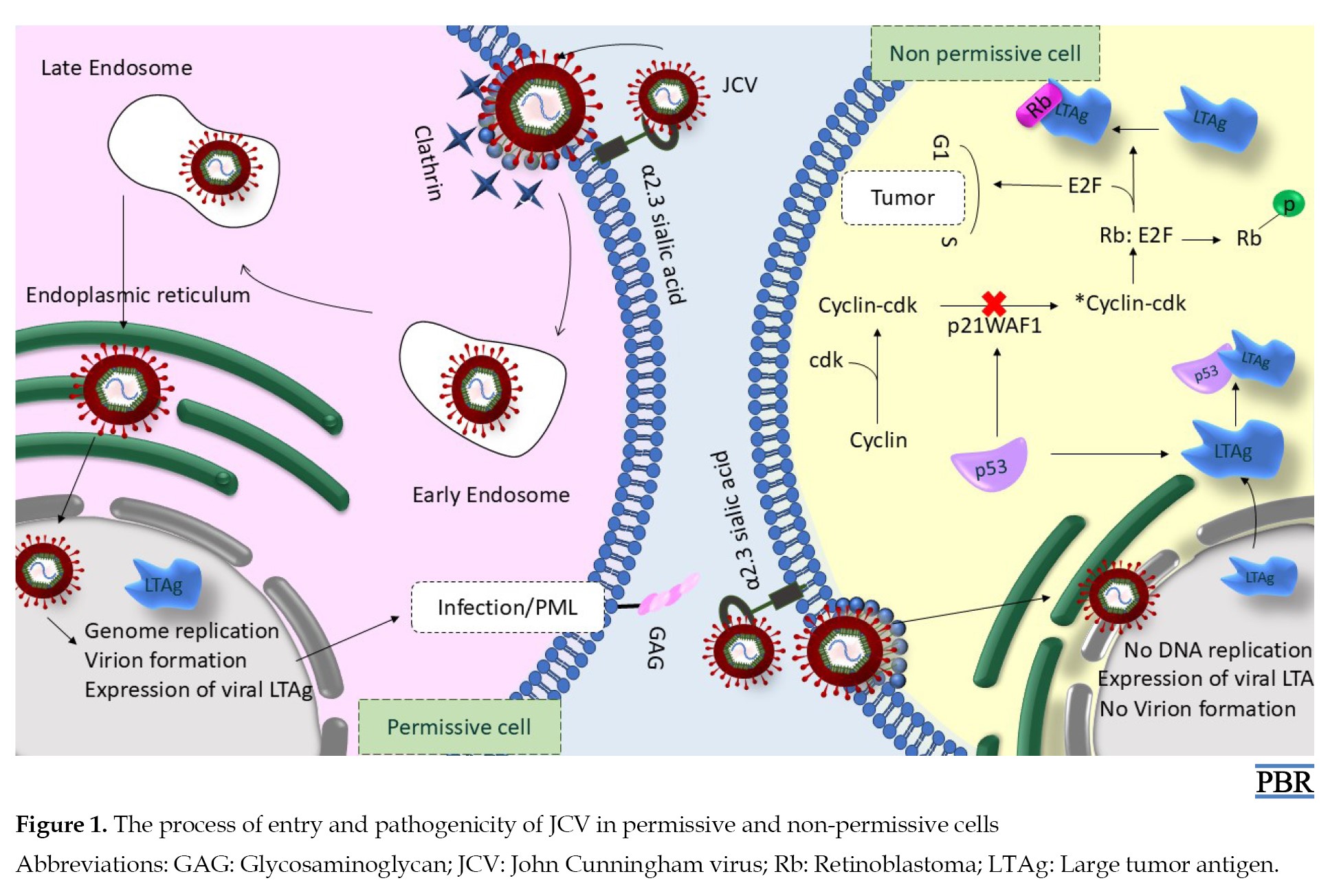
Cancer is a dangerous disease and is the second leading cause of mortality worldwide. It is estimated that 20% of all cancers globally are associated with infectious agents. Approximately 12% of these cases may be attributed to oncogenic virus infections. Viral genes can modulate the physiological machinery of infected cells, ultimately leading to cellular transformation and cancer development. Despite the prevalence of oncogenic viruses, understanding and managing virus-induced cancers remains challenging [24, 30, 31]. JCV is also classified as an oncogenic virus, thus possessing the potential to cause cellular transformation and cancer [32, 33]. The entry of this virus into non-permissive cells, which do not support viral DNA replication, can result in incomplete infection or cellular transformation (oncogenesis) [34]. In transformed cells, JCV plays a significant role in anchorage-dependent growth, rapid division, prolonged lifespan, increased ploidy, unstable multicentric chromosomes, genomic instability, impaired DNA repair, and increased micronuclei formation [14]. The oncogenic potential of JCV is primarily dependent on LTAg; stAg and the agnoprotein appear to collaborate with LTAg in cellular transformation [6]. LTAg regulates and controls the cell cycle in cancer cells by targeting signaling pathways, such as p53, β-catenin, IRS, Rb, TGF-β1, protein kinase B/phosphoinositide-3-kinase, and adenosine monophosphate-activated protein kinase [14]. According to one model, the interaction between LTAg and p53 prevents p53 from enhancing transcription of p21/WAF-1, an inhibitor of cyclin-dependent kinases, including cyclins A and E. The reduced activity of G1/S cyclins results in pRb remaining hypophosphorylated and active, which in turn retains the S phase-specific transcription factor E2F [35]. LTAg can also interact with pRb to release E2F from the pRb: E2F complex, allowing E2F to exert its effects on cell proliferation by promoting the transcription of S-phase genes. In other words, pRb normally inhibits E2F, preventing the transition from G1 to S phase of the cell cycle. When pRb is inactivated by LTAg, E2F is released, resulting in increased cellular proliferation. In summary, the release of E2F from pRb by LTAg activates the expression of P14 alternative reading frame (ARF), which subsequently stabilizes p53. However, LTAg binds to and inactivates p53, thereby obstructing its function in response to DNA damage or P14 ARF production [24]. Existing evidence indicates that JCV DNA and its encoded products are present in tissues affected by brain cancer (BC), oral cancer, EC, GC, CRC, BC, cervical cancer, pancreatic cancer, and liver cancer (LC). Therefore, JCV can be considered a potential risk factor for cancer development and should be further investigated for its role in early prevention, diagnosis, and treatment of cancers [6, 14, 36] (Figure 1).
Examination of cancers associated with the JCV in humans
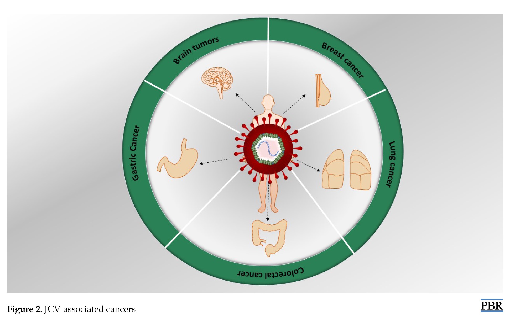
In this section, we explore some types of cancers associated with JCV (Figure 2).
The role of the JCV in brain tumors
Current evidence suggests that JCV plays a significant role in the development of various CNS tumors, including astrocytomas, glioblastomas, neuroblastomas, and medulloblastomas. LTAg is a key regulatory protein of JCV associated with brain tumor formation in humans and can be detected by immunohistochemistry (IHC) in 50% of human BT [37, 38]. The first systematic investigation was conducted in 1996 on brain tumor tissues to detect JCV DNA and proteins in a patient with oligodendroglioma in the absence of PML, JCV DNA was confirmed using polymerase chain reaction (PCR). JCV RNA and LTAg protein were identified in tumor tissue using primer extension analysis and western blotting, respectively. In another study, 57 to 83 percent of 85 glioma samples tested positive for JCV [7, 24, 39].
The association between the JCV and GC
GC is a significant global issue with a high mortality rate, causing approximately 769,000 deaths annually. It ranks fifth in terms of prevalence and fourth in mortality worldwide. Studies suggest that JCV may be involved in the onset of GC. In one study, JCV sequences were isolated from 54.84% of GC tissue samples and 32.25% of non-cancerous tissue samples [40]. IHC analysis revealed the presence of LTAg protein in the nuclear compartment, indicating that JCV could play an important role in the transformation of gastric cells into malignancy [41]. Furthermore, another study found LTAg expression in 49% of 90 GC tissue samples, suggesting that LTAg significantly influences gastric carcinogenesis through genetic and epigenetic changes [42].
The role of the JCV in CRC
CRC is the most common malignancy with high mortality rates worldwide. The association between JCV and colon and rectal cancer has recently been reported. Studies have confirmed the presence of JCV DNA in 10% of 120 samples. A meta-analysis indicated that JCV presence in colorectal tissues increases the likelihood of developing CRC by 4.70 times. Thus, JCV may act as an oncogenic virus that increases the risk of CRC, supporting the hypothesis of the role of viral factors in cancer pathogenesis [43-45].
Examination of lung cancer associated with the JCV
Recently, the relationship between LC and JCV has been highlighted. In a study involving 13 surgically resected lung tumor samples, nested PCR revealed that 7 samples were positive for JCV, indicating the potential impact of this virus on LC [34]. Another study also demonstrated the presence of the JCV genome, LTAg, VP proteins, and agnoprotein using nested PCR, while IHC was performed to assess the relationship between p53 and β-catenin in LC, alongside LTAg. The results showed that LTAg was detected in 25 of 62 LC samples, with IHC revealing a significant correlation between LTAg and p53, as well as nuclear β-catenin. This study suggests a link between JCV and LC, positing that LTAg may contribute to oncogenesis by inactivating p53 and disrupting the Wnt signaling pathway regulation in LCs [46].
The JCV and breast cancer
BC is the most common malignancy among women worldwide, influenced by a wide range of internal and external factors that can initiate, promote, and progress the disease. JCV may also be associated with this cancer. In a study of tissues from patients with BC, LTAg from JCV was detected in 23% of the samples. LTAg affects p53 wild-type by stabilizing β-catenin and also leads to chromosomal instability. These processes activate G2 checkpoint pathways mediated by ataxia telangiectasia mutated (ATM) and RAD3-related (ATR), resulting in G2 cell cycle arrest [47]. Additionally, another study found JCV DNA in 23% of primary BC samples using PCR. These results were correlated with clinical, pathological, and viral parameters [48, 49].
The JCV and EC
In recent years, the incidence of esophageal adenocarcinoma has significantly increased in industrialized countries. A study discussing the relationship between JCV and EC reported that JCV was isolated from 85% of normal esophageal biopsies and 100% of carcinoma samples. Furthermore, using IHC, LTAg was identified in 53% of samples, agnoprotein in 42%, and the tumor suppressor protein p53 in 58%. These results provide evidence of JCV infection of gastrointestinal cells and highlight its potential role in the development of upper gastrointestinal carcinomas [50].
Conclusion
Emerging evidence regarding the role of JCV in cancer development underscores the complex interplay between viral infections and tumorigenesis. While JCV typically remains asymptomatic in immunocompetent individuals, its potential to disrupt essential cellular pathways in immunocompromised populations raises significant concerns about its oncogenic characteristics. Understanding the mechanisms through which JCV contributes to tumorigenesis is crucial for advancing our knowledge of virus-related carcinogenesis. The ability of JCV to integrate with host DNA and alter cellular function indicates substantial potential for oncogenic activity. This emphasizes the necessity for further research to elucidate these complex interactions and their implications for cancer development. Uncovering the oncogenic mechanisms of JCV may inform more effective prevention and treatment options for patients with viral diseases and malignancies. Ultimately, a deeper understanding of JCV’s role in cancer may pave the way for new interventions that could significantly improve public health outcomes in cancer prevention and management.
Ethical Considerations
Compliance with ethical guidelines
There were no ethical considerations to be considered in this research.
Funding
This research did not receive any grant from funding agencies in the public, commercial, or non-profit sectors.
Authors' contributions
Conceptualization, methodology, investigation, and writing the original draft: Abolfazl Jafari-Sales and Kosar Hosseini-Karkaj; Supervision, data collection, data analysis, review and editing: Mehrdad Pashazadeh and Ali Salimi Jeda.
Conflict of interest
The authors declared no conflict of interest.
Acknowledgments
The authors gratefully acknowledge the academic support provided by the Faculty of Medical Sciences, Tabriz Medical Sciences Branch, Islamic Azad University, Tabriz, Iran.
Full-Text: (62 Views)
Introduction
The John Cunningham virus (JCV) belongs to the Polyomaviridae family and contains a double-stranded DNA [1-4]. The Polyomaviridae family comprises over 100 members, primarily infecting mammals and birds [5]. JCV was first isolated in 1971 from the brain of a patient with progressive multifocal leukoencephalopathy (PML). In most cases, individuals become infected with this virus during childhood or adolescence, and by adulthood, approximately 50-80% of them are seropositive. Currently, the exact modes of JCV transmission are not well defined; however, the presence of viral DNA in B cells and stromal cells of the tonsils and oropharynx supports the hypothesis of respiratory transmission. Furthermore, JCV has been identified in raw sewage and a high percentage of natural tissue samples obtained from the upper and lower gastrointestinal tract of humans, indicating that this virus may be transmitted through the consumption of contaminated water or food. Additionally, JCV has been reported in various other tissues, including the spleen, lymph nodes, lungs, bone marrow, brain, B lymphocytes, and kidneys. JCV exhibits limited and specific tissue tropism, primarily infecting the kidneys, oligodendrocytes, and astrocytes in the central nervous system (CNS) [6-9]. JCV infection is often latent or chronic, and infected individuals may be asymptomatic [3]. After primary infection in immunocompetent individuals, the virus migrates to the kidneys and undergoes continuous replication. Ultimately, JCV is excreted in the urine. Due to its asymptomatic nature, JCV infection is rarely detected in blood samples [10]. In immunocompromised individuals, JCV becomes neurotropic and infectious; infection can occur through rearrangement of the noncoding control region (NCCR) and mutations in the VP1 capsid protein [11]. Widespread viral infections can lead to the development of cancer in humans. All oncoviruses induce tumors via metabolic pathways. The mechanism by which oncoviruses cause cancer varies based on their genetic material (DNA or ribonucleic acid [RNA]) [12]. According to studies, JCV is a neurotropic human virus associated with neurological diseases and capable of infecting various cell types. Recently, JCV genomic sequences have been identified in various human malignancies, including brain tumors, colorectal cancer (CRC), gastric cancer (GC), and esophageal cancer (EC) [13, 14]. This article provides a comprehensive and multidimensional examination of the effects of JCV on human health. Its primary focus is on the mechanisms of entry and pathogenicity of this virus, its oncogenic potential, and the complex relationships between JCV and various cancers. This study aimed to analyze the role of JCV in cancer onset and progression and to evaluate its clinical implications for public health. This study may enhance understanding of interactions between JCV and the host immune system and pave the way for the development of new preventive and therapeutic strategies.
Structural characteristics of the JCV: A genome examination
The JCV capsid is icosahedral and lacks an envelope. The genome of this virus consists of a single circular, double-stranded DNA molecule approximately 5,100 base pairs in length [15-17]. The viral genome encodes cellular histones H2A, H2B, H3, and H4, which ultimately form a minichromosome that structurally resembles the host cell’s chromatin. Typically, viral particles do not contain linker histones; however, JCV acquires them upon entering the host cell [6]. The genome is primarily divided into two coding regions based on the timing of protein expression: A late coding region and a regulatory region (RR). The RR encodes the large tumor antigen (LTAg) and small tumor antigen (stAg). In contrast, the late region encodes the structural proteins VP1, VP2, and VP3, the regulatory agnoprotein, and two microRNAs (miRNAs) processed from a precursor microRNA (pre-miRNA) known as pre-miRNA J-1. Because miRNAs are fully complementary to LTAg, they play a significant role in regulating LTAg transcript stability. The NCCR separates the late genomic region and RR, also known as the hypervariable RR. This region includes the origin of replication and regulatory areas for early and late transcription. Notably, the NCCR contains host-specific DNA and binding sites for transcription factors. The highest sequence diversity characterizes the NCCR and determines viral replication efficiency and cellular tropism. The JCV capsid consists of three proteins: VP1, VP2, and VP3, with VP1 being the most critical component. The capsid structure contains 72 pentamers, each composed of five VP1 molecules and one VP2 or VP3 molecule. Only VP1 is positioned on the capsid surface, thereby determining receptor specificity [3, 16, 18-20]. LTAg and its variants have distinct functions and interact with both host and viral proteins as well as DNA. T proteins can facilitate viral replication by directing host cells into S phase, regulating transcription from both host and viral genomes, and directly participating in viral DNA replication [19]. The agnoprotein is also likely involved in processes, such as replication, viral transcription, viroporin function, and the transport of new virions from the nucleus. Furthermore, agnoprotein may bind to various cellular factors, including p53, Y-box binding protein 1, Ku70, fasciculation and elongation protein zeta-1, heterochromatin protein 1α, protein phosphatase 2A, AP-3, proliferating cell nuclear antigen, and α-soluble N-ethylmaleimide-sensitive factor attachment proteins. VP proteins are also crucial for lifecycle events, including attachment to host cell receptors, entry, and penetration [5, 21].
Mechanism of entry and pathogenicity of the JCV
The entry of JCV into the human body begins with the interaction between VP1 and the pentasaccharide NeuNAc-α2,6-Gal-β1,4-GlcNAc-β1,3-Gal-β1,4-Glc, followed by engagement with sialic acid associated with α-2,3. The virus enters the host cell by binding to sialic acid, subsequently initiating clathrin-mediated endocytosis. Primary and late endosomes serve as sites for JCV after it enters cells. The potential for JCV to be transported within the cytoplasm to reach the nucleus is facilitated by various cellular compartments; at this stage, transcription of the viral genome is considered more critical than viral DNA replication because the early gene products of this virus, such as LTAg, stAg, and LTAg splice variants, are essential for initiating the lytic phase. The primary product LTAg enables viral DNA replication by binding to the viral replication origin, a process that depends on the availability of host DNA polymerase, replication protein A, host enzymes, and cofactors expressed during the S phase of the cell cycle. According to existing studies, JCV entry into glial cells may be facilitated by the serotonin receptor 5HT2AR [3, 6, 21, 22]. This virus induces cell growth by expressing oncoproteins and T antigens while disrupting apoptosis since it requires the host cell’s DNA synthetic machinery for replication [23]. Transforming growth factor beta 1 (TGF-β1) and tumor necrosis factor alpha stimulate JCV replication, whereas interferon-gamma suppresses this process [14]. Additionally, JCV interacts with cellular modulators, such as the retinoblastoma protein (pRB) and p53, to regulate multiple cellular processes, ultimately promoting cell cycle progression [21]. Viral end products are released through host cell lysis. Another potential route of JCV infection in a cell suggests that entry into non-permissive cells that do not support viral replication may lead to cellular transformation or oncogenesis. The primary LTAg protein is a key factor in cellular transformation and tumor development [6]. Complex membrane networks in host cells, such as the plasma membrane, endolysosomes, and the endoplasmic reticulum (ER), play a significant role in initiating and advancing viral infections through specialized retro-translocation machinery that moves ER components into the cytosol. Polyomaviruses represent a unique category that penetrates the ER membrane during their entry into host cells (Figure 1) [22].
The John Cunningham virus (JCV) belongs to the Polyomaviridae family and contains a double-stranded DNA [1-4]. The Polyomaviridae family comprises over 100 members, primarily infecting mammals and birds [5]. JCV was first isolated in 1971 from the brain of a patient with progressive multifocal leukoencephalopathy (PML). In most cases, individuals become infected with this virus during childhood or adolescence, and by adulthood, approximately 50-80% of them are seropositive. Currently, the exact modes of JCV transmission are not well defined; however, the presence of viral DNA in B cells and stromal cells of the tonsils and oropharynx supports the hypothesis of respiratory transmission. Furthermore, JCV has been identified in raw sewage and a high percentage of natural tissue samples obtained from the upper and lower gastrointestinal tract of humans, indicating that this virus may be transmitted through the consumption of contaminated water or food. Additionally, JCV has been reported in various other tissues, including the spleen, lymph nodes, lungs, bone marrow, brain, B lymphocytes, and kidneys. JCV exhibits limited and specific tissue tropism, primarily infecting the kidneys, oligodendrocytes, and astrocytes in the central nervous system (CNS) [6-9]. JCV infection is often latent or chronic, and infected individuals may be asymptomatic [3]. After primary infection in immunocompetent individuals, the virus migrates to the kidneys and undergoes continuous replication. Ultimately, JCV is excreted in the urine. Due to its asymptomatic nature, JCV infection is rarely detected in blood samples [10]. In immunocompromised individuals, JCV becomes neurotropic and infectious; infection can occur through rearrangement of the noncoding control region (NCCR) and mutations in the VP1 capsid protein [11]. Widespread viral infections can lead to the development of cancer in humans. All oncoviruses induce tumors via metabolic pathways. The mechanism by which oncoviruses cause cancer varies based on their genetic material (DNA or ribonucleic acid [RNA]) [12]. According to studies, JCV is a neurotropic human virus associated with neurological diseases and capable of infecting various cell types. Recently, JCV genomic sequences have been identified in various human malignancies, including brain tumors, colorectal cancer (CRC), gastric cancer (GC), and esophageal cancer (EC) [13, 14]. This article provides a comprehensive and multidimensional examination of the effects of JCV on human health. Its primary focus is on the mechanisms of entry and pathogenicity of this virus, its oncogenic potential, and the complex relationships between JCV and various cancers. This study aimed to analyze the role of JCV in cancer onset and progression and to evaluate its clinical implications for public health. This study may enhance understanding of interactions between JCV and the host immune system and pave the way for the development of new preventive and therapeutic strategies.
Structural characteristics of the JCV: A genome examination
The JCV capsid is icosahedral and lacks an envelope. The genome of this virus consists of a single circular, double-stranded DNA molecule approximately 5,100 base pairs in length [15-17]. The viral genome encodes cellular histones H2A, H2B, H3, and H4, which ultimately form a minichromosome that structurally resembles the host cell’s chromatin. Typically, viral particles do not contain linker histones; however, JCV acquires them upon entering the host cell [6]. The genome is primarily divided into two coding regions based on the timing of protein expression: A late coding region and a regulatory region (RR). The RR encodes the large tumor antigen (LTAg) and small tumor antigen (stAg). In contrast, the late region encodes the structural proteins VP1, VP2, and VP3, the regulatory agnoprotein, and two microRNAs (miRNAs) processed from a precursor microRNA (pre-miRNA) known as pre-miRNA J-1. Because miRNAs are fully complementary to LTAg, they play a significant role in regulating LTAg transcript stability. The NCCR separates the late genomic region and RR, also known as the hypervariable RR. This region includes the origin of replication and regulatory areas for early and late transcription. Notably, the NCCR contains host-specific DNA and binding sites for transcription factors. The highest sequence diversity characterizes the NCCR and determines viral replication efficiency and cellular tropism. The JCV capsid consists of three proteins: VP1, VP2, and VP3, with VP1 being the most critical component. The capsid structure contains 72 pentamers, each composed of five VP1 molecules and one VP2 or VP3 molecule. Only VP1 is positioned on the capsid surface, thereby determining receptor specificity [3, 16, 18-20]. LTAg and its variants have distinct functions and interact with both host and viral proteins as well as DNA. T proteins can facilitate viral replication by directing host cells into S phase, regulating transcription from both host and viral genomes, and directly participating in viral DNA replication [19]. The agnoprotein is also likely involved in processes, such as replication, viral transcription, viroporin function, and the transport of new virions from the nucleus. Furthermore, agnoprotein may bind to various cellular factors, including p53, Y-box binding protein 1, Ku70, fasciculation and elongation protein zeta-1, heterochromatin protein 1α, protein phosphatase 2A, AP-3, proliferating cell nuclear antigen, and α-soluble N-ethylmaleimide-sensitive factor attachment proteins. VP proteins are also crucial for lifecycle events, including attachment to host cell receptors, entry, and penetration [5, 21].
Mechanism of entry and pathogenicity of the JCV
The entry of JCV into the human body begins with the interaction between VP1 and the pentasaccharide NeuNAc-α2,6-Gal-β1,4-GlcNAc-β1,3-Gal-β1,4-Glc, followed by engagement with sialic acid associated with α-2,3. The virus enters the host cell by binding to sialic acid, subsequently initiating clathrin-mediated endocytosis. Primary and late endosomes serve as sites for JCV after it enters cells. The potential for JCV to be transported within the cytoplasm to reach the nucleus is facilitated by various cellular compartments; at this stage, transcription of the viral genome is considered more critical than viral DNA replication because the early gene products of this virus, such as LTAg, stAg, and LTAg splice variants, are essential for initiating the lytic phase. The primary product LTAg enables viral DNA replication by binding to the viral replication origin, a process that depends on the availability of host DNA polymerase, replication protein A, host enzymes, and cofactors expressed during the S phase of the cell cycle. According to existing studies, JCV entry into glial cells may be facilitated by the serotonin receptor 5HT2AR [3, 6, 21, 22]. This virus induces cell growth by expressing oncoproteins and T antigens while disrupting apoptosis since it requires the host cell’s DNA synthetic machinery for replication [23]. Transforming growth factor beta 1 (TGF-β1) and tumor necrosis factor alpha stimulate JCV replication, whereas interferon-gamma suppresses this process [14]. Additionally, JCV interacts with cellular modulators, such as the retinoblastoma protein (pRB) and p53, to regulate multiple cellular processes, ultimately promoting cell cycle progression [21]. Viral end products are released through host cell lysis. Another potential route of JCV infection in a cell suggests that entry into non-permissive cells that do not support viral replication may lead to cellular transformation or oncogenesis. The primary LTAg protein is a key factor in cellular transformation and tumor development [6]. Complex membrane networks in host cells, such as the plasma membrane, endolysosomes, and the endoplasmic reticulum (ER), play a significant role in initiating and advancing viral infections through specialized retro-translocation machinery that moves ER components into the cytosol. Polyomaviruses represent a unique category that penetrates the ER membrane during their entry into host cells (Figure 1) [22].
Mechanisms of JCV-mediated oncogenesis
The activity of the early gene product, LTag, is central to the oncogenic potential of JC polyomavirus. This multifunctional phosphoprotein is a primary viral oncoprotein essential for viral DNA replication in host cells. LTag orchestrates viral replication by binding to and inactivating key tumor suppressor proteins, most notably p53 and the retinoblastoma family proteins (pRb, p107, p130). This inactivation disrupts critical cell cycle checkpoints and apoptotic pathways, promoting uncontrolled cellular proliferation [24, 25]. Furthermore, LTag directly modulates viral replication by targeting host DNA damage response pathways. It has been demonstrated that LTag induces G2 cell cycle arrest, creating a favorable environment for viral genome amplification. This is achieved through its interaction with and activation of the ATM and ATR kinase signaling pathways, which are central to the DNA damage-induced G2/M checkpoint. By co-opting these pathways, LTag ensures the host cell remains in a state conducive to viral replication, simultaneously increasing genomic instability and the accumulation of mutations, a hallmark of cancer development [14, 26].
In addition to the large T antigen, JC polyomavirus encodes a late auxiliary protein called agnoprotein. Though small, this highly multifunctional protein plays a crucial regulatory role in the viral life cycle by interacting with and modulating numerous host cellular systems. Agnoprotein localizes to key cellular compartments, including the nucleus, perinuclear region, plasma membrane, and mitochondria, exerting pleiotropic effects [27]. Specifically, agnoprotein-host interactions significantly impair fundamental cellular processes, such as protein synthesis and degradation, by inhibiting mRNA maturation and nuclear export, thereby suppressing host protein synthesis and interfering with the ubiquitin-proteasome system, which disrupts regulated protein degradation and may lead to the accumulation of oncogenic factors [25]. Furthermore, it plays a direct role in viral trafficking by disrupting cytoskeletal dynamics and vesicular transport, thereby facilitating viral assembly and egress. Additionally, agnoprotein localizes to mitochondria, inducing oxidative stress and disrupting metabolic functions, and its interactions with the ER and Golgi apparatus interfere with protein trafficking and secretory pathways [25, 28]. These disruptions contribute to substantial cellular stress and genomic instability, collectively fostering a chronic state that promotes the acquisition of pro-oncogenic mutations and uncontrolled cell growth, thereby compounding the oncogenic effects of the large T antigen [29].
The relationship between the JCV and cancer in humans
Cancer is a dangerous disease and is the second leading cause of mortality worldwide. It is estimated that 20% of all cancers globally are associated with infectious agents. Approximately 12% of these cases may be attributed to oncogenic virus infections. Viral genes can modulate the physiological machinery of infected cells, ultimately leading to cellular transformation and cancer development. Despite the prevalence of oncogenic viruses, understanding and managing virus-induced cancers remains challenging [24, 30, 31]. JCV is also classified as an oncogenic virus, thus possessing the potential to cause cellular transformation and cancer [32, 33]. The entry of this virus into non-permissive cells, which do not support viral DNA replication, can result in incomplete infection or cellular transformation (oncogenesis) [34]. In transformed cells, JCV plays a significant role in anchorage-dependent growth, rapid division, prolonged lifespan, increased ploidy, unstable multicentric chromosomes, genomic instability, impaired DNA repair, and increased micronuclei formation [14]. The oncogenic potential of JCV is primarily dependent on LTAg; stAg and the agnoprotein appear to collaborate with LTAg in cellular transformation [6]. LTAg regulates and controls the cell cycle in cancer cells by targeting signaling pathways, such as p53, β-catenin, IRS, Rb, TGF-β1, protein kinase B/phosphoinositide-3-kinase, and adenosine monophosphate-activated protein kinase [14]. According to one model, the interaction between LTAg and p53 prevents p53 from enhancing transcription of p21/WAF-1, an inhibitor of cyclin-dependent kinases, including cyclins A and E. The reduced activity of G1/S cyclins results in pRb remaining hypophosphorylated and active, which in turn retains the S phase-specific transcription factor E2F [35]. LTAg can also interact with pRb to release E2F from the pRb: E2F complex, allowing E2F to exert its effects on cell proliferation by promoting the transcription of S-phase genes. In other words, pRb normally inhibits E2F, preventing the transition from G1 to S phase of the cell cycle. When pRb is inactivated by LTAg, E2F is released, resulting in increased cellular proliferation. In summary, the release of E2F from pRb by LTAg activates the expression of P14 alternative reading frame (ARF), which subsequently stabilizes p53. However, LTAg binds to and inactivates p53, thereby obstructing its function in response to DNA damage or P14 ARF production [24]. Existing evidence indicates that JCV DNA and its encoded products are present in tissues affected by brain cancer (BC), oral cancer, EC, GC, CRC, BC, cervical cancer, pancreatic cancer, and liver cancer (LC). Therefore, JCV can be considered a potential risk factor for cancer development and should be further investigated for its role in early prevention, diagnosis, and treatment of cancers [6, 14, 36] (Figure 1).
Examination of cancers associated with the JCV in humans
In this section, we explore some types of cancers associated with JCV (Figure 2).
The role of the JCV in brain tumors
Current evidence suggests that JCV plays a significant role in the development of various CNS tumors, including astrocytomas, glioblastomas, neuroblastomas, and medulloblastomas. LTAg is a key regulatory protein of JCV associated with brain tumor formation in humans and can be detected by immunohistochemistry (IHC) in 50% of human BT [37, 38]. The first systematic investigation was conducted in 1996 on brain tumor tissues to detect JCV DNA and proteins in a patient with oligodendroglioma in the absence of PML, JCV DNA was confirmed using polymerase chain reaction (PCR). JCV RNA and LTAg protein were identified in tumor tissue using primer extension analysis and western blotting, respectively. In another study, 57 to 83 percent of 85 glioma samples tested positive for JCV [7, 24, 39].
The association between the JCV and GC
GC is a significant global issue with a high mortality rate, causing approximately 769,000 deaths annually. It ranks fifth in terms of prevalence and fourth in mortality worldwide. Studies suggest that JCV may be involved in the onset of GC. In one study, JCV sequences were isolated from 54.84% of GC tissue samples and 32.25% of non-cancerous tissue samples [40]. IHC analysis revealed the presence of LTAg protein in the nuclear compartment, indicating that JCV could play an important role in the transformation of gastric cells into malignancy [41]. Furthermore, another study found LTAg expression in 49% of 90 GC tissue samples, suggesting that LTAg significantly influences gastric carcinogenesis through genetic and epigenetic changes [42].
The role of the JCV in CRC
CRC is the most common malignancy with high mortality rates worldwide. The association between JCV and colon and rectal cancer has recently been reported. Studies have confirmed the presence of JCV DNA in 10% of 120 samples. A meta-analysis indicated that JCV presence in colorectal tissues increases the likelihood of developing CRC by 4.70 times. Thus, JCV may act as an oncogenic virus that increases the risk of CRC, supporting the hypothesis of the role of viral factors in cancer pathogenesis [43-45].
Examination of lung cancer associated with the JCV
Recently, the relationship between LC and JCV has been highlighted. In a study involving 13 surgically resected lung tumor samples, nested PCR revealed that 7 samples were positive for JCV, indicating the potential impact of this virus on LC [34]. Another study also demonstrated the presence of the JCV genome, LTAg, VP proteins, and agnoprotein using nested PCR, while IHC was performed to assess the relationship between p53 and β-catenin in LC, alongside LTAg. The results showed that LTAg was detected in 25 of 62 LC samples, with IHC revealing a significant correlation between LTAg and p53, as well as nuclear β-catenin. This study suggests a link between JCV and LC, positing that LTAg may contribute to oncogenesis by inactivating p53 and disrupting the Wnt signaling pathway regulation in LCs [46].
The JCV and breast cancer
BC is the most common malignancy among women worldwide, influenced by a wide range of internal and external factors that can initiate, promote, and progress the disease. JCV may also be associated with this cancer. In a study of tissues from patients with BC, LTAg from JCV was detected in 23% of the samples. LTAg affects p53 wild-type by stabilizing β-catenin and also leads to chromosomal instability. These processes activate G2 checkpoint pathways mediated by ataxia telangiectasia mutated (ATM) and RAD3-related (ATR), resulting in G2 cell cycle arrest [47]. Additionally, another study found JCV DNA in 23% of primary BC samples using PCR. These results were correlated with clinical, pathological, and viral parameters [48, 49].
The JCV and EC
In recent years, the incidence of esophageal adenocarcinoma has significantly increased in industrialized countries. A study discussing the relationship between JCV and EC reported that JCV was isolated from 85% of normal esophageal biopsies and 100% of carcinoma samples. Furthermore, using IHC, LTAg was identified in 53% of samples, agnoprotein in 42%, and the tumor suppressor protein p53 in 58%. These results provide evidence of JCV infection of gastrointestinal cells and highlight its potential role in the development of upper gastrointestinal carcinomas [50].
Conclusion
Emerging evidence regarding the role of JCV in cancer development underscores the complex interplay between viral infections and tumorigenesis. While JCV typically remains asymptomatic in immunocompetent individuals, its potential to disrupt essential cellular pathways in immunocompromised populations raises significant concerns about its oncogenic characteristics. Understanding the mechanisms through which JCV contributes to tumorigenesis is crucial for advancing our knowledge of virus-related carcinogenesis. The ability of JCV to integrate with host DNA and alter cellular function indicates substantial potential for oncogenic activity. This emphasizes the necessity for further research to elucidate these complex interactions and their implications for cancer development. Uncovering the oncogenic mechanisms of JCV may inform more effective prevention and treatment options for patients with viral diseases and malignancies. Ultimately, a deeper understanding of JCV’s role in cancer may pave the way for new interventions that could significantly improve public health outcomes in cancer prevention and management.
Ethical Considerations
Compliance with ethical guidelines
There were no ethical considerations to be considered in this research.
Funding
This research did not receive any grant from funding agencies in the public, commercial, or non-profit sectors.
Authors' contributions
Conceptualization, methodology, investigation, and writing the original draft: Abolfazl Jafari-Sales and Kosar Hosseini-Karkaj; Supervision, data collection, data analysis, review and editing: Mehrdad Pashazadeh and Ali Salimi Jeda.
Conflict of interest
The authors declared no conflict of interest.
Acknowledgments
The authors gratefully acknowledge the academic support provided by the Faculty of Medical Sciences, Tabriz Medical Sciences Branch, Islamic Azad University, Tabriz, Iran.
References
- Takahashi K, Sekizuka T, Fukumoto H, Nakamichi K, Suzuki T, Sato Y, et al. Deep-sequence identification and role in virus replication of a JC virus quasispecies in patients with progressive multifocal leukoencephalopathy. J Virol. 2016; 91(1):e01335-16. [DOI:10.1128/JVI.01335-16] [PMID]
- Kartau M, Verkkoniemi-Ahola A, Paetau A, Palomäki M, Janes R, Ristola M, et al. The incidence and predisposing factors of John Cunningham virus-induced progressive multifocal leukoencephalopathy in Southern Finland: A population-based study. Open Forum Infect Dis. 2019; 6(2):ofz024. [DOI:10.1093/ofid/ofz024] [PMID]
- Suleman M, Khan TA, Ejaz H, Maroof S, Alshammari A, Albekairi NA, et al. Structural vaccinology, molecular simulation and immune simulation approaches to design multi-epitopes vaccine against John Cunningham virus. Microb Pathog. 2024; 189:106572. [DOI:10.1016/j.micpath.2024.106572] [PMID]
- Vilibic-Cavlek T, Bogdanic M, Peric T, Radmanic L, Antolasic L, Milasincic L, et al. Prevalence of JC polyomavirus in patients with neuroinvasive disease of unknown etiology in Croatia. Medicina. 2023; 60(1):69. [DOI:10.3390/medicina60010069] [PMID]
- Zou W, Imperiale MJ. Biology of Polyomavirus miRNA. Front Microbiol. 2021; 12:662892. [DOI:10.3389/fmicb.2021.662892] [PMID]
- Delbue S, Comar M, Ferrante P. Review on the role of the human polyomavirus JC in the development of tumors. Infect Agent Cancer. 2017; 12:10. [DOI:10.1186/s13027-017-0122-0] [PMID]
- Maginnis MS, Atwood WJ. JC virus: an oncogenic virus in animals and humans? Semin Cancer Biol. 2009; 19(4):261-9. [DOI:10.1016/j.semcancer.2009.02.013] [PMID]
- Dwyer C, Sharmin S, Kalincik T. Rates of John Cunningham virus seroconversion greatly reduced in natalizumab-treated patients during COVID-19-related lockdowns. Eur J Neurol. 2024; 31(1):e16059. [DOI:10.1111/ene.16059] [PMID]
- Del Valle L, Khalili K. Induction of brain tumors by the archetype strain of human neurotropic JCPyV in a transgenic mouse model. Viruses. 2021; 13(2):162. [DOI:10.3390/v13020162] [PMID]
- Sgarlata E, Chisari CG, Toscano S, Finocchiaro C, Lo Fermo S, Millefiorini E, et al. Changes in John Cunningham virus index in multiple sclerosis patients treated with different disease-modifying therapies. Curr Neuropharmacol. 2022; 20(10):1978-87. [DOI:10.2174/1570159X19666211111123202] [PMID]
- Flores J, Anguiano O, Rivas-Alonso V, González-Conchillos H, Pérez-Saldivar M, Sotelo J, et al. Mutations in the John Cunningham virus vp1 gene could predispose to the development of progressive multifocal leukoencephalopathy in multiple sclerosis patients undergoing treatment with natalizumab. Mult Scler Relat Disord. 2021; 56:103266. [DOI:10.1016/j.msard.2021.103266] [PMID]
- Bhaskar Kalarani I, Thasneem K, Veerabathiran R. Chapter 5 - Oncoviruses: Future prospects of molecular mechanisms and therapeutic strategies. In: Ennaji MM, editor. Oncogenic Viruses. Cambridge: Academic Press; 2023. [DOI:10.1016/B978-0-12-824156-1.00012-1]
- Mohammed Ali SH, Mohammed Al-Alwany SH, Khalil Hussein A. Molecular Interplay of John Cunningham virus with interleukin 1 beta in colorectal carcinomatous tissues from a group of Iraqi patients. Arch Razi Inst. 2022; 77(6):2299-306. [DOI:10.22092/ARI.2022.358617.2267] [PMID]
- Zheng HC, Xue H, Zhang CY. The oncogenic roles of JC polyomavirus in cancer. Front Oncol. 2022; 12:976577. [DOI:10.3389/fonc.2022.976577] [PMID]
- Ikegaya H. Geographical identification of cadavers by human parasites. Forensic Sci Int Genet. 2008; 2(2):83-90. [DOI:10.1016/j.fsigen.2007.10.184] [PMID]
- Butic AB, Spencer SA, Shaheen SK, Lukacher AE. Polyomavirus wakes up and chooses neurovirulence. Viruses. 2023; 15(10):2112. [DOI:10.3390/v15102112] [PMID]
- Mettenleiter TC. Breaching the barrier-the nuclear envelope in virus infection. J Mol Biol. 2016; 428(10 Pt A):1949-61. [DOI:10.1016/j.jmb.2015.10.001] [PMID]
- Cortese I, Reich DS, Nath A. Progressive multifocal leukoencephalopathy and the spectrum of JC virus-related disease. Nat Rev Neurol. 2021; 17(1):37-51. [DOI:10.1038/s41582-020-00427-y] [PMID]
- Ferenczy MW, Marshall LJ, Nelson CD, Atwood WJ, Nath A, Khalili K, et al. Molecular biology, epidemiology, and pathogenesis of progressive multifocal leukoencephalopathy, the JC virus-induced demyelinating disease of the human brain. Clin Microbiol Rev. 2012; 25(3):471-506. [DOI:10.1128/CMR.05031-11] [PMID]
- Torres C. Evolution and molecular epidemiology of polyomaviruses. Infect Genet Evol. 2020; 79:104150. [DOI:10.1016/j.meegid.2019.104150] [PMID]
- Mouliou DS. John Cunningham virus and progressive multifocal leukoencephalopathy: A falsely played diagnosis. Diseases. 2024; 12(5):100. [DOI:10.3390/diseases12050100] [PMID]
- Bhattacharjee S, Chattaraj S. Entry, infection, replication, and egress of human polyomaviruses: An update. Can J Microbiol. 2017; 63(3):193-211. [DOI:10.1139/cjm-2016-0519] [PMID]
- Imperiale MJ. The human polyomaviruses: An overview. In: Khalili K, Stoner GL, editors. Human polyomaviruses: Molecular and clinical perspectives. Hoboken: Wiley; 2001. [Link]
- Ahye N, Bellizzi A, May D, Wollebo HS. The role of the jc virus in central nervous system tumorigenesis. Int J Mol Sci. 2020; 21(17):6236. [DOI:10.3390/ijms21176236] [PMID]
- Link A, Shin SK, Nagasaka T, Balaguer F, Koi M, Jung B, et al. JC virus mediates invasion and migration in colorectal metastasis. Plos One. 2009; 4(12):e8146. [DOI:10.1371/journal.pone.0008146] [PMID]
- Ajuh ET, Wu Z, Kraus E, Weissbach FH, Bethge T, Gosert R, et al. Novel human polyomavirus noncoding control regions differ in bidirectional gene expression according to host cell, large T-antigen expression, and clinically occurring rearrangements. J Virol. 2018; 92(7):e02231-17. [DOI:10.1128/JVI.02231-17] [PMID]
- Khalili K, White MK, Sawa H, Nagashima K, Safak M. The agnoprotein of polyomaviruses: A multifunctional auxiliary protein. J Cell Physiol. 2005; 204(1):1-7. [DOI:10.1002/jcp.20266] [PMID]
- Saribas AS, Coric P, Hamazaspyan A, Davis W, Axman R, White MK, et al. Emerging from the unknown: structural and functional features of agnoprotein of polyomaviruses. J Cell Physiol. 2016; 231(10):2115-27. [DOI:10.1002/jcp.25329] [PMID]
- Ricciardiello L, Baglioni M, Giovannini C, Pariali M, Cenacchi G, Ripalti A, et al. Induction of chromosomal instability in colonic cells by the human polyomavirus JC virus. Cancer Res. 2003; 63(21):7256-62. [PMID]
- Mushtaq M, Darekar S, Kashuba E. DNA Tumor Viruses and Cell Metabolism. Oxid Med Cell Longev. 2016; 2016:6468342. [DOI:10.1155/2016/6468342] [PMID]
- He J, Liu L, Tang F, Zhou Y, Liu H, Lu C, et al. Paradoxical effects of DNA tumor virus oncogenes on epithelium-derived tumor cell fate during tumor progression and chemotherapy response. Signal Transduct Target Ther. 2021; 6(1):408. [DOI:10.1038/s41392-021-00787-x] [PMID]
- Moens U, Van Ghelue M, Ehlers B. Are human polyomaviruses co-factors for cancers induced by other oncoviruses? Rev Med Virol. 2014; 24(5):343-60. [DOI:10.1002/rmv.1798] [PMID]
- White MK, Khalili K. Expression of JC virus regulatory proteins in human cancer: Potential mechanisms for tumourigenesis. Eur J Cancer. 2005; 41(16):2537-48. [DOI:10.1016/j.ejca.2005.08.019] [PMID]
- Sinagra E, Raimondo D, Gallo E, Stella M, Cottone M, Rossi F, et al. JC virus and lung adenocarcinoma: Fact or myth? Anticancer Res. 2017; 37(6):3311. [DOI:10.21873/anticanres.11699]
- Reiss K, Khalili K. Viruses and cancer: Lessons from the human polyomavirus, JCV. Oncogene. 2003; 22(42):6517-23. [DOI:10.1038/sj.onc.1206959] [PMID]
- Ksiaa F, Ziadi S, Mokni M, Korbi S, Trimeche M. The presence of JC virus in gastric carcinomas correlates with patient's age, intestinal histological type and aberrant methylation of tumor suppressor genes. Mod Pathol. 2010; 23(4):522-30. [DOI:10.1038/modpathol.2009.184] [PMID]
- Noch E, Sariyer IK, Gordon J, Khalili K. JC virus T-antigen regulates glucose metabolic pathways in brain tumor cells. Plos one. 2012; 7(4):e35054. [DOI:10.1371/journal.pone.0035054] [PMID]
- Muñoz-Mármol AM, Mola G, Ruiz-Larroya T, Fernández-Vasalo A, Vela E, Mate JL, et al. Rarity of JC virus DNA sequences and early proteins in human gliomas and medulloblastomas: the controversial role of JC virus in human neurooncogenesis. Neuropathol Appl Neurobiol. 2006; 32(2):131-40. [DOI:10.1111/j.1365-2990.2006.00711.x] [PMID]
- White MK, Gordon J, Reiss K, Del Valle L, Croul S, Giordano A, et al. Human polyomaviruses and brain tumors. Brain Res Brain Res Rev. 2005; 50(1):69-85. [DOI:10.1016/j.brainresrev.2005.04.007] [PMID]
- Soltani S, Farahani A, Shahbahrami R, Shateri Z, Emadi MS, Pakzad R, et al. Investigation of Epstein-barr virus, cytomegalovirus, human herpesvirus 6, and polyoma viruses (JC virus, BK virus) among gastric cancer patients: A cross sectional study. Health Sci Rep. 2024; 7(4):e2043. [DOI:10.1002/hsr2.2043] [PMID]
- Izi S, Youssefi M, Rahmani F, Roshan NM, Yari A, Avval FZ. Detection of JC Polyomavirus tumor antigen in gastric carcinoma: A report from Iran. Iran J Microbiol. 2018; 10(4):266. [PMID]
- Yamaoka S, Yamamoto H, Nosho K, Taniguchi H, Adachi Y, Sasaki S, et al. Genetic and epigenetic characteristics of gastric cancers with JC virus T-antigen. World J Gastroenterol. 2009; 15(44):5579-85 [DOI:10.3748/wjg.15.5579] [PMID]
- Haghi Navand A, Teimoori A, Makvandi M, Nisi N, Seyedian SS, Ranjbari N, et al. Study on JV virus in patients with colon cancer type adenocarcinoma. Asian Pac J Cancer Prev. 2019; 20(4):1147-51. [DOI:10.31557/APJCP.2019.20.4.1147] [PMID]
- Prezioso C, Pietropaolo V, Moens U, Ciotti M. JC polyomavirus: A short review of its biology, its association with progressive multifocal leukoencephalopathy, and the diagnostic value of different methods to manifest its activity or presence. Expert Rev Mol Diagn. 2023; 23(2):143-57. [DOI:10.1080/14737159.2023.2179394] [PMID]
- Shin SK, Li MS, Fuerst F, Hotchkiss E, Meyer R, Kim IT, et al. Oncogenic T-antigen of JC virus is present frequently in human gastric cancers. Cancer. 2006; 107(3):481-8. [DOI:10.1002/cncr.22028] [PMID]
- Abdel-Aziz HO, Murai Y, Hong M, Kutsuna T, Takahashi H, Nomoto K, et al. Detection of the JC virus genome in lung cancers: possible role of the T-antigen in lung oncogenesis. Appl Immunohistochem Mol Morphol. 2007; 15(4):394-400. [DOI:10.1097/01.pai.0000213126.96590.64] [PMID]
- Alibek K, Kakpenova A, Mussabekova A, Sypabekova M, Karatayeva N. Role of viruses in the development of breast cancer. Infect Agent Cancer. 2013; 8:32.[DOI:10.1186/1750-9378-8-32] [PMID]
- Hachana M, Amara K, Ziadi S, Gacem RB, Korbi S, Trimeche M. Investigation of human JC and BK polyomaviruses in breast carcinomas. Breast Cancer Res Treat. 2012; 133(3):969-77. [DOI:10.1007/s10549-011-1876-5] [PMID]
- James LM, Georgopoulos AP. Breast cancer, viruses, and human leukocyte antigen (HLA). Sci Rep. 2024; 14(1):16179. [DOI:10.1038/s41598-024-65707-9] [PMID]
- Del Valle L, White MK, Enam S, Piña Oviedo S, Bromer MQ, et al. Detection of JC virus DNA sequences and expression of viral T antigen and agnoprotein in esophageal carcinoma. Cancer. 2005; 103(3):516-27. [DOI:10.1002/cncr.20806] [PMID]
Type of Study: Review article |
Subject:
Cellular and Molecular Biology
| Rights and permissions | |
 |
This work is licensed under a Creative Commons Attribution-NonCommercial 4.0 International License. |


This is an open access article distributed under the terms of the Creative Commons Attribution License (CC-By-NC), which permits use, distribution, and reproduction in any medium, provided the original work is properly cited and is not used for commercial purposes.
Contact Information