

Volume 11, Issue 3 (2025)
Pharm Biomed Res 2025, 11(3): 189-200 |
Back to browse issues page


Download citation:
BibTeX | RIS | EndNote | Medlars | ProCite | Reference Manager | RefWorks
Send citation to:


BibTeX | RIS | EndNote | Medlars | ProCite | Reference Manager | RefWorks
Send citation to:
Mohammadi S, Ostadrahimi N, Rezaei A. Anti-aging Effects of Salvia officinalis Extract via Telomerase Modulation and Oxidative Stress Reduction. Pharm Biomed Res 2025; 11 (3) :189-200
URL: http://pbr.mazums.ac.ir/article-1-675-en.html
URL: http://pbr.mazums.ac.ir/article-1-675-en.html


1- Department of Anatomy and Cell Biology, Faculty of Medicine, Mashhad University of Medical Sciences, Mashhad, Iran. & Applied Biomedical Research Center, Mashhad University of Medical Sciences, Mashhad, Iran.
2- Department of Anatomy and Cell Biology, Faculty of Medicine, Mashhad University of Medical Sciences, Mashhad, Iran.
2- Department of Anatomy and Cell Biology, Faculty of Medicine, Mashhad University of Medical Sciences, Mashhad, Iran.
Full-Text [PDF 792 kb]
(237 Downloads)
| Abstract (HTML) (977 Views)
CAT activity
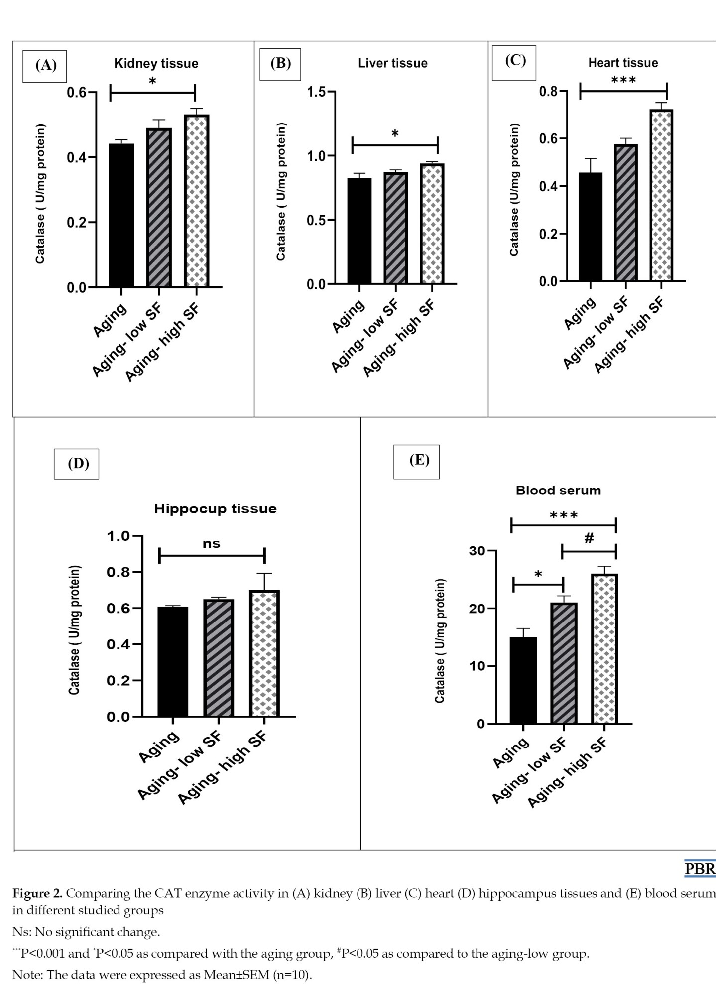
Based on the results, CAT activity was higher in the group that received 200 mg/kg extract compared to the aging group in the kidney, liver (P<0.05), and heart tissues (P<0.001). Insignificant change was found in CAT activity in the hippocampus tissue between the groups. In the blood serum, CAT activity in the aging group was reduced compared to the group was received 100 mg/kg extract (P<0.05) and 200 mg/kg extract (P<0.001). In addition, CAT activity in the group that received 100 mg/kg extract was lower compared to the group that received 200 mg/kg extract in the blood serum (P<0.05) (Figure 2).
Thiol concentration
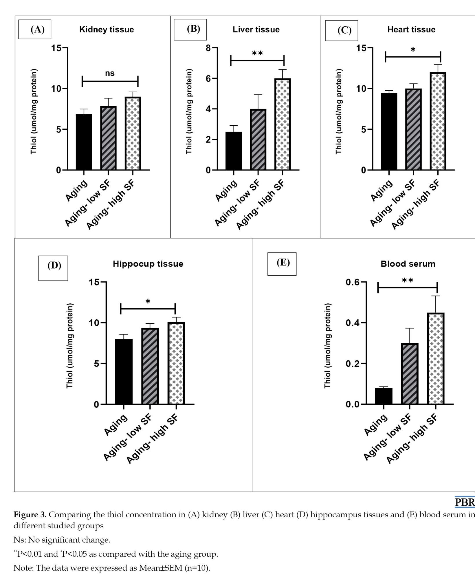
The thiol concentration in the group receiving 200 mg/kg extract considerably increased compared to the aging group in the heart, hippocampus (P<0.05), liver tissues, and blood serum (P<0.01). In contrast, an insignificant difference was found between groups in the kidney tissues (Figure 3).
Urea and creatinine level
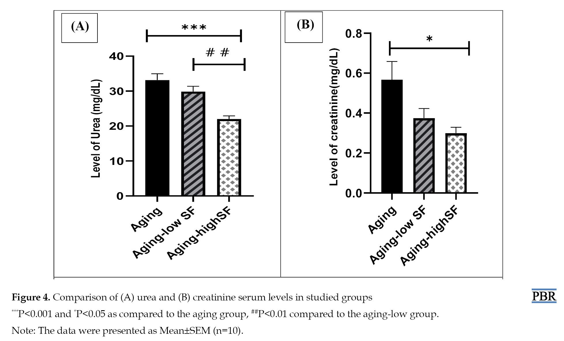
According to the results, a significant decrease was found in the level of urea in the group that received 200 mg/kg extract compared the aging group (P<0.001) and the group that received 100 mg/kg extract (P<0.01) (Figure 4A).
Discussion
Aging is an intricate process that increases mortality and decreases physiological function [25]. Although the aging process is poorly understood, the accumulation of free radicals released during mitochondrial metabolism during aging leads to cellular toxicity and damage to the mitochondrial and nuclear DNA and cellular membrane structure. Besides, antioxidants are known as scavengers of free radicals, and their anti-aging properties include their anti-inflammatory impact and delay or prevention of diabetes, cancer, and brain disorders. Antioxidants effectively reduce blood pressure and the development of atherosclerosis [26]. S. officinalis is a crucial pharmaceutical herb for many purposes [27]. Based on previous studies, S. officinalis has strong antioxidant activity [28]. Accordingly, in this study, we examined the impact of S. officinalis as an antioxidant in aged tissues.
The aging process is associated with an imbalance between pro-oxidant and antioxidant molecules, which increases oxidative stress. Due to the enhancement of oxidative stress, intracellular ROS levels increase and damage proteins, lipids, and DNA [29]. Antioxidant mechanisms play a crucial role in protecting cells from ROS. CAT, a hemoprotein, is involved in the detoxification of H2O2 [30]. Thiol groups, as sulfhydryl groups-containing antioxidants, release hydrogen into the environment under oxidative stress, which binds to excess oxygen and deactivates ROS [31]. Based on previous studies, in the aging group, the MDA level significantly increased, while CAT activity significantly reduced compared to other groups [30, 32]. In the current research, the MDA level in the aging group that received a high dose of S. officinalis significantly decreased in liver, kidney, and heart tissues as compared to the aging group. The thiol concentration in the aging-high S. officinalis group in the liver, heart, hippocampus tissues, and blood serum was higher than in the aging group. Also, the CAT activity in the aging group receiving a high dose of S. officinalis has notably increased compared to the aging group in liver, heart, and kidney tissues. Regarding blood serum, CAT activity in both of the S. officinalis groups had a significant enhancement compared to the aging group. Our results were confirmed by Kolac et al., research reported that S. officinalis is effective against oxidative stress, increases the CAT activity and decreases MDA levels in the inflammation group in the kidney, liver, and lung tissues [12]. This study found an insignificant difference in CAT activity in hippocampal tissue between the different groups. This contradicts Osman’s study, which proved that the extract elevated CAT activity in the brain [33].
Function reduction of multiple organs is evident in aging. Especially in the liver, aging causes hepatic steatosis and progressive inflammation. In natural aging rat models, deterioration of liver morphology and function has been reported. ALT and AST enzymes are vital biomarkers of liver function that are involved in the catabolism of amino acids and bile production. The increase in these enzymes in the serum is due to damage to the hepatocyte membrane. According to previous research, an increment in the level of ALT and AST has been observed in the aging group [34, 35]. Based on the present research findings, high and low doses of S. officinalis lead to a significant reduction in the ALT level in the aging group. However, this change was not significant at the AST level. Our results partially agree with a previous study that indicated S. officinalis has decreased the AST and ALT dose-dependently [36].
The impairment of kidney function is associated with aging. Aging can cause tubular atrophy, interstitial fibrosis, and glomerulosclerosis. Besides, the high serum urea and creatinine levels are demonstrated as remarkable kidney dysfunction during aging [37]. In the current study, using S. officinalis in high doses has significantly decreased the serum urea and creatinine level in aging. In addition, the urea level in the aging-high SF group was lower compared to that in the aging-low group. These results were also in accordance with the previous reports which showed that S. officinalis effectively reduces the level of urea and creatinine in a dose-dependent manner [35]. Telomerase enzyme compensates for telomere erosion. Tissue aging and age-related diseases are associated with telomere shortening and subsequent cell senescence. Tsoukalas’s study demonstrated that telomerase activity and telomerase reverse transcriptase expression were significantly reduced in 21-month-old rats as compared to 6-month-old ones in the brain cortex and cerebellum. It has been suggested that the telomerase reverse transcriptase expression is age-dependent [38]. According to the present study, administrating S. officinalis in high and low doses has increased the telomerase activity in 20-month-old rats.
Our results showed that S. officinalis administration has beneficial effects on tissue aging impairment. Although we emphasized these effects in aged male rats, these may be temporary and suppressed, and they can differ in females. Therefore, further studies with changing the duration of administration and comparing males and females are suggested.
S. officinalis contains compounds that prevent oxidative damage to cells. This helps slow down the aging process. S. officinalis’s anti-inflammatory compounds can reduce chronic inflammation, one factor that accelerates aging. Phytochemicals found in S. officinalis include flavonoids, such as apigenin, luteolin, and dimethoxyapgenin, which have antioxidant and anti-inflammatory properties. Terpenoids, including camphene, borneol, and camphor, are known to be anti-inflammatory. Volatile oils include compounds, such as cineole, terbinol, and verdane, which have antimicrobial and anti-inflammatory properties.
Conclusion
Based on current results, aging leads to oxidative stress in tissues, which disrupts kidney and liver function. Aging can also change the telomerase enzyme activity. Using S. officinalis as an antioxidant agent, especially at a high dose (200 mg/kg) for 2 weeks, may alter the adverse impacts of aging in tissues.
Ethical Considerations
Compliance with ethical guidelines
This study was approved by the Research Ethical Committee of Mashhad University of Medical Sciences, Mashhad, Iran (Code: IR.MUMS.MEDICAL.REC.1400.531). Related procedures were performed according to the National Institute of Health Guide for the Care and Use of Laboratory Animals.
Funding
This study was sponsored by the Research Vice-Chancellor of Mashhad University of Medical Sciences, Mashhad, Iran (Project No.: 970988).
Authors' contributions
Conceptualization and supervision: Shabnam Mohammadi; Methodology and data collection: Abolfazl Rezaei; Analysis and writing the original draft: Negar Ostad-Rahimi; Review and editing: All authors.
Conflict of interest
The authors declared no conflict of interest.
Acknowledgments
The authors are grateful to Research Vice-Chancellor of Mashhad University of Medical Sciences, Mashhad, Iran.
Full-Text: (281 Views)
Introduction
According to forecasts by the United Nations (UN), by 2050, one out of every six people will be over 65 years old, and the number of 80-year-olds will triple. Moreover, aging as a risk factor can lead to most chronic illnesses in humans, including cancer, neurodegenerative diseases, and cardiovascular diseases [1]. Aging is known as a progressive loss of physiological integrity, which causes functional impairment and raises vulnerability to death. Additionally, aging includes complex physiological processes that are not well understood. The most well-known theory in the aging process is Denham Harman, which defines the accumulation of free radicals produced in mitochondrial metabolism as leading to cellular toxicity [2]. Previous studies indicated that in many organisms, oxidative stress damage increases with aging [3]. Reactive oxygen species (ROS) are composed of various chemical species, including superoxide anion (O2−), hydroxyl radical, and hydrogen peroxide (H2O2). The ROS’s uncontrolled production is related to increased damage in nucleic acids, proteins, lipids, and carbohydrates; also, it can change the metabolic activities as an effective factor and finally lead to different pathogenesis and aging [2, 4]. Also, due to the role of mitochondria in apoptosis, mitochondrial oxidative stress plays a significant role in enhancing apoptosis during aging [5]. Based on studies, aging not only reduces the activity of catalase (CAT), superoxide dismutase (SOD), glutathione peroxidase (GPx), glutathione reductase (GR), and glutathione-S-transferase (GSTs) but also increases the level of malondialdehyde (MDA). In other words, the lower activity of antioxidant enzymes following aging demonstrates impaired antioxidant defense in the aging organism. Furthermore, the intensity of peroxidative lipid structure and oxidant and antioxidant balance are disrupted in aging. Moreover, telomere shortening is known as an aging biomarker, making cells susceptible to certain diseases. Based on the evidence, oxidative stress is associated with telomere shortening during aging. Antioxidants can be effective in reducing the rate of telomere shortening during aging [6]. Aging is a crucial factor affecting the increase in aspartate aminotransferase (AST), alanine aminotransferase (ALT), urea, and creatinine levels. Insufficient antioxidants may enhance oxidative damage due to aging. Accordingly, dietary supplementation with antioxidants can prevent the development of chronic diseases by reducing the adverse effects of lipid peroxidation, free radicals, and ROS [7, 8].
Salvia officinalis, an aromatic and medicinal plant, is a Labiatae/Lamiaceae family member. Although its origin is the Middle East and Mediterranean regions, it is now found worldwide. The antioxidant properties of S. officinalis have been previously reported. Also, its therapeutic potential is undeniable [9, 10]. S. officinalis has been utilized as an anti-cholinesterase, anti-inflammatory, antidiabetic, and anticancer [11]. Also, S. officinalis not only increases the activity of antioxidant enzymes but also effectively reduces lipid peroxidation products [12].
Therefore, the present study was conducted to identify the influence of administrating an herbal extract of S. officinalis on biochemical factors and oxidative stress indices in major organs of 20-month-old rats.
Materials and Methods
Preparation of S. officinalis extract
To prepare the S. officinalis extract, herbarium collected and approved the fresh S. officinalis. Then it was dried and powdered in the shade. A total of 100 g of plant powder was poured into an Erlenmeyer flask with half a liter of 50% alcohol. Absolute alcohol is used as a solvent. The Erlenmeyer was covered by aluminum foil and placed in a shaker incubator for 48 hours. The residue was filtered with Whatman filter paper, and the solvent was removed by rotary evaporation. The mixture was then placed in an oven at 50 °C to convert it to a dried extract for solvent removal. The required doses were prepared with physiological serum and stored in a sterile container in the refrigerator [13].
Animal issues
All rats were kept under standard conditions (12 h dark and light cycle, 50% relative humidity, at a temperature of 22-24 °C, and free access to drinking water and food). In this experimental study, the animal house of Mashhad University of Medical Sciences provided 30 aged male rats (20-month-old). To investigate the effect of S. officinalis, 30 aged rats were randomly divided into three groups of six rats each as follows:
1-Aging group received normal saline.
2-Aging-low S. officinalis group: Aged rats received 100 mg/kg extract of S. officinalis by oral gavage daily for 2 weeks.
3-Aging-high S. officinalis group: Aged rats received a 200 mg /kg extract of S. officinalis by oral gavage daily for 2 weeks [14, 15].
In this study, normal saline was used as a solvent. Finally, all aged rats were anesthetized with xylazine and ketamine, and the heart, liver, hippocampus, and right kidney tissues were collected for oxidative stress assessment. Also, blood samples were obtained from the cardiac apex. After separating the blood serum, the amounts of urea, creatinine, liver enzymes, telomerase enzyme, and oxidants and antioxidants in the serum of aged rats were measured.
Biochemical assessment
A phosphate-buffered solution was used to homogenize tissues. Then, homogenized tissues and blood serum were used to evaluate the MDA concentration, the total thiol groups, and CAT activity [16]. The blood samples were further centrifuged at 5000 rmp for 15 mins to isolate the blood serum. Then, serum was stored at -80 °C and used to evaluate liver enzymes, urea, creatinine, and the levels of oxidants and antioxidants [17].
MDA measurement
This study used the thiobarbituric acid reactive substances (TBARS) method to measure the MDA level. MDA is the terminal product of lipid peroxidation. When MDA interacts with thiobarbituric acid, it generates a red complex with an optical density peak at 532 nm. A spectrophotometer was used to measure the absorbance at 535 nm. The MDA level was then estimated utilizing the Equation 1 [18].
1. C(M)=Absorbance/1.56×105
Thiol concentration measurement
This study utilized a dithiol nitrobenzoic acid (DTNB) reagent to evaluate the thiol groups. Its reaction with S. officinalis groups of thiol forms a yellow complex of 1, 3, 5-trinitrobenzene with a peak absorbance at 412 nm. Further, 1 mL tromethamine-ethylenediaminetetraacetic acid topical (Tris-EDTA) buffer was added to 50 μL of serum; absorbance and tissues were then read at 412 nm against tromethamine-ethylenediaminetetraacetic acid topical (Tris-EDTA) buffer alone (A1). Then 20 μL DTNB reagent was added to the solution, and the sample absorbance was measured again (A2) after 10 minutes. The DTNB reagents’ absorbance was considered blank (B). The Equation 2 was used to calculate total thiol concentration (mM) [19].
2. Total thiol concentration (mM)=(A2-A1-B)×1.07/0.05×13.6
CAT activity measurement
CAT activity was measured utilizing the Aebi׳s method. This technique decomposes H2O2 at a 240 nm wavelength. The process was initiated by adding 30 mM H2O2 to an adequate size of tissue homogeneity in the 50 mM sodium phosphate buffer (pH=7). Further, the absorbance was measured in 3 minutes at 240 nm. The specific activity was finally obtained in per milligram of protein per minute (units/mg protein/min) [20].
Liver enzymes evaluation
Liver enzymes, including ALT and AST, were evaluated per the kit protocol (Pars Azmon, Iran) [21].
Urea and creatinine level evaluation
Urea and creatinine were measured to evaluate kidney function. Colorimetric diagnostic kits were used to measure the urea and creatinine levels. Measurements were performed based on the manufacturer’s recommendations (Pars Azmon, Iran) [22, 23].
Telomerase enzyme measurement
Blood serum was analyzed based on the guidelines of the enzyme linked immunosorbent assay (ELISA) kit (Elab Science) for the telomerase enzyme activity measurement [24]. A total of 100 μL of serum was first added to the plate. The samples were incubated for 90 minutes at 37 °C. One-hundred μL of biotinlated detection Ab was added to the plate and incubated for one h at 37 °C. After 3 times washing, 100 μL of horseradish peroxidase (HPR) conjugate was added to the plate and incubated at 37 °C for 30 minutes. Following washing, 90 μL of substrate solution was added and incubated for 15 minutes at 37 °C. Finally, 50 μL of stop solution was added and read at 450 nm wavelength.
Statistical analysis
The GraphPad Prism software, version 8.0 was utilized for statistical analysis. The one-way analysis of variance (ANOVA) and Tukey’s post-hoc test were used to compare significant differences between groups. Statistical significant was set at P<0.05.
Results
Biochemical assessment
MDA concentration
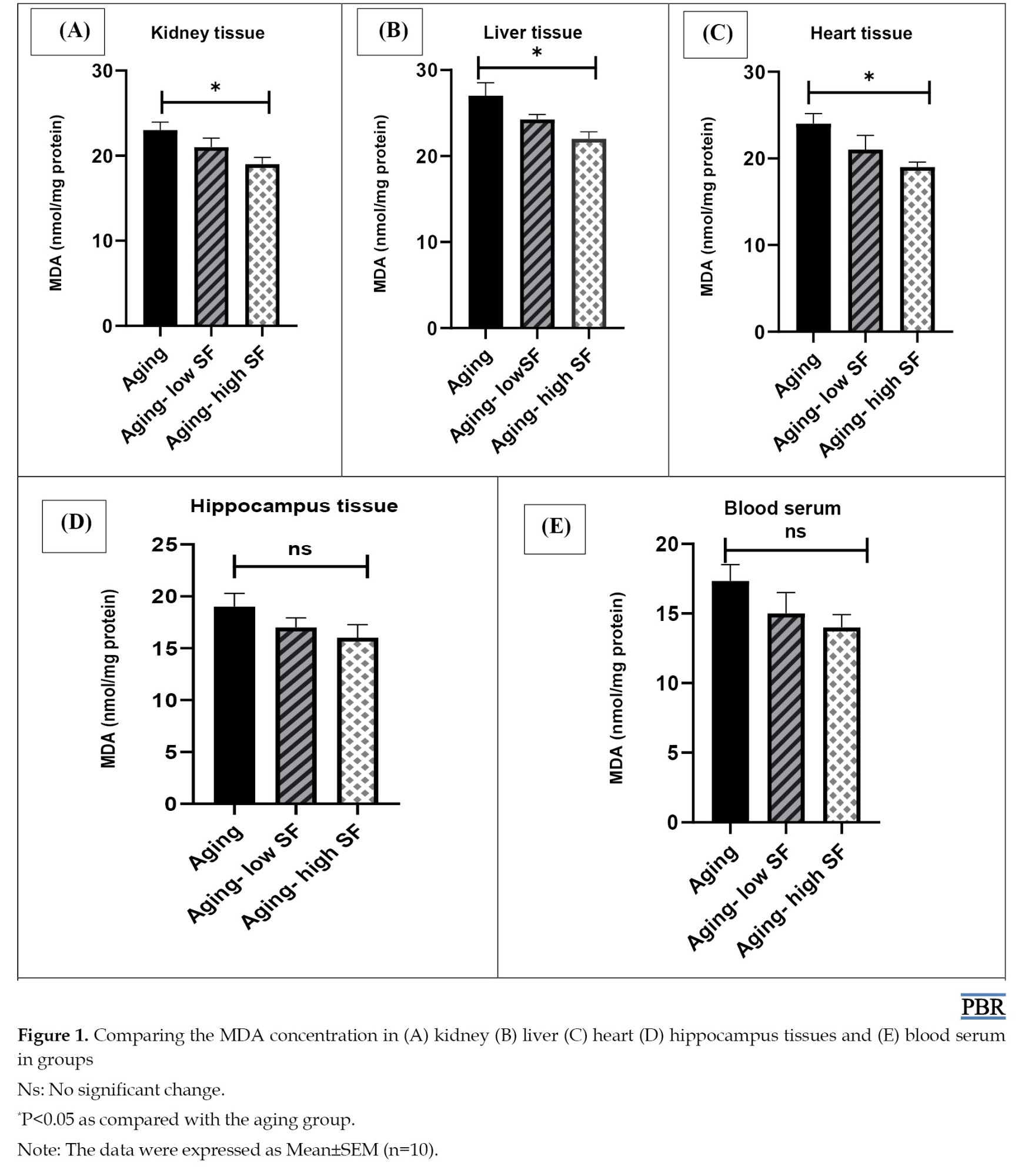
Based on the MDA concentration measurement in the liver, kidney, and heart tissues, the MDA level in the aging group was considerably higher than the group that received 200 mg/kg of the extract (P<0.05). While in the hippocampus tissue and blood serum, an insignificant difference was found in the MDA concentration between the groups (Figure 1).
According to forecasts by the United Nations (UN), by 2050, one out of every six people will be over 65 years old, and the number of 80-year-olds will triple. Moreover, aging as a risk factor can lead to most chronic illnesses in humans, including cancer, neurodegenerative diseases, and cardiovascular diseases [1]. Aging is known as a progressive loss of physiological integrity, which causes functional impairment and raises vulnerability to death. Additionally, aging includes complex physiological processes that are not well understood. The most well-known theory in the aging process is Denham Harman, which defines the accumulation of free radicals produced in mitochondrial metabolism as leading to cellular toxicity [2]. Previous studies indicated that in many organisms, oxidative stress damage increases with aging [3]. Reactive oxygen species (ROS) are composed of various chemical species, including superoxide anion (O2−), hydroxyl radical, and hydrogen peroxide (H2O2). The ROS’s uncontrolled production is related to increased damage in nucleic acids, proteins, lipids, and carbohydrates; also, it can change the metabolic activities as an effective factor and finally lead to different pathogenesis and aging [2, 4]. Also, due to the role of mitochondria in apoptosis, mitochondrial oxidative stress plays a significant role in enhancing apoptosis during aging [5]. Based on studies, aging not only reduces the activity of catalase (CAT), superoxide dismutase (SOD), glutathione peroxidase (GPx), glutathione reductase (GR), and glutathione-S-transferase (GSTs) but also increases the level of malondialdehyde (MDA). In other words, the lower activity of antioxidant enzymes following aging demonstrates impaired antioxidant defense in the aging organism. Furthermore, the intensity of peroxidative lipid structure and oxidant and antioxidant balance are disrupted in aging. Moreover, telomere shortening is known as an aging biomarker, making cells susceptible to certain diseases. Based on the evidence, oxidative stress is associated with telomere shortening during aging. Antioxidants can be effective in reducing the rate of telomere shortening during aging [6]. Aging is a crucial factor affecting the increase in aspartate aminotransferase (AST), alanine aminotransferase (ALT), urea, and creatinine levels. Insufficient antioxidants may enhance oxidative damage due to aging. Accordingly, dietary supplementation with antioxidants can prevent the development of chronic diseases by reducing the adverse effects of lipid peroxidation, free radicals, and ROS [7, 8].
Salvia officinalis, an aromatic and medicinal plant, is a Labiatae/Lamiaceae family member. Although its origin is the Middle East and Mediterranean regions, it is now found worldwide. The antioxidant properties of S. officinalis have been previously reported. Also, its therapeutic potential is undeniable [9, 10]. S. officinalis has been utilized as an anti-cholinesterase, anti-inflammatory, antidiabetic, and anticancer [11]. Also, S. officinalis not only increases the activity of antioxidant enzymes but also effectively reduces lipid peroxidation products [12].
Therefore, the present study was conducted to identify the influence of administrating an herbal extract of S. officinalis on biochemical factors and oxidative stress indices in major organs of 20-month-old rats.
Materials and Methods
Preparation of S. officinalis extract
To prepare the S. officinalis extract, herbarium collected and approved the fresh S. officinalis. Then it was dried and powdered in the shade. A total of 100 g of plant powder was poured into an Erlenmeyer flask with half a liter of 50% alcohol. Absolute alcohol is used as a solvent. The Erlenmeyer was covered by aluminum foil and placed in a shaker incubator for 48 hours. The residue was filtered with Whatman filter paper, and the solvent was removed by rotary evaporation. The mixture was then placed in an oven at 50 °C to convert it to a dried extract for solvent removal. The required doses were prepared with physiological serum and stored in a sterile container in the refrigerator [13].
Animal issues
All rats were kept under standard conditions (12 h dark and light cycle, 50% relative humidity, at a temperature of 22-24 °C, and free access to drinking water and food). In this experimental study, the animal house of Mashhad University of Medical Sciences provided 30 aged male rats (20-month-old). To investigate the effect of S. officinalis, 30 aged rats were randomly divided into three groups of six rats each as follows:
1-Aging group received normal saline.
2-Aging-low S. officinalis group: Aged rats received 100 mg/kg extract of S. officinalis by oral gavage daily for 2 weeks.
3-Aging-high S. officinalis group: Aged rats received a 200 mg /kg extract of S. officinalis by oral gavage daily for 2 weeks [14, 15].
In this study, normal saline was used as a solvent. Finally, all aged rats were anesthetized with xylazine and ketamine, and the heart, liver, hippocampus, and right kidney tissues were collected for oxidative stress assessment. Also, blood samples were obtained from the cardiac apex. After separating the blood serum, the amounts of urea, creatinine, liver enzymes, telomerase enzyme, and oxidants and antioxidants in the serum of aged rats were measured.
Biochemical assessment
A phosphate-buffered solution was used to homogenize tissues. Then, homogenized tissues and blood serum were used to evaluate the MDA concentration, the total thiol groups, and CAT activity [16]. The blood samples were further centrifuged at 5000 rmp for 15 mins to isolate the blood serum. Then, serum was stored at -80 °C and used to evaluate liver enzymes, urea, creatinine, and the levels of oxidants and antioxidants [17].
MDA measurement
This study used the thiobarbituric acid reactive substances (TBARS) method to measure the MDA level. MDA is the terminal product of lipid peroxidation. When MDA interacts with thiobarbituric acid, it generates a red complex with an optical density peak at 532 nm. A spectrophotometer was used to measure the absorbance at 535 nm. The MDA level was then estimated utilizing the Equation 1 [18].
1. C(M)=Absorbance/1.56×105
Thiol concentration measurement
This study utilized a dithiol nitrobenzoic acid (DTNB) reagent to evaluate the thiol groups. Its reaction with S. officinalis groups of thiol forms a yellow complex of 1, 3, 5-trinitrobenzene with a peak absorbance at 412 nm. Further, 1 mL tromethamine-ethylenediaminetetraacetic acid topical (Tris-EDTA) buffer was added to 50 μL of serum; absorbance and tissues were then read at 412 nm against tromethamine-ethylenediaminetetraacetic acid topical (Tris-EDTA) buffer alone (A1). Then 20 μL DTNB reagent was added to the solution, and the sample absorbance was measured again (A2) after 10 minutes. The DTNB reagents’ absorbance was considered blank (B). The Equation 2 was used to calculate total thiol concentration (mM) [19].
2. Total thiol concentration (mM)=(A2-A1-B)×1.07/0.05×13.6
CAT activity measurement
CAT activity was measured utilizing the Aebi׳s method. This technique decomposes H2O2 at a 240 nm wavelength. The process was initiated by adding 30 mM H2O2 to an adequate size of tissue homogeneity in the 50 mM sodium phosphate buffer (pH=7). Further, the absorbance was measured in 3 minutes at 240 nm. The specific activity was finally obtained in per milligram of protein per minute (units/mg protein/min) [20].
Liver enzymes evaluation
Liver enzymes, including ALT and AST, were evaluated per the kit protocol (Pars Azmon, Iran) [21].
Urea and creatinine level evaluation
Urea and creatinine were measured to evaluate kidney function. Colorimetric diagnostic kits were used to measure the urea and creatinine levels. Measurements were performed based on the manufacturer’s recommendations (Pars Azmon, Iran) [22, 23].
Telomerase enzyme measurement
Blood serum was analyzed based on the guidelines of the enzyme linked immunosorbent assay (ELISA) kit (Elab Science) for the telomerase enzyme activity measurement [24]. A total of 100 μL of serum was first added to the plate. The samples were incubated for 90 minutes at 37 °C. One-hundred μL of biotinlated detection Ab was added to the plate and incubated for one h at 37 °C. After 3 times washing, 100 μL of horseradish peroxidase (HPR) conjugate was added to the plate and incubated at 37 °C for 30 minutes. Following washing, 90 μL of substrate solution was added and incubated for 15 minutes at 37 °C. Finally, 50 μL of stop solution was added and read at 450 nm wavelength.
Statistical analysis
The GraphPad Prism software, version 8.0 was utilized for statistical analysis. The one-way analysis of variance (ANOVA) and Tukey’s post-hoc test were used to compare significant differences between groups. Statistical significant was set at P<0.05.
Results
Biochemical assessment
MDA concentration
Based on the MDA concentration measurement in the liver, kidney, and heart tissues, the MDA level in the aging group was considerably higher than the group that received 200 mg/kg of the extract (P<0.05). While in the hippocampus tissue and blood serum, an insignificant difference was found in the MDA concentration between the groups (Figure 1).
CAT activity
Based on the results, CAT activity was higher in the group that received 200 mg/kg extract compared to the aging group in the kidney, liver (P<0.05), and heart tissues (P<0.001). Insignificant change was found in CAT activity in the hippocampus tissue between the groups. In the blood serum, CAT activity in the aging group was reduced compared to the group was received 100 mg/kg extract (P<0.05) and 200 mg/kg extract (P<0.001). In addition, CAT activity in the group that received 100 mg/kg extract was lower compared to the group that received 200 mg/kg extract in the blood serum (P<0.05) (Figure 2).
Thiol concentration
The thiol concentration in the group receiving 200 mg/kg extract considerably increased compared to the aging group in the heart, hippocampus (P<0.05), liver tissues, and blood serum (P<0.01). In contrast, an insignificant difference was found between groups in the kidney tissues (Figure 3).
Urea and creatinine level
According to the results, a significant decrease was found in the level of urea in the group that received 200 mg/kg extract compared the aging group (P<0.001) and the group that received 100 mg/kg extract (P<0.01) (Figure 4A).
Also, the creatinine level in the group that received 200 mg/kg extract was lower compared to the aging group (P<0.05) (Figure 4B).
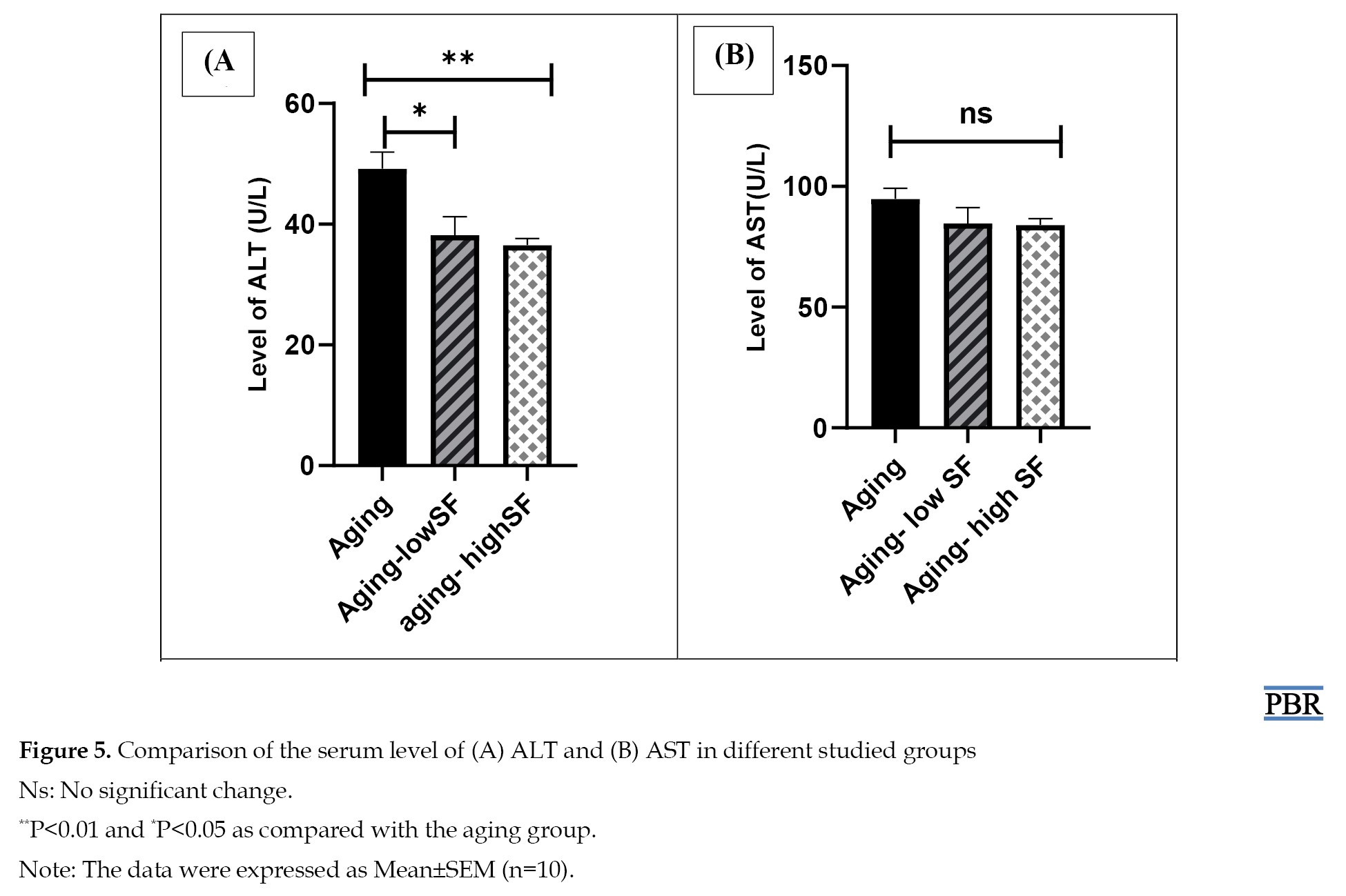
ALT and AST level
The results of liver enzyme measurement showed that the ALT level in the group receiving 200 mg/kg extract (P<0.01) and 100 mg/kg extract (P<0.05) was notably reduced compared to the aging group (Figure 5A).
ALT and AST level
The results of liver enzyme measurement showed that the ALT level in the group receiving 200 mg/kg extract (P<0.01) and 100 mg/kg extract (P<0.05) was notably reduced compared to the aging group (Figure 5A).
An insignificant difference was found in the AST level between groups (Figure 5B).
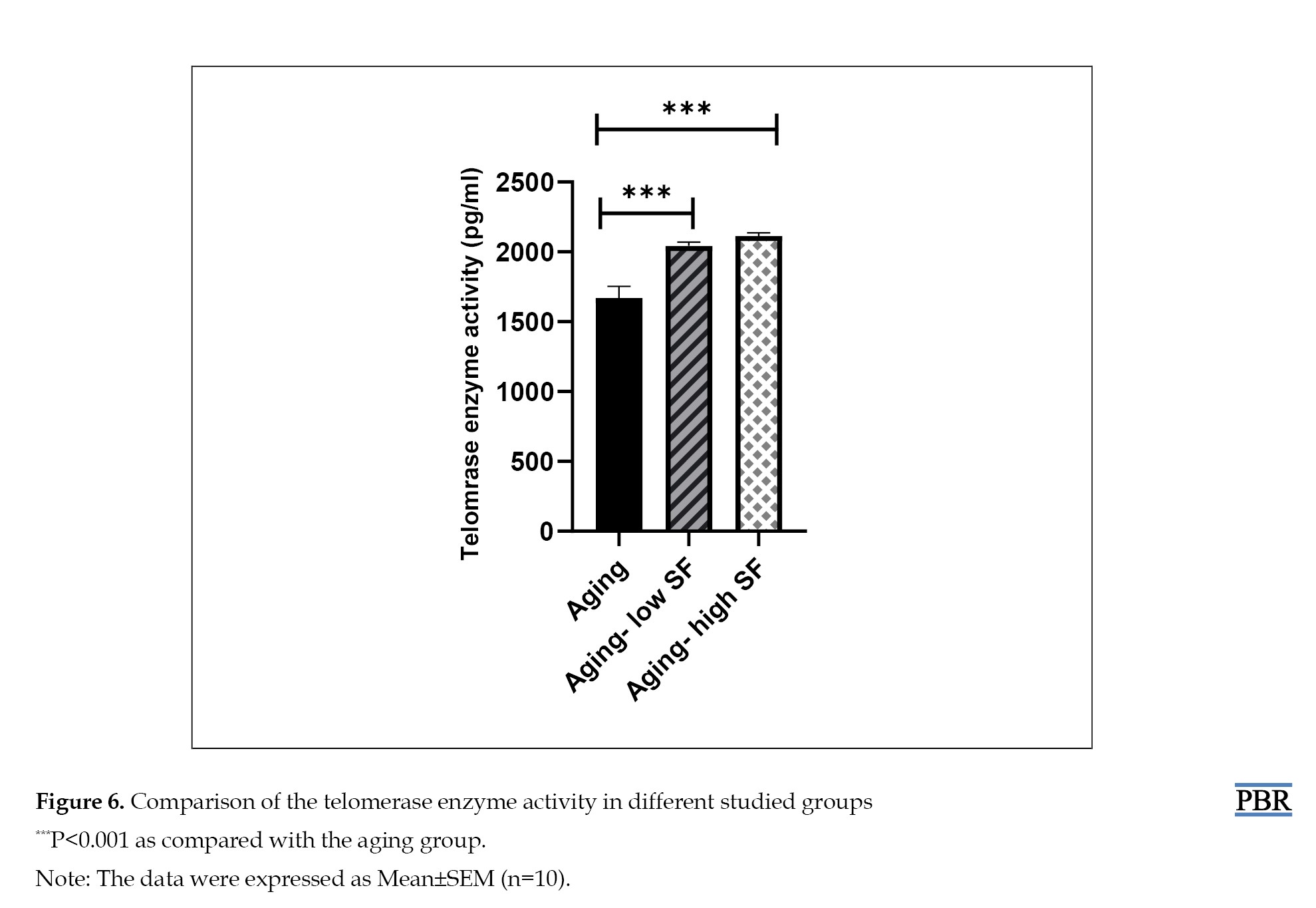
Telomerase enzyme activity
The results indicated that the activity of telomerase enzyme was considerably higher in the groups that received 200 mg/kg and 100 mg/kg extract compared to the aging group (P<0.001) (Figure 6).
Telomerase enzyme activity
The results indicated that the activity of telomerase enzyme was considerably higher in the groups that received 200 mg/kg and 100 mg/kg extract compared to the aging group (P<0.001) (Figure 6).
Discussion
Aging is an intricate process that increases mortality and decreases physiological function [25]. Although the aging process is poorly understood, the accumulation of free radicals released during mitochondrial metabolism during aging leads to cellular toxicity and damage to the mitochondrial and nuclear DNA and cellular membrane structure. Besides, antioxidants are known as scavengers of free radicals, and their anti-aging properties include their anti-inflammatory impact and delay or prevention of diabetes, cancer, and brain disorders. Antioxidants effectively reduce blood pressure and the development of atherosclerosis [26]. S. officinalis is a crucial pharmaceutical herb for many purposes [27]. Based on previous studies, S. officinalis has strong antioxidant activity [28]. Accordingly, in this study, we examined the impact of S. officinalis as an antioxidant in aged tissues.
The aging process is associated with an imbalance between pro-oxidant and antioxidant molecules, which increases oxidative stress. Due to the enhancement of oxidative stress, intracellular ROS levels increase and damage proteins, lipids, and DNA [29]. Antioxidant mechanisms play a crucial role in protecting cells from ROS. CAT, a hemoprotein, is involved in the detoxification of H2O2 [30]. Thiol groups, as sulfhydryl groups-containing antioxidants, release hydrogen into the environment under oxidative stress, which binds to excess oxygen and deactivates ROS [31]. Based on previous studies, in the aging group, the MDA level significantly increased, while CAT activity significantly reduced compared to other groups [30, 32]. In the current research, the MDA level in the aging group that received a high dose of S. officinalis significantly decreased in liver, kidney, and heart tissues as compared to the aging group. The thiol concentration in the aging-high S. officinalis group in the liver, heart, hippocampus tissues, and blood serum was higher than in the aging group. Also, the CAT activity in the aging group receiving a high dose of S. officinalis has notably increased compared to the aging group in liver, heart, and kidney tissues. Regarding blood serum, CAT activity in both of the S. officinalis groups had a significant enhancement compared to the aging group. Our results were confirmed by Kolac et al., research reported that S. officinalis is effective against oxidative stress, increases the CAT activity and decreases MDA levels in the inflammation group in the kidney, liver, and lung tissues [12]. This study found an insignificant difference in CAT activity in hippocampal tissue between the different groups. This contradicts Osman’s study, which proved that the extract elevated CAT activity in the brain [33].
Function reduction of multiple organs is evident in aging. Especially in the liver, aging causes hepatic steatosis and progressive inflammation. In natural aging rat models, deterioration of liver morphology and function has been reported. ALT and AST enzymes are vital biomarkers of liver function that are involved in the catabolism of amino acids and bile production. The increase in these enzymes in the serum is due to damage to the hepatocyte membrane. According to previous research, an increment in the level of ALT and AST has been observed in the aging group [34, 35]. Based on the present research findings, high and low doses of S. officinalis lead to a significant reduction in the ALT level in the aging group. However, this change was not significant at the AST level. Our results partially agree with a previous study that indicated S. officinalis has decreased the AST and ALT dose-dependently [36].
The impairment of kidney function is associated with aging. Aging can cause tubular atrophy, interstitial fibrosis, and glomerulosclerosis. Besides, the high serum urea and creatinine levels are demonstrated as remarkable kidney dysfunction during aging [37]. In the current study, using S. officinalis in high doses has significantly decreased the serum urea and creatinine level in aging. In addition, the urea level in the aging-high SF group was lower compared to that in the aging-low group. These results were also in accordance with the previous reports which showed that S. officinalis effectively reduces the level of urea and creatinine in a dose-dependent manner [35]. Telomerase enzyme compensates for telomere erosion. Tissue aging and age-related diseases are associated with telomere shortening and subsequent cell senescence. Tsoukalas’s study demonstrated that telomerase activity and telomerase reverse transcriptase expression were significantly reduced in 21-month-old rats as compared to 6-month-old ones in the brain cortex and cerebellum. It has been suggested that the telomerase reverse transcriptase expression is age-dependent [38]. According to the present study, administrating S. officinalis in high and low doses has increased the telomerase activity in 20-month-old rats.
Our results showed that S. officinalis administration has beneficial effects on tissue aging impairment. Although we emphasized these effects in aged male rats, these may be temporary and suppressed, and they can differ in females. Therefore, further studies with changing the duration of administration and comparing males and females are suggested.
S. officinalis contains compounds that prevent oxidative damage to cells. This helps slow down the aging process. S. officinalis’s anti-inflammatory compounds can reduce chronic inflammation, one factor that accelerates aging. Phytochemicals found in S. officinalis include flavonoids, such as apigenin, luteolin, and dimethoxyapgenin, which have antioxidant and anti-inflammatory properties. Terpenoids, including camphene, borneol, and camphor, are known to be anti-inflammatory. Volatile oils include compounds, such as cineole, terbinol, and verdane, which have antimicrobial and anti-inflammatory properties.
Conclusion
Based on current results, aging leads to oxidative stress in tissues, which disrupts kidney and liver function. Aging can also change the telomerase enzyme activity. Using S. officinalis as an antioxidant agent, especially at a high dose (200 mg/kg) for 2 weeks, may alter the adverse impacts of aging in tissues.
Ethical Considerations
Compliance with ethical guidelines
This study was approved by the Research Ethical Committee of Mashhad University of Medical Sciences, Mashhad, Iran (Code: IR.MUMS.MEDICAL.REC.1400.531). Related procedures were performed according to the National Institute of Health Guide for the Care and Use of Laboratory Animals.
Funding
This study was sponsored by the Research Vice-Chancellor of Mashhad University of Medical Sciences, Mashhad, Iran (Project No.: 970988).
Authors' contributions
Conceptualization and supervision: Shabnam Mohammadi; Methodology and data collection: Abolfazl Rezaei; Analysis and writing the original draft: Negar Ostad-Rahimi; Review and editing: All authors.
Conflict of interest
The authors declared no conflict of interest.
Acknowledgments
The authors are grateful to Research Vice-Chancellor of Mashhad University of Medical Sciences, Mashhad, Iran.
References
- Cai Y, Song W, Li J, Jing Y, Liang C, Zhang L, et al. The landscape of aging. Sci China Life Sci. 2022; 65(12):2354-454. [DOI:10.1007/s11427-022-2161-3] [PMID]
- Guillaumet-Adkins A, Yañez Y, Peris-Diaz MD, Calabria I, Palanca-Ballester C, Sandoval J. Epigenetics and Oxidative Stress in Aging. Oxid Med Cell Longev. 2017; 2017:9175806. [DOI:10.1155/2017/9175806] [PMID]
- Salmon AB, Richardson A, Pérez VI. Update on the oxidative stress theory of aging: Does oxidative stress play a role in aging or healthy aging? Free Radic Biol Med. 2010; 48(5):642-55. [DOI:10.1016/j.freeradbiomed.2009.12.015] [PMID]
- Newsholme P, Rebelato E, Abdulkader F, Krause M, Carpinelli A, Curi R. Reactive oxygen and nitrogen species generation, antioxidant defenses, and β-cell function: A critical role for amino acids. J Endocrinol. 2012; 214(1):11-20. [DOI:10.1530/JOE-12-0072] [PMID]
- Yamaguchi R, Perkins G. Dynamics of mitochondrial structure during apoptosis and the enigma of Opa1. Biochim Biophys Acta. 2009; 1787(8):963-72. [DOI:10.1016/j.bbabio.2009.02.005] [PMID]
- Gavia-García G, Rosado-Pérez J, Arista-Ugalde TL, Aguiñiga-Sánchez I, Santiago-Osorio E, Mendoza-Núñez VM. Telomere Length and Oxidative Stress and Its Relation with Metabolic Syndrome Components in the Aging. Biology (Basel). 2021; 10(4):253. [DOI:10.3390/biology10040253] [PMID]
- Kozakiewicz M, Kornatowski M, Krzywińska O, Kędziora-Kornatowska K. Changes in the blood antioxidant defense of advanced age people. Clin Interv Aging. 2019; 14:763-71.[DOI:10.2147/CIA.S201250] [PMID]
- Ramesh T, Kim SW, Sung JH, Hwang SY, Sohn SH, Yoo SK, et al. Effect of fermented Panax ginseng extract (GINST) on oxidative stress and antioxidant activities in major organs of aged rats. Exp Gerontol. 2012; 47(1):77-84. [DOI:10.1016/j.exger.2011.10.007] [PMID]
- Ghorbani A, Esmaeilizadeh M. Pharmacological properties of Salvia officinalis and its components. J Tradit Complement Med. 2017; 7(4):433-40. [DOI:10.1016/j.jtcme.2016.12.014] [PMID]
- Zare, H. Effects of salvia officinalis extract on the breast cancer cell line. SciMed J. 2019; 1(1):25-9. [DOI:10.28991/SciMedJ-2019-0101-4]
- El Euch SK, Hassine D, Cazaux S, Bouzouita N, Bouajila J. Salvia officinalis essential oil: Chemical analysis and evaluation of anti-enzymatic and antioxidant bioactivities. S Afr J Bot. 2019; 120:253-60. [DOI:10.1016/j.sajb.2018.07.010]
- Kolac UK, Ustuner MC, Tekin N, Ustuner D, Colak E, Entok E. The Anti-Inflammatory and antioxidant effects of salvia officinalis on lipopolysaccharide-induced inflammation in rats. J Med Food. 2017; 20(12):1193-1200. [DOI:10.1089/jmf.2017.0035] [PMID]
- Vafaei A, Mohammadi S, Fazel A, Soukhtanloo M, Mohammadipour A, Beheshti F. Effects of carob (Ceratonia siliqua) on sperm quality, testicular structure, testosterone level and oxidative stress in busulfan-induced infertile mice. Pharm Sci. 2018; 24(2):104-11. [DOI:10.15171/PS.2018.16]
- Bagheri Y, Keshtmand Z, Rahbarghazi R, Gharamaleki MN, Barati A, Bagheri S, et al. Salvia officinalis hydroalcoholic extract improved reproduction capacity and behavioral activity in rats exposed to immobilization stress. Anim Sci J. 2020; 91(1):e13382. [DOI:10.1111/asj.13382] [PMID]
- Dinel AL, Lucas C, Guillemet D, Layé S, Pallet V, Joffre C. Chronic supplementation with a mix of salvia officinalis and salvia lavandulaefolia improves morris water maze learning in normal adult C57Bl/6J mice. Nutrients. 2020; 12(6):1777. [DOI:10.3390/nu12061777] [PMID]
- Jalilvand N, Hosseini M, Beheshti F, Ebrahimzadeh-Bideskan A. Protective effect of PPARγ agonist pioglitazone, on testicular tissue and sperm parameters in hypothyroid rats. Toxin Rev. 2019; 40(3):267-76. [DOI:10.1080/15569543.2018.1564775]
- Pallio G, Micali A, Benvenga S, Antonelli A, Marini HR, Puzzolo D, et al. Myo-inositol in the protection from cadmium-induced toxicity in mice kidney: An emerging nutraceutical challenge. Food Chem Toxicol. 2019; 132:110675. [DOI:10.1016/j.fct.2019.110675] [PMID]
- Osatd-Rahimi N, Saburi E, Karimi S, Boustan A, Ebrahimzadeh-Bideskan A. The therapeutic effect of melatonin on female offspring ovarian reserve and quality in BALB/c mice after exposing their mother to methamphetamine during pregnancy and lactation. Iran J Basic Med Sci. 2023; 26(2):208-15. [PMID]
- Yazdi HB, Hojati V, Shiravi A, Hosseinian S, Vaezi G, Hadjzadeh MA. Liver dysfunction and oxidative stress in streptozotocin-induced diabetic rats: Protective role of artemisia turanica. J Pharmacopuncture. 2019; 22(2):109-14. [DOI:10.3831/KPI.2019.22.014] [PMID]
- Gaharwar US, Meena R, Rajamani P. Iron oxide nanoparticles induced cytotoxicity, oxidative stress and DNA damage in lymphocytes. J Appl Toxicol. 2017; 37(10):1232-44. [DOI:10.1002/jat.3485] [PMID]
- Pars Azmon Co. [GPT (ALAT) quantitative detection kit in serum or plasma by photometric method (Persian)]. Karaj: Pars Azmon Co: 2020. [Link]
- Pars Azmon Co. [UV urea quantitative detection kit in serum, plasma and urine using photometric method (Persian)]. Karaj: Pars Azmon Co: 2020. [Link]
- Pars Azmon Co. [Creatinine quantitative detection kit in serum, plasma or urine using photometric method (Persian)]. Karaj: Pars Azmon Co: 2020. [Link]
- Fahmy MA, Diab KA, Abdel-Samie NS, Omara EA, Hassan ZM. Carbon tetrachloride induced hepato/renal toxicity in experimental mice: Antioxidant potential of Egyptian Salvia officinalis L essential oil. Environ Sci Pollut Res Int. 2018; 25(28):27858-76. [DOI:10.1007/s11356-018-2820-6] [PMID]
- AlDehaini DMB, Al-Bustan SA, Ali ME, Malalla ZHA, Sater M, Giha HA. Shortening of the leucocytes' telomeres length in T2DM independent of age and telomerase activity. Acta Diabetol. 2020; 57(11):1287-95. [DOI:10.1007/s00592-020-01550-4] [PMID]
- Halim M, Halim A. The effects of inflammation, aging and oxidative stress on the pathogenesis of diabetes mellitus (type 2 diabetes). Diabetes Metab Syndr. 2019; 13(2):1165-72. [DOI:10.1016/j.dsx.2019.01.040] [PMID]
- Uwa LM. The anti-aging efficacy of antioxidants. Curr Trends Biomedical Eng Biosci. 2017; 7(4):555716. [DOI:10.19080/CTBEB.2017.07.555716]
- Vosoughi N, Gomarian M, Pirbalouti AG, Khaghani S, Malekpoor F. Essential oil composition and total phenolic, flavonoid contents, and antioxidant activity of sage (Salvia officinalis L.) extract under chitosan application and irrigation frequencies. Ind Crops Prod. 2018; 117:366-74. [DOI:10.1016/j.indcrop.2018.03.021]
- Khiya Z, Oualcadi Y, Gamar A, Berrekhis F, Zair T, Hilali F. Correlation of total polyphenolic content with antioxidant activity of hydromethanolic extract and their fractions of the Salvia officinalis leaves from different regions of Morocco. J Chem. 2021; 1-11. [DOI:10.1155/2021/8585313]
- Barrera G, Pizzimenti S, Daga M, Dianzani C, Arcaro A, Cetrangolo GP, et al. Lipid peroxidation-derived aldehydes, 4-hydroxynonenal and malondialdehyde in aging-related disorders. Antioxidants (Basel). 2018; 7(8):102. [DOI:10.3390/antiox7080102] [PMID]
- Zhang Y, Chen X, Yang L, Zu Y, Lu Q. Effects of rosmarinic acid on liver and kidney antioxidant enzymes, lipid peroxidation and tissue ultrastructure in aging mice. Food Funct. 2015; 6(3):927-31. [DOI:10.1039/C4FO01051E] [PMID]
- Sener S, Akbas A, Kilinc F, Baran P, Erel O, Aktas A. Thiol/disulfide homeostasis as a marker of oxidative stress in rosacea: A controlled spectrophotometric study. Cutan Ocul Toxicol. 2019; 38(1):55-8. [DOI:10.1080/15569527.2018.1517124] [PMID]
- Ahangarpour A, Najimi SA, Farbood Y. Effects of Vitex agnus-castus fruit on sex hormones and antioxidant indices in a d-galactose-induced aging female mouse model. J Chin Med Assoc. 2016; 79(11):589-96. [DOI:10.1016/j.jcma.2016.05.006] [PMID]
- Osman N, Abd El-Azime AS. Salvia officinalis L.(Sage) ameliorates radiation-induced oxidative brain damage in rats. Arab. J Nucl Sci Appl. 2013; 46(1):297-304. [Link]
- Han X, Bao X, Lou Q, Xie X, Zhang M, Zhou S, et al. Nicotinamide riboside exerts protective effect against aging-induced NAFLD-like hepatic dysfunction in mice. PeerJ. 2019; 7:e7568. [DOI:10.7717/peerj.7568] [PMID]
- Rashwan HM, Mohammed HE, El-Nekeety AA, Hamza ZK, Abdel-Aziem SH, Hassan NS, et al. Bioactive phytochemicals from Salvia officinalis attenuate cadmium-induced oxidative damage and genotoxicity in rats. Environ Sci Pollut Res Int. 2021; 28(48):68498-512. [DOI:10.1007/s11356-021-15407-y] [PMID]
- Jedidi S, Aloui F, Selmi S, Selmi H, Sammari H, Ayari A, et al. Antioxidant properties of salvia officinalis decoction extract and mechanism of its protective effects on ethanol-induced liver and kidney injuries. J Med Food. 2022; 25(5):546-56. [DOI:10.1089/jmf.2021.0134] [PMID]
- Bahri F, Khaksari M, Movahedinia S, Shafiei B, Rajizadeh MA, Nazari-Robati M. Improving SIRT1 by trehalose supplementation reduces oxidative stress, inflammation, and histopathological scores in the kidney of aged rats. J Food Biochem. 2021; 45(10):e13931. [DOI:10.1111/jfbc.13931] [PMID]
- Tsoukalas D, Buga AM, Docea AO, Sarandi E, Mitrut R, Renieri E, et al. Reversal of brain aging by targeting telomerase: A nutraceutical approach. Int J Mol Med. 2021; 48(5):199. [DOI:10.3892/ijmm.2021.5032] [PMID]
Type of Study: Original Research |
Subject:
Traditional Medicine
| Rights and permissions | |
 |
This work is licensed under a Creative Commons Attribution-NonCommercial 4.0 International License. |

This is an open access article distributed under the terms of the Creative Commons Attribution License (CC-By-NC), which permits use, distribution, and reproduction in any medium, provided the original work is properly cited and is not used for commercial purposes.
Contact Information
