

Volume 11, Issue 3 (2025)
Pharm Biomed Res 2025, 11(3): 227-236 |
Back to browse issues page


Download citation:
BibTeX | RIS | EndNote | Medlars | ProCite | Reference Manager | RefWorks
Send citation to:


BibTeX | RIS | EndNote | Medlars | ProCite | Reference Manager | RefWorks
Send citation to:
Tahir A, Kobi K A, Usman M A, Abubakar N, Yunusa S. Pharmacological Assessment of Indigofera hochstetteri: Anti-inflammatory and Analgesic Potential. Pharm Biomed Res 2025; 11 (3) :227-236
URL: http://pbr.mazums.ac.ir/article-1-674-en.html
URL: http://pbr.mazums.ac.ir/article-1-674-en.html


1- Department of Pharmacology, Faculty of Basic Medical Sciences, Bauchi State University, Gadau, Nigeria.
2- Department of Pharmacology and Therapeutics, Faculty of Basic Clinical Sciences, College of Health Sciences, Usmanu Danfodiyo University, Sokoto 840001, Nigeria
3- Department of Pharmacology and Therapeutics, Faculty of Basic Clinical Sciences, College of Health Sciences, Usmanu Danfodiyo University, Sokoto, Nigeria.
Full-Text [PDF 813 kb]
(232 Downloads)
| Abstract (HTML) (874 Views)
2. Hot plate test
The results of the hot plate-induced nociception in mice (Table 3) showed that the I. hochstetteri whole-plant extract significantly delayed the reaction times to pain for over 90 min of the study.
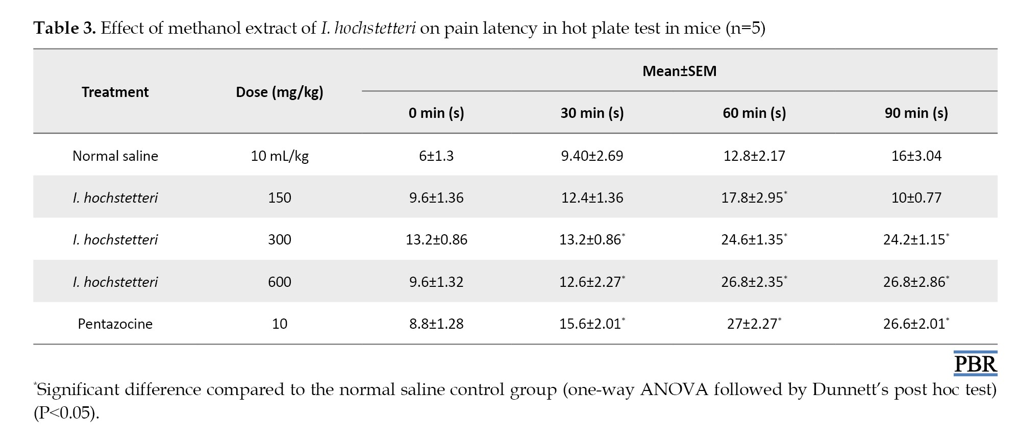
The result at 600 mg/kg was comparable to the standard drug (pentazocine).
Discussion
Pain and inflammation are the major symptoms of most common disease conditions in humans; however, the current conventional drugs are known to have several side effects. I. hochstetteri is a well-known plant in Africa, with a long history of utilization in treating diseases. Preliminary phytochemical screening of the methanol extract revealed the presence of glycosides, steroids/terpenes, flavonoids, and saponins, while tannins, alkaloids, and anthraquinones were absent. The presence of terpenes, flavonoids, saponins, alkaloids and tannins was confirmed in previous studies [21]. This disparity could be due to differences in solvents and extraction methods.
In an acute toxicity study, the results suggested that the methanol extract of I. hochstetteri has a relatively moderate acute toxicity profile, with an LD50 of approximately 2,200 mg/kg [22]. Lower doses up to 1000 mg/kg did not result in mortality, indicating a higher threshold for acute toxicity at these levels. However, doses exceeding 1600 mg/kg begin to show significant toxicity, with complete mortality at 2900 mg/kg and 5000 mg/kg (Table 1).
The formalin-induced paw edema model is a valuable tool in pharmacological research for studying acute inflammatory responses and pain mechanisms. Formalin injection into the hind paw produces a biphasic response: An early neurogenic phase, followed by a later tissue-mediated phase. Upon injection, formalin triggers acute inflammation characterized by increased vascular permeability and plasma extravasation. This process induces the release of inflammatory mediators, such as substance P, prostanoids, serotonin, and histamine, all contributing to edema formation. A neurogenic component involving transient receptor potential ankyrin 1 (TRPA1) receptors is also involved. The rapid initiation of edema is closely linked to early nociception, which relies on primary afferent neurons and axonal reflexes. The down-regulation of the inflammatory response, especially in the later stages dominated by tissue-mediated components, is primarily controlled via supraspinal. This down-regulation involves descending neuronal pathways and potentially a secondary humoral component [23-26].
The results of the anti-inflammatory effects of the methanol extract of I. hochstetteri on formalin-induced paw edema in mice showed that at the lower dose of 150 mg/kg, the extract produced a slight decrease in paw size from 4.45±0.11 mm at 0 minutes to 4.21±0.12 mm at 90 minutes. This reduction was significantly only at 90 minutes (P<0.05). At the dose of 300 mg/kg, a significant reduction in paw edema was recorded from 30 minutes (4.59±0.04 mm) and continuing through 60 minutes (4.39±0.04 mm) and 90 minutes (4.12±0.02 mm). The highest dose tested (600 mg/kg) significantly reduced the paw edema at all measured time points. Indomethacin (10 mg/kg) as the positive control also significantly reduced paw edema at all time points (Table 2). These results show that I. hochstetteri exhibits a dose-dependent anti-inflammatory effect in formalin-induced paw edema in mice. As expected, indomethacin was the most effective in reducing inflammation, showing a substantial decrease in paw size throughout the study. However, the efficacy of I. hochstetteri at higher doses approached that of indomethacin, suggesting its potential as a viable natural alternative for inflammation management.
Xylene-induced ear edema is a classical model for acute inflammatory tests used to evaluate the anti-inflammatory potential of drugs and natural products. Xylene exposure causes the upregulation and expression of cyclooxygenase-2 (COX-2), a pro-inflammatory enzyme that catalyzes prostaglandins (PGs) production, contributing to edema formation, and increases myeloperoxidase activity in the ear tissue [26]. Xylene causes a significant increase in the levels of inflammatory mediators, such as nuclear factor kappa B (NF-κB) p65, tumor necrosis factor α (TNF-α), and interleukin-1β (IL-1β) in the ear, as well as induces oxidative stress [26]. Therefore, compounds that inhibit the upregulation of COX-2, NF-κB, pro-inflammatory cytokines, and oxidative stress markers can attenuate the xylene-induced ear edema.
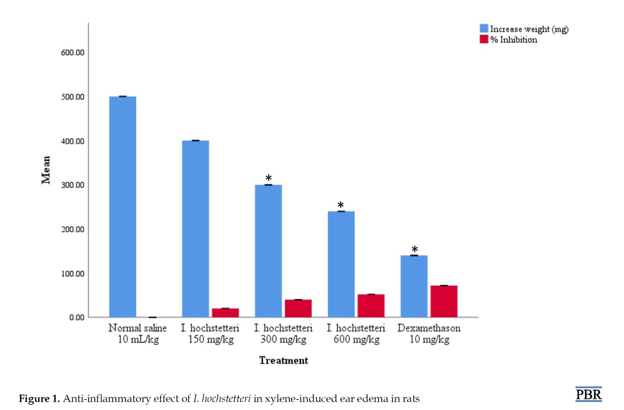
In this study, treatment with 150 mg/kg of the extract resulted in a decrease in ear weight of 400±0.31 mg, corresponding to a 20% inhibition of edema. At 300 and 600 mg/kg doses, the extract significantly reduced ear weight, achieving a 40% and 52% inhibition of inflammation, respectively. This result was statistically significant (P<0.05), demonstrating a dose-dependent and strong anti-inflammatory potential of I. hochstetteri at higher doses. The progressive decrease in ear edema with increasing extract doses suggests that higher concentrations of the bioactive compounds in I. hochstetteri are more effective at mitigating inflammation. As a positive control, dexamethasone, a well-known corticosteroid, substantially decreased ear weight, corresponding to 72% inhibition. This marked reduction (P<0.05) underscores the high efficacy of dexamethasone in reducing inflammation, serving as a benchmark for the efficacy of I. hochstetteri (Figure 1). Comparing the efficacy of I. hochstetteri with dexamethasone, it is evident that although the plant extract is less potent than the synthetic corticosteroid, it still offers considerable anti-inflammatory benefits. This is particularly relevant given the side effects associated with long-term corticosteroid use.
The above anti-inflammatory effects observed can be attributed to the phytochemicals found. The recorded phytochemicals (flavonoids, terpenes, saponins, and glycosides) have been reported to possess anti-inflammatory properties via multiple mechanisms, including targeting various inflammatory pathways, modulating inflammatory cytokines, inhibiting inflammatory and antioxidant pathways [27-35]. Furthermore, glycosides inhibited leukocyte function [34], while terpenes and saponins modulated the activity of immune cells [30, 31, 36].
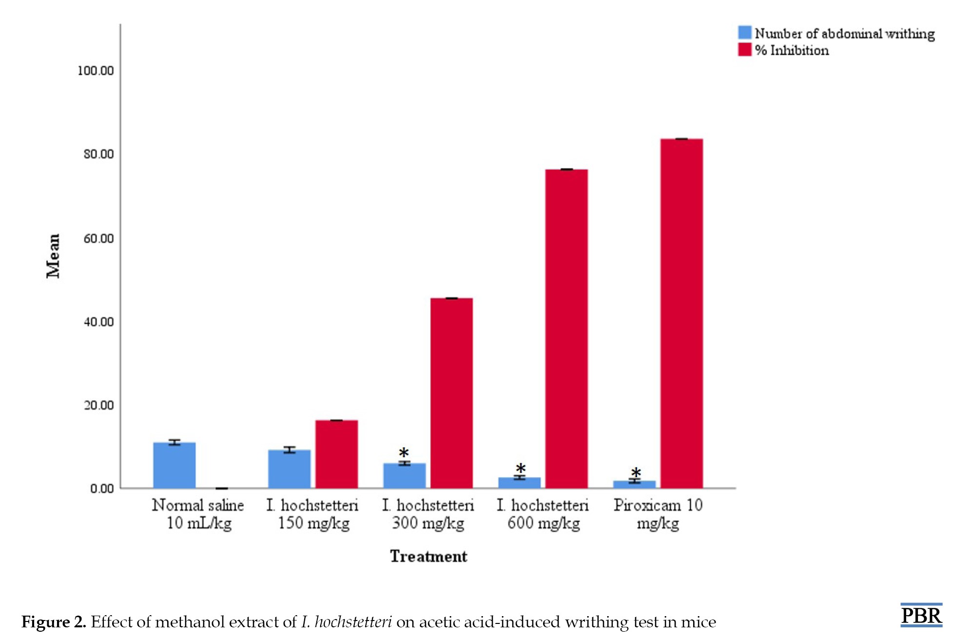
Acetic acid-induced writhing and hot plate tests were used to evaluate the analgesic effects of the extracts. Injected intraperitoneally of acetic acid induces pain by irritating the peritoneal cavity via a peripheral mechanism involving the COX pathway, causing the release of various inflammatory mediators, including PGs (particularly PG E2 [PGE2] and PG I2 [PGI2]), bradykinin, substance P, and histamine [37, 38]. The characteristic writhing or stretching behavior observed in rodents following the intraperitoneal injection of acetic acid indicates the presence of pain, and a reduction in the number of writhes indicates the analgesic effect of the tested substance [38]. In this study, administration of 150 mg/kg of I. hochstetteri resulted in a slight reduction in writhes, corresponding to a 16.3% inhibition. Although this dose showed some analgesic effect, it was not statistically significant. The extract at 300 mg/kg and 600 mg/kg, produced a significant reduction (P<0.05) in writhing, which corresponded to 45.5% and 76.3% inhibition, respectively. As a positive control, piroxicam significantly reduced the number of writhes, representing an 83.6% inhibition. The results show a clear dose-dependent analgesic effect of the methanol extract of I. hochstetteri in the acetic acid-induced writhing test. The significant reduction in the number of abdominal writhes at 300 mg/kg and 600 mg/kg indicates that I. hochstetteri has strong peripheral analgesic properties (Figure 2).
The hot plate test is a model of centrally mediated nociception widely used to assess the analgesic effects of compounds by measuring the response to a thermal nociceptive stimulus. The procedure involves placing a rodent on a hot plate maintained at a constant temperature and recording the latency to a nociceptive response, such as paw licking, paw flicking, or jumping. Longer latencies indicate less sensitivity to pain, thus an analgesic effect [39]. The results shown in Table 3 present the data on the effect of the methanol extract of the whole plant I. hochstetteri on pain latency in mice using the hot plate test. Mice treated with normal saline showed increasing pain latency over time, reflecting the natural adaptation to the hot plate stimulus without any analgesic intervention. The 150 mg/kg extract resulted in a significant pain latency only at 60 minutes, suggesting a temporary analgesic effect; however, at 300 and 600 mg/kg, there is a marked increase in pain latency at 30, 60, and 90 minutes. Therefore, the methanol extract of I. hochstetteri demonstrated significant analgesic effects in the hot plate test, with higher doses providing sustained pain relief comparable to the standard analgesic, pentazocine.
The observed analgesic effects could be attributed to the phytochemicals present in the extract. Terpenes, glycosides, saponins, and flavonoids have been reported to elicit analgesic effects by inhibiting inflammatory enzymes (such as COX-1, COX-2, and lipoxygenases [LOX]), modulating inflammatory pathways (such as NF-κB and mitogen-activated protein kinase [MAPK]), and depleting endogenous neurotransmitters [32, 40-42]. In addition, saponins interact with arachidonic acid metabolism, glycosides, terpenes, and flavonoids, which inhibit ion channels involved in pain signaling (voltage-gated ion channels, such as sodium, potassium, and calcium channels), and glycosides and flavonoids exert antioxidant properties [32, 40, 41, 43, 44].
Conclusion
The whole-plant methanol extract of I. hochstetteri showed significant potential as a natural anti-inflammatory and analgesic agent at the doses investigated. The study’s findings highlight the extract’s efficacy in reducing inflammation and alleviating pain in animal models, suggesting that it could be a viable alternative to traditional NSAIDs and opioids. The presence of active phytochemicals, such as glycosides, terpenes, flavonoids, and saponins supports its traditional use and indicates its therapeutic potential. Further research should investigate the exact mechanisms of action underlying the anti-inflammatory and analgesic effects of I. hochstetteri. The isolation and characterization of the specific bioactive compounds responsible for the observed effects could lead to developing new, targeted anti-inflammatory and analgesic therapies. Also, extended toxicity studies are necessary to assess the long-term safety profile of I. hochstetteri, particularly at higher doses.
Ethical Considerations
Compliance with ethical guidelines
The study’s ethical protocol was approved by the Faculty of Basic Medical Sciences’ Research and Ethics Committee (FBMSREC), Bauchi State University Gadau, Nigeria (Code: BASUG/FBMS/REC/VOL.07/0103).
Funding
This research did not receive any grant from funding agencies in the public, commercial, or non-profit sectors.
Authors' contributions
Conceptualization: Albashir Tahir, Musab Abba Usman, and Suleiman Yunusa; Methodology: Albashir Tahir and Suleiman Yunusa; Software: Albashir Tahir and Nura Abubakar; Validation: Albashir Tahir and Nura Abubakar; Writing the original draft: Albashir Tahir and Khadija Abdullahi Kobi; Review and editing: Albashir Tahir, Nura Abubakar and Suleiman Yunusa; Supervision: Musab Abba Usman and Suleiman Yunusa; Project administration: Albashir Tahir and Khadija Abdullahi Kobi; Final approval: All authors.
Conflict of interest
The authors declared no conflict of interest.
Acknowledgments
The authors thank the Laboratory staff of the Department of Pharmacology, Bauchi State University, Gadau, Nigeria, for their technical support.
References
Full-Text: (173 Views)
Introduction
Inflammation and pain are fundamental physiological responses that are critical components of the body's defense mechanism. Despite these protective roles, chronic pain can become pathological, often associated with depression, anxiety, and sleep disturbances [1]. Chronic inflammation, on the other hand, is a common underlying factor in many diseases, including cardiovascular diseases, cancer, autoimmune diseases, and metabolic disorders, substantially contributing to health burden [2].
Non-steroidal anti-inflammatory drugs (NSAIDs) and corticosteroids are often employed in the treatment of inflammation, while NSAIDs and opioids are used for pain management. However, prolonged use of NSAIDs can lead to gastrointestinal adverse effects, increased risk of cardiovascular events, and renal impairment, especially in patients with pre-existing kidney conditions, limiting their safety for chronic use [3]. The most pressing issues with opioid use include a wide range of adverse effects, potential for addiction and abuse, tolerance, and withdrawal symptoms, making them a less-than-ideal solution for chronic pain management [4, 5]. Long-term use of corticosteroids, which are potent anti-inflammatory agents, includes weight gain, osteoporosis, osteonecrosis, diabetes, hypertension, myopathy, cataracts, glaucoma, immunosuppression, psychiatric disturbances, gastrointestinal, endocrine, and dermatological adverse effects [6].
The search for better and safer alternatives to conventional treatments for inflammation and pain has led to renewed interest in plant-based therapies. Plants have been used for medicinal purposes for centuries, and modern research has increasingly validated their efficacy and safety [7]. Therefore, as studies continue to uncover the therapeutic potential of various plants, this study aimed to evaluate the anti-inflammatory and analgesic potential of Indigofera hochstetteri.
I. hochstetteri is a plant species belonging to the Indigofera genus within the family Fabaceae, the third largest family of land plants in terms of species, with about 19000 known species and 751 genera [8]. I. hochstetteri is found in various regions, including Kenya, Tanzania, Uganda, Chad, Congo, Djibouti, Eritrea, Ethiopia, Socotra, Somalia, Sudan, Algeria, Egypt, Angola, Zambia, Benin, Burkina, Mali, Mauritania, Nigeria, Niger, Senegal, Cameroon, Zaire, Oman, Saudi Arabia, Yemen, India, and Pakistan [9, 10]. Different parts of plants in the Indigofera genus have been widely employed in the treatment of pain and inflammatory conditions, swellings, arthritis, wounds, skin diseases, gastrointestinal disorders, respiratory diseases, malaria, menstrual cramps, dysmenorrhea, infertility, uterine fibroids, premature ejaculation, diabetes, epilepsy, liver diseases, eczema, scabies, abscess, leprosy, hepatitis, worms, gonorrhea, HIV-related symptoms, diphtheria, tuberculosis, snake bites, epistaxis, and jaundice [11].
Pharmacological studies of crude extracts and purified fractions of different Indigofera species have reported various properties, including analgesic, anti-arthritic, antidiabetic and antidyslipidemic, antidiarrheal, anti-inflammatory, antipyretic, antioxidant, antifungal, antitrypanosomal, antiplasmodial, antimicrobial, anthelmintic, antifilarial, antiulcerogenic and gastroprotective, antiviral, hepatoprotective, hypotensive, immunomodulatory, insecticidal, anticancer, anticonvulsant, neuroprotective, nephroprotective, reproprotective, genoprotective, wound-healing, and skin rejuvenating activities [11-15]. Despite the widespread usage of these plants, I. hochstetteri remains largely unexplored, resulting in a paucity of data on its medicinal potential.
Materials and Methods
Experimental animals
Swiss albino mice and Wistar rats weighing 15-38 g and 150-200 g of either sex were obtained from the National Veterinary Research Institute, Vom, Plateau State. The animals were transported humanely and housed at the Animal House of the Department of Pharmacology, Bauchi State University, Gadau, Nigeria. The animals were fed with a standard pellet diet and water ad libitum. They were kept under standard conditions (12-hour light and 12-hour dark cycle) in a propylene cage at room temperature.
Plant collection and identification
The whole plant of I. hochstetteri was collected in July 2019 at Samaru village in Zaria local government area of Kaduna state. It was identified at the collection point and further authenticated at the Herbarium Unit of the Department of Biological Sciences, Ahmadu Bello University, Zaria (voucher specimen number: 900153).
Preparation and extraction procedure
The I. hochstetteri plant was air-dried, reduced to a powder, labeled, and stored at room temperature for subsequent use. A total of 4, 162 g of the powdered plant was extracted using 70% v/v methanol using the maceration method for 7 days. The extract was evaporated in vacuo using a rotary evaporator to yield a dark brown residue (396 g), subsequently referred to as the methanol plant extract.
Preliminary phytochemical screening
The qualitative phytochemical constituents of the methanol plant extract were screened using a standard protocol [16].
Acute toxicity studies
The acute toxicity profile of I. hochstetteri was evaluated intraperitoneally in mice as Lorke (1983) described [17]. Nine mice of either sex were divided into three groups containing three mice each in the first phase and administered 10, 100, and 1000 mg/kg of the extract. In the second phase, three animals received 1600, 2900, and 5000 mg/kg of the extract based on the outcomes of the phase one study. The median lethal dose was calculated using the Equation 1:
1. LD50=√((Minimal) lethal dose×Maximal nonlethal dose)
Anti-inflammatory study
1. Formalin-induced paw edema in mice
The chronic anti-inflammatory effect of the methanol extract of I. hochstetteri was evaluated using formalin-induced paw edema as described by Eddouks et al. (2012) [18]. Twenty-five mice were divided into five groups of five animals. Thirty minutes before formalin injection, animals in group one were administered 10 mL/kg normal saline, while group five received Indomethacin at 10 mg/kg. Groups two, three, and four were administered graded doses of the extract at 150, 300, and 600 mg/kg, respectively, after which inflammation was induced by sub-plantar injection of 20 mL of freshly prepared 2% formalin in the right hind paw. Paw thickness was measured using a digital caliper at 0, 30, 60, and 90 minutes, and increases in paw thickness were recorded.
2. Xylene-induced ear edema test in rats
The method described by Hosseinzadeh and Younesi (2002) [19] was used to assess the acute anti-inflammatory effect of I. hochstetteri in rats. The animals were divided into five groups consisting of five animals each. Groups one and five served as the negative and standard control groups, respectively. Animals in group one were administered 10 ml/kg of normal saline, while those in group five received dexamethasone at 10 mg/kg. Groups two, three, and four were given graded doses of the extract at 150, 300, and 600 mg/kg, respectively. Thirty minutes after the intraperitoneal injection of the extract, 0.03 mL of xylene was applied to the anterior and posterior surfaces of the right ear. The left ear was considered a control, and 15 minutes later, the animals were euthanized. Both ears were removed and weighed, and the increase in weight caused by the irritant was calculated by taking the difference between the weights of the untreated left and treated right ear sections.
Analgesic study
1. Acetic acid-induced writhing test
For the writhing test, twenty-five mice were randomly divided into five groups of five mice. Group one was treated with normal saline 10 mL/kg, groups two, three, and four were treated with graded doses of the extract at 150, 300, and 600 mg/kg, respectively, and group five was treated with piroxicam at 10 mg/kg. All drugs were administered intraperitoneally, and 30 minutes later, each mouse was injected intraperitoneally with 10 ml/kg of 0.6% acetic acid. The animals were placed in a transparent cage and observed for 10 minutes for abdominal writhing (constriction of the abdominal muscle and stretching of the hind and limb count). The percentage inhibition of abdominal writhing was calculated using the Equation 2:
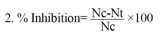
Where Nc and Nt equal the number of abdominal writhings in the control and test groups, respectively.
2. Hot plate test
The central analgesic effect of the extract was evaluated using a hot plate test as described by [20]. Before the test, mice were placed on a hot plate maintained at a temperature of 50-55 °C, and only mice exhibiting an initial nociceptive response within the first 20 seconds were included in the study. Twenty-five mice were randomly grouped into five groups, n=5. Group one was treated with 10 ml/kg of normal saline, groups two, three, and four were treated intraperitoneally with 150, 300, and 600 mg/kg of the extract, respectively, while group five was treated with Pentazocine at 10 mg/kg. All drug administrations were done via the intraperitoneal route. Thirty minutes later, animals were placed on a hot plate maintained at 50-55 °C, and the reaction time (latency) to pain response indicated through jumping or paw licking was recorded using a stopwatch at 0, 30, 60, and 90 minutes. A cut-off time of 20 s was set to reduce skin injury.
Statistical analysis
All data obtained were analyzed using one-way analysis of variance (ANOVA) followed by the Dunnet post hoc and expressed as MEAN±SEM, and percentages. Differences were considered significant at P˂0.05. Analyses were conducted using SPSS software, version 22.
Results
Percentage yield and phytoconstituents of the extract
The extraction of approximately 4,162 kg of I. hochstetteri’s whole plants yielded 396 g of the methanol extract, corresponding to a yield of 9.5%. Qualitative phytochemical assessment of the extract detected the presence of flavonoids, glycosides, steroids/terpenes, and saponins, while tannins, alkaloids, and anthraquinones were absent.
Median lethal dose value (LD50)
In phase I, no mortality was observed in mice administered 10, 100, and 1000 mg/kg doses. However, in phase II, mortality was observed at higher doses: One mouse died at 2900 mg/kg, and another at 5000 mg/kg. The 1600 mg/kg dose did not result in mortality in the mice tested (Table 1).
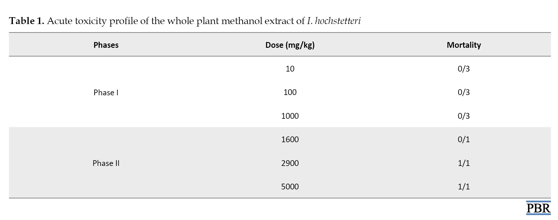
The calculated lethal dose (LD50) of the extract was approximately 2,200 mg/kg.
Methanol extract of I. hochstetteri exhibits anti-inflammatory action
1. Formalin-induced paw edema in mice
The whole plant methanol extract of I. hochstetteri at 300 and 600 mg/kg doses significantly reduced formalin-induced paw edema in mice at 30, 60, and 90 minutes. The extract at the 150 mg/kg dose only produced a significant reduction in paw edema at 90 minutes (Table 2).

2. Xylene-induced ear edema test
Figure 1 shows the effects of I.
Inflammation and pain are fundamental physiological responses that are critical components of the body's defense mechanism. Despite these protective roles, chronic pain can become pathological, often associated with depression, anxiety, and sleep disturbances [1]. Chronic inflammation, on the other hand, is a common underlying factor in many diseases, including cardiovascular diseases, cancer, autoimmune diseases, and metabolic disorders, substantially contributing to health burden [2].
Non-steroidal anti-inflammatory drugs (NSAIDs) and corticosteroids are often employed in the treatment of inflammation, while NSAIDs and opioids are used for pain management. However, prolonged use of NSAIDs can lead to gastrointestinal adverse effects, increased risk of cardiovascular events, and renal impairment, especially in patients with pre-existing kidney conditions, limiting their safety for chronic use [3]. The most pressing issues with opioid use include a wide range of adverse effects, potential for addiction and abuse, tolerance, and withdrawal symptoms, making them a less-than-ideal solution for chronic pain management [4, 5]. Long-term use of corticosteroids, which are potent anti-inflammatory agents, includes weight gain, osteoporosis, osteonecrosis, diabetes, hypertension, myopathy, cataracts, glaucoma, immunosuppression, psychiatric disturbances, gastrointestinal, endocrine, and dermatological adverse effects [6].
The search for better and safer alternatives to conventional treatments for inflammation and pain has led to renewed interest in plant-based therapies. Plants have been used for medicinal purposes for centuries, and modern research has increasingly validated their efficacy and safety [7]. Therefore, as studies continue to uncover the therapeutic potential of various plants, this study aimed to evaluate the anti-inflammatory and analgesic potential of Indigofera hochstetteri.
I. hochstetteri is a plant species belonging to the Indigofera genus within the family Fabaceae, the third largest family of land plants in terms of species, with about 19000 known species and 751 genera [8]. I. hochstetteri is found in various regions, including Kenya, Tanzania, Uganda, Chad, Congo, Djibouti, Eritrea, Ethiopia, Socotra, Somalia, Sudan, Algeria, Egypt, Angola, Zambia, Benin, Burkina, Mali, Mauritania, Nigeria, Niger, Senegal, Cameroon, Zaire, Oman, Saudi Arabia, Yemen, India, and Pakistan [9, 10]. Different parts of plants in the Indigofera genus have been widely employed in the treatment of pain and inflammatory conditions, swellings, arthritis, wounds, skin diseases, gastrointestinal disorders, respiratory diseases, malaria, menstrual cramps, dysmenorrhea, infertility, uterine fibroids, premature ejaculation, diabetes, epilepsy, liver diseases, eczema, scabies, abscess, leprosy, hepatitis, worms, gonorrhea, HIV-related symptoms, diphtheria, tuberculosis, snake bites, epistaxis, and jaundice [11].
Pharmacological studies of crude extracts and purified fractions of different Indigofera species have reported various properties, including analgesic, anti-arthritic, antidiabetic and antidyslipidemic, antidiarrheal, anti-inflammatory, antipyretic, antioxidant, antifungal, antitrypanosomal, antiplasmodial, antimicrobial, anthelmintic, antifilarial, antiulcerogenic and gastroprotective, antiviral, hepatoprotective, hypotensive, immunomodulatory, insecticidal, anticancer, anticonvulsant, neuroprotective, nephroprotective, reproprotective, genoprotective, wound-healing, and skin rejuvenating activities [11-15]. Despite the widespread usage of these plants, I. hochstetteri remains largely unexplored, resulting in a paucity of data on its medicinal potential.
Materials and Methods
Experimental animals
Swiss albino mice and Wistar rats weighing 15-38 g and 150-200 g of either sex were obtained from the National Veterinary Research Institute, Vom, Plateau State. The animals were transported humanely and housed at the Animal House of the Department of Pharmacology, Bauchi State University, Gadau, Nigeria. The animals were fed with a standard pellet diet and water ad libitum. They were kept under standard conditions (12-hour light and 12-hour dark cycle) in a propylene cage at room temperature.
Plant collection and identification
The whole plant of I. hochstetteri was collected in July 2019 at Samaru village in Zaria local government area of Kaduna state. It was identified at the collection point and further authenticated at the Herbarium Unit of the Department of Biological Sciences, Ahmadu Bello University, Zaria (voucher specimen number: 900153).
Preparation and extraction procedure
The I. hochstetteri plant was air-dried, reduced to a powder, labeled, and stored at room temperature for subsequent use. A total of 4, 162 g of the powdered plant was extracted using 70% v/v methanol using the maceration method for 7 days. The extract was evaporated in vacuo using a rotary evaporator to yield a dark brown residue (396 g), subsequently referred to as the methanol plant extract.
Preliminary phytochemical screening
The qualitative phytochemical constituents of the methanol plant extract were screened using a standard protocol [16].
Acute toxicity studies
The acute toxicity profile of I. hochstetteri was evaluated intraperitoneally in mice as Lorke (1983) described [17]. Nine mice of either sex were divided into three groups containing three mice each in the first phase and administered 10, 100, and 1000 mg/kg of the extract. In the second phase, three animals received 1600, 2900, and 5000 mg/kg of the extract based on the outcomes of the phase one study. The median lethal dose was calculated using the Equation 1:
1. LD50=√((Minimal) lethal dose×Maximal nonlethal dose)
Anti-inflammatory study
1. Formalin-induced paw edema in mice
The chronic anti-inflammatory effect of the methanol extract of I. hochstetteri was evaluated using formalin-induced paw edema as described by Eddouks et al. (2012) [18]. Twenty-five mice were divided into five groups of five animals. Thirty minutes before formalin injection, animals in group one were administered 10 mL/kg normal saline, while group five received Indomethacin at 10 mg/kg. Groups two, three, and four were administered graded doses of the extract at 150, 300, and 600 mg/kg, respectively, after which inflammation was induced by sub-plantar injection of 20 mL of freshly prepared 2% formalin in the right hind paw. Paw thickness was measured using a digital caliper at 0, 30, 60, and 90 minutes, and increases in paw thickness were recorded.
2. Xylene-induced ear edema test in rats
The method described by Hosseinzadeh and Younesi (2002) [19] was used to assess the acute anti-inflammatory effect of I. hochstetteri in rats. The animals were divided into five groups consisting of five animals each. Groups one and five served as the negative and standard control groups, respectively. Animals in group one were administered 10 ml/kg of normal saline, while those in group five received dexamethasone at 10 mg/kg. Groups two, three, and four were given graded doses of the extract at 150, 300, and 600 mg/kg, respectively. Thirty minutes after the intraperitoneal injection of the extract, 0.03 mL of xylene was applied to the anterior and posterior surfaces of the right ear. The left ear was considered a control, and 15 minutes later, the animals were euthanized. Both ears were removed and weighed, and the increase in weight caused by the irritant was calculated by taking the difference between the weights of the untreated left and treated right ear sections.
Analgesic study
1. Acetic acid-induced writhing test
For the writhing test, twenty-five mice were randomly divided into five groups of five mice. Group one was treated with normal saline 10 mL/kg, groups two, three, and four were treated with graded doses of the extract at 150, 300, and 600 mg/kg, respectively, and group five was treated with piroxicam at 10 mg/kg. All drugs were administered intraperitoneally, and 30 minutes later, each mouse was injected intraperitoneally with 10 ml/kg of 0.6% acetic acid. The animals were placed in a transparent cage and observed for 10 minutes for abdominal writhing (constriction of the abdominal muscle and stretching of the hind and limb count). The percentage inhibition of abdominal writhing was calculated using the Equation 2:
Where Nc and Nt equal the number of abdominal writhings in the control and test groups, respectively.
2. Hot plate test
The central analgesic effect of the extract was evaluated using a hot plate test as described by [20]. Before the test, mice were placed on a hot plate maintained at a temperature of 50-55 °C, and only mice exhibiting an initial nociceptive response within the first 20 seconds were included in the study. Twenty-five mice were randomly grouped into five groups, n=5. Group one was treated with 10 ml/kg of normal saline, groups two, three, and four were treated intraperitoneally with 150, 300, and 600 mg/kg of the extract, respectively, while group five was treated with Pentazocine at 10 mg/kg. All drug administrations were done via the intraperitoneal route. Thirty minutes later, animals were placed on a hot plate maintained at 50-55 °C, and the reaction time (latency) to pain response indicated through jumping or paw licking was recorded using a stopwatch at 0, 30, 60, and 90 minutes. A cut-off time of 20 s was set to reduce skin injury.
Statistical analysis
All data obtained were analyzed using one-way analysis of variance (ANOVA) followed by the Dunnet post hoc and expressed as MEAN±SEM, and percentages. Differences were considered significant at P˂0.05. Analyses were conducted using SPSS software, version 22.
Results
Percentage yield and phytoconstituents of the extract
The extraction of approximately 4,162 kg of I. hochstetteri’s whole plants yielded 396 g of the methanol extract, corresponding to a yield of 9.5%. Qualitative phytochemical assessment of the extract detected the presence of flavonoids, glycosides, steroids/terpenes, and saponins, while tannins, alkaloids, and anthraquinones were absent.
Median lethal dose value (LD50)
In phase I, no mortality was observed in mice administered 10, 100, and 1000 mg/kg doses. However, in phase II, mortality was observed at higher doses: One mouse died at 2900 mg/kg, and another at 5000 mg/kg. The 1600 mg/kg dose did not result in mortality in the mice tested (Table 1).
The calculated lethal dose (LD50) of the extract was approximately 2,200 mg/kg.
Methanol extract of I. hochstetteri exhibits anti-inflammatory action
1. Formalin-induced paw edema in mice
The whole plant methanol extract of I. hochstetteri at 300 and 600 mg/kg doses significantly reduced formalin-induced paw edema in mice at 30, 60, and 90 minutes. The extract at the 150 mg/kg dose only produced a significant reduction in paw edema at 90 minutes (Table 2).
2. Xylene-induced ear edema test
Figure 1 shows the effects of I.
hochstetteri on xylene-induced acute inflammation in rats. Compared to the control group, the inhibition produced by the extract at 300 and 600 mg/kg doses was significant (P<0.05), with percentage inhibitions of 40% and 52%, respectively. Dexamethasone treatment (10 mg/kg) inhibited ear edema with an inhibition rate of 72% (P<0.05).
Methanol extract of I. hochstetteri exhibits both peripheral and central analgesia
1. Acetic acid-induced writhing test
The methanol extract of I. hochstetteri (300 and 600 mg/kg) significantly reduced the number of abdominal writhings in mice. The analgesic effect of the extract was dose-dependent (Figure 2).
2. Hot plate test
The results of the hot plate-induced nociception in mice (Table 3) showed that the I. hochstetteri whole-plant extract significantly delayed the reaction times to pain for over 90 min of the study.
The result at 600 mg/kg was comparable to the standard drug (pentazocine).
Discussion
Pain and inflammation are the major symptoms of most common disease conditions in humans; however, the current conventional drugs are known to have several side effects. I. hochstetteri is a well-known plant in Africa, with a long history of utilization in treating diseases. Preliminary phytochemical screening of the methanol extract revealed the presence of glycosides, steroids/terpenes, flavonoids, and saponins, while tannins, alkaloids, and anthraquinones were absent. The presence of terpenes, flavonoids, saponins, alkaloids and tannins was confirmed in previous studies [21]. This disparity could be due to differences in solvents and extraction methods.
In an acute toxicity study, the results suggested that the methanol extract of I. hochstetteri has a relatively moderate acute toxicity profile, with an LD50 of approximately 2,200 mg/kg [22]. Lower doses up to 1000 mg/kg did not result in mortality, indicating a higher threshold for acute toxicity at these levels. However, doses exceeding 1600 mg/kg begin to show significant toxicity, with complete mortality at 2900 mg/kg and 5000 mg/kg (Table 1).
The formalin-induced paw edema model is a valuable tool in pharmacological research for studying acute inflammatory responses and pain mechanisms. Formalin injection into the hind paw produces a biphasic response: An early neurogenic phase, followed by a later tissue-mediated phase. Upon injection, formalin triggers acute inflammation characterized by increased vascular permeability and plasma extravasation. This process induces the release of inflammatory mediators, such as substance P, prostanoids, serotonin, and histamine, all contributing to edema formation. A neurogenic component involving transient receptor potential ankyrin 1 (TRPA1) receptors is also involved. The rapid initiation of edema is closely linked to early nociception, which relies on primary afferent neurons and axonal reflexes. The down-regulation of the inflammatory response, especially in the later stages dominated by tissue-mediated components, is primarily controlled via supraspinal. This down-regulation involves descending neuronal pathways and potentially a secondary humoral component [23-26].
The results of the anti-inflammatory effects of the methanol extract of I. hochstetteri on formalin-induced paw edema in mice showed that at the lower dose of 150 mg/kg, the extract produced a slight decrease in paw size from 4.45±0.11 mm at 0 minutes to 4.21±0.12 mm at 90 minutes. This reduction was significantly only at 90 minutes (P<0.05). At the dose of 300 mg/kg, a significant reduction in paw edema was recorded from 30 minutes (4.59±0.04 mm) and continuing through 60 minutes (4.39±0.04 mm) and 90 minutes (4.12±0.02 mm). The highest dose tested (600 mg/kg) significantly reduced the paw edema at all measured time points. Indomethacin (10 mg/kg) as the positive control also significantly reduced paw edema at all time points (Table 2). These results show that I. hochstetteri exhibits a dose-dependent anti-inflammatory effect in formalin-induced paw edema in mice. As expected, indomethacin was the most effective in reducing inflammation, showing a substantial decrease in paw size throughout the study. However, the efficacy of I. hochstetteri at higher doses approached that of indomethacin, suggesting its potential as a viable natural alternative for inflammation management.
Xylene-induced ear edema is a classical model for acute inflammatory tests used to evaluate the anti-inflammatory potential of drugs and natural products. Xylene exposure causes the upregulation and expression of cyclooxygenase-2 (COX-2), a pro-inflammatory enzyme that catalyzes prostaglandins (PGs) production, contributing to edema formation, and increases myeloperoxidase activity in the ear tissue [26]. Xylene causes a significant increase in the levels of inflammatory mediators, such as nuclear factor kappa B (NF-κB) p65, tumor necrosis factor α (TNF-α), and interleukin-1β (IL-1β) in the ear, as well as induces oxidative stress [26]. Therefore, compounds that inhibit the upregulation of COX-2, NF-κB, pro-inflammatory cytokines, and oxidative stress markers can attenuate the xylene-induced ear edema.
In this study, treatment with 150 mg/kg of the extract resulted in a decrease in ear weight of 400±0.31 mg, corresponding to a 20% inhibition of edema. At 300 and 600 mg/kg doses, the extract significantly reduced ear weight, achieving a 40% and 52% inhibition of inflammation, respectively. This result was statistically significant (P<0.05), demonstrating a dose-dependent and strong anti-inflammatory potential of I. hochstetteri at higher doses. The progressive decrease in ear edema with increasing extract doses suggests that higher concentrations of the bioactive compounds in I. hochstetteri are more effective at mitigating inflammation. As a positive control, dexamethasone, a well-known corticosteroid, substantially decreased ear weight, corresponding to 72% inhibition. This marked reduction (P<0.05) underscores the high efficacy of dexamethasone in reducing inflammation, serving as a benchmark for the efficacy of I. hochstetteri (Figure 1). Comparing the efficacy of I. hochstetteri with dexamethasone, it is evident that although the plant extract is less potent than the synthetic corticosteroid, it still offers considerable anti-inflammatory benefits. This is particularly relevant given the side effects associated with long-term corticosteroid use.
The above anti-inflammatory effects observed can be attributed to the phytochemicals found. The recorded phytochemicals (flavonoids, terpenes, saponins, and glycosides) have been reported to possess anti-inflammatory properties via multiple mechanisms, including targeting various inflammatory pathways, modulating inflammatory cytokines, inhibiting inflammatory and antioxidant pathways [27-35]. Furthermore, glycosides inhibited leukocyte function [34], while terpenes and saponins modulated the activity of immune cells [30, 31, 36].
Acetic acid-induced writhing and hot plate tests were used to evaluate the analgesic effects of the extracts. Injected intraperitoneally of acetic acid induces pain by irritating the peritoneal cavity via a peripheral mechanism involving the COX pathway, causing the release of various inflammatory mediators, including PGs (particularly PG E2 [PGE2] and PG I2 [PGI2]), bradykinin, substance P, and histamine [37, 38]. The characteristic writhing or stretching behavior observed in rodents following the intraperitoneal injection of acetic acid indicates the presence of pain, and a reduction in the number of writhes indicates the analgesic effect of the tested substance [38]. In this study, administration of 150 mg/kg of I. hochstetteri resulted in a slight reduction in writhes, corresponding to a 16.3% inhibition. Although this dose showed some analgesic effect, it was not statistically significant. The extract at 300 mg/kg and 600 mg/kg, produced a significant reduction (P<0.05) in writhing, which corresponded to 45.5% and 76.3% inhibition, respectively. As a positive control, piroxicam significantly reduced the number of writhes, representing an 83.6% inhibition. The results show a clear dose-dependent analgesic effect of the methanol extract of I. hochstetteri in the acetic acid-induced writhing test. The significant reduction in the number of abdominal writhes at 300 mg/kg and 600 mg/kg indicates that I. hochstetteri has strong peripheral analgesic properties (Figure 2).
The hot plate test is a model of centrally mediated nociception widely used to assess the analgesic effects of compounds by measuring the response to a thermal nociceptive stimulus. The procedure involves placing a rodent on a hot plate maintained at a constant temperature and recording the latency to a nociceptive response, such as paw licking, paw flicking, or jumping. Longer latencies indicate less sensitivity to pain, thus an analgesic effect [39]. The results shown in Table 3 present the data on the effect of the methanol extract of the whole plant I. hochstetteri on pain latency in mice using the hot plate test. Mice treated with normal saline showed increasing pain latency over time, reflecting the natural adaptation to the hot plate stimulus without any analgesic intervention. The 150 mg/kg extract resulted in a significant pain latency only at 60 minutes, suggesting a temporary analgesic effect; however, at 300 and 600 mg/kg, there is a marked increase in pain latency at 30, 60, and 90 minutes. Therefore, the methanol extract of I. hochstetteri demonstrated significant analgesic effects in the hot plate test, with higher doses providing sustained pain relief comparable to the standard analgesic, pentazocine.
The observed analgesic effects could be attributed to the phytochemicals present in the extract. Terpenes, glycosides, saponins, and flavonoids have been reported to elicit analgesic effects by inhibiting inflammatory enzymes (such as COX-1, COX-2, and lipoxygenases [LOX]), modulating inflammatory pathways (such as NF-κB and mitogen-activated protein kinase [MAPK]), and depleting endogenous neurotransmitters [32, 40-42]. In addition, saponins interact with arachidonic acid metabolism, glycosides, terpenes, and flavonoids, which inhibit ion channels involved in pain signaling (voltage-gated ion channels, such as sodium, potassium, and calcium channels), and glycosides and flavonoids exert antioxidant properties [32, 40, 41, 43, 44].
Conclusion
The whole-plant methanol extract of I. hochstetteri showed significant potential as a natural anti-inflammatory and analgesic agent at the doses investigated. The study’s findings highlight the extract’s efficacy in reducing inflammation and alleviating pain in animal models, suggesting that it could be a viable alternative to traditional NSAIDs and opioids. The presence of active phytochemicals, such as glycosides, terpenes, flavonoids, and saponins supports its traditional use and indicates its therapeutic potential. Further research should investigate the exact mechanisms of action underlying the anti-inflammatory and analgesic effects of I. hochstetteri. The isolation and characterization of the specific bioactive compounds responsible for the observed effects could lead to developing new, targeted anti-inflammatory and analgesic therapies. Also, extended toxicity studies are necessary to assess the long-term safety profile of I. hochstetteri, particularly at higher doses.
Ethical Considerations
Compliance with ethical guidelines
The study’s ethical protocol was approved by the Faculty of Basic Medical Sciences’ Research and Ethics Committee (FBMSREC), Bauchi State University Gadau, Nigeria (Code: BASUG/FBMS/REC/VOL.07/0103).
Funding
This research did not receive any grant from funding agencies in the public, commercial, or non-profit sectors.
Authors' contributions
Conceptualization: Albashir Tahir, Musab Abba Usman, and Suleiman Yunusa; Methodology: Albashir Tahir and Suleiman Yunusa; Software: Albashir Tahir and Nura Abubakar; Validation: Albashir Tahir and Nura Abubakar; Writing the original draft: Albashir Tahir and Khadija Abdullahi Kobi; Review and editing: Albashir Tahir, Nura Abubakar and Suleiman Yunusa; Supervision: Musab Abba Usman and Suleiman Yunusa; Project administration: Albashir Tahir and Khadija Abdullahi Kobi; Final approval: All authors.
Conflict of interest
The authors declared no conflict of interest.
Acknowledgments
The authors thank the Laboratory staff of the Department of Pharmacology, Bauchi State University, Gadau, Nigeria, for their technical support.
References
- Stretanski MF, Kopitnik NL, Matha A, Conermann T. Chronic pain. [Updated 2025 Jun 23]. In: StatPearls [Internet]. Treasure Island (FL): StatPearls Publishing; 2025 Jan-. Available from: [Link]
- Pahwa R, Goyal A, Jialal I. Chronic inflammation. 2023 Aug 7. In: StatPearls [Internet]. Treasure Island (FL): StatPearls Publishing; 2025. [PMID]
- Saad J, Mathew D. Nonsteroidal anti-inflammatory drugs toxicity. 2025 Sep 15. In: StatPearls [Internet]. Treasure Island (FL): StatPearls Publishing; 2025. [PMID]
- Crockett SD, Greer KB, Heidelbaugh JJ, Falck-Ytter Y, Hanson BJ, Sultan S, et al. American Gastroenterological Association Institute Guideline on the Medical Management of Opioid-Induced Constipation. Gastroenterology. 2019; 156(1):218-26. [DOI:10.1053/j.gastro.2018.07.016] [PMID]
- Cohen B, Ruth LJ, Preuss CV. Opioid Analgesics. 2023 Apr 29. In: StatPearls [Internet]. Treasure Island (FL): StatPearls Publishing; 2025. [PMID]
- Hodgens A, Sharman T. Corticosteroids. 2023 May 1. In: StatPearls [Internet]. Treasure Island (FL): StatPearls Publishing; 2025 Jan. [PMID]
- Mensah MLK, Komlaga G, Forkuo A, Firempong C, Anning A, Dickson RA. Toxicity and Safety Implications of Herbal Medicines Used in Africa. In: Builders P, editor. Herbal Medicine. London: IntechOpen; 2019. [DOI:10.5772/intechopen.72437]
- Christenhusz MJM, Byng JW. The number of known plants species in the world and its annual increase. Phytotaxa. 2016; 261:201-17. [DOI:10.11646/phytotaxa.261.3.1]
- Govaerts R. World checklist of vascular plants (WCVP) -version 12. Melbourne: Royal Botanic Gardens, Kew Research Repository; 2023. [Link]
- Open Herbarium. Indigofera hochstetter. [Internet]. 2024 [Updated 2024 May 25]. Available from: [Link]
- Gerometta E, Grondin I, Smadja J, Frederich M, Gauvin-Bialecki A. A review of traditional uses, phytochemistry and pharmacology of the genus Indigofera. J Ethnopharmacol. 2020; 253:112608. [DOI:10.1016/j.jep.2020.112608] [PMID]
- Prabhu N, Harshini D, Gowsalya A, Rekha J, Rajamehala M, Karthick PJ, et al. Evaluation of Genoprotective Activity of Indigofera tinctoria using Allium cepa Root. J Pharm Res Int. 2021; 33(60B):2958-72. [DOI:10.9734/jpri/2021/v33i60B34965]
- Haider MS, Imran I. Pharmacological investigation of activities pertaining to modulation of gastrointestinal, respiratory and cardiovascular parameters by Indigofera argentea in experimental models. Pak J Pharm Sci. 2020; 33(5(Supplementary)):2257-67. [PMID]
- Rakwa EE, Koubala BB, Mando BN, Djongra M, Nveikoueing F, Ndjonka D. Antifilarial Activity of the Methanolic Extract of Indigofera tinctoria (Fabaceae) on Bovine Parasites (Onchocerca ochengi). J Parasitol Res. 2022; 2022:7828551. [DOI:10.1155/2022/7828551] [PMID]
- Bhat SA, Zargar MI, Wani SUD, Mohiuddin I, Masoodi MH, Shakeel F, et al. In-vitro evaluation of Indigofera heterantha extracts for antibacterial, antifungal and anthelmintic activities. J Pharm Health Care Sci. 2024; ;10(1):7. [DOI:10.1186/s40780-024-00328-y] [PMID]
- Evans WC, Trease GE, Evans D. Trease and Evans’ pharmacognosy. Edinburgh ; New York: WB Saunders; 2002. [Link]
- Lorke D. A new approach to practical acute toxicity testing. Arch Toxicol. 1983; 54:275–87. [DOI:10.1007/BF01234480]
- Eddouks M, Chattopadhyay D, Zeggwagh NA. Animal models as tools to investigate antidiabetic and anti-inflammatory plants. Evid Based Complement Alternat Med. 2012; 2012:142087. [DOI:10.1155/2012/142087] [PMID]
- Hosseinzadeh H, Younesi HM. Antinociceptive and anti-inflammatory effects of Crocus sativus L. stigma and petal extracts in mice. BMC Pharmacol. 2002; 2:7. [PMID]
- Eddy NB, Leimbach D. Synthetic analgesics. II. Dithienylbutenyl- and dithienylbutylamines. J Pharmacol Exp Ther. 1953; 107(3):385-93. [DOI:10.1016/S0022-3565(25)05180-8]
- Atta E, Al faifi T, El-Shabasy A. Chemotaxonomic and morphological classification of six Indigofera species in Jazan region, KSA. J Saudi Chem Soc. 2022; 26(3):101476. [10.1016/j.jscs.2022.101476]
- Hodge HC, Sterner JH. Tabulation of toxicity classes. Am Ind Hyg Assoc Q. 1949; 10(4):93-6. [DOI:10.1080/00968204909344159] [PMID]
- Wheeler-Aceto H, Cowan A. Neurogenic and tissue-mediated components of formalin-induced edema: Evidence for supraspinal regulation. Agents Actions. 1991; 34(1-2):264-9. [DOI:10.1007/BF01993299] [PMID]
- Damas J, Liégeois JF. The inflammatory reaction induced by formalin in the rat paw. Naunyn Schmiedebergs Arch Pharmacol. 1999; 359(3):220-7. [DOI:10.1007/pl00005345] [PMID]
- McNamara CR, Mandel-Brehm J, Bautista DM, Siemens J, Deranian KL, Zhao M, Hayward NJ, et al. TRPA1 mediates formalin-induced pain. Proc Natl Acad Sci U S A. 2007; (33):13525-30. [DOI:10.1073/pnas.0705924104] [PMID]
- Soliman SM, Teaima MH, Rashwan KO, Ali BM, Jasti BR, El-Nabarawi MA, et al. The deleterious effect of xylene-induced ear edema in rats: Protective role of dexketoprofen trometamol transdermal invasomes via inhibiting the oxidative stress/NF-κB/COX-2 pathway. Int J Pharm. 2023; 631:122525. [DOI:10.1016/j.ijpharm.2022.122525] [PMID]
- Al-Khayri JM, Sahana GR, Nagella P, Joseph BV, Alessa FM, Al-Mssallem MQ. Flavonoids as potential anti-inflammatory molecules: A review. Molecules. 2022; 27(9):2901. [DOI:10.3390/molecules27092901] [PMID]
- Ahmadi M, Bekeschus S, Weltmann KD, von Woedtke T, Wende K. Non-steroidal anti-inflammatory drugs: Recent advances in the use of synthetic COX-2 inhibitors. RSC Med Chem. 2022; 13(5):471-96. [DOI:10.1039/d1md00280e] [PMID]
- Ju Z, Li M, Xu J, Howell DC, Li Z, Chen FE. Recent development on COX-2 inhibitors as promising anti-inflammatory agents: The past 10 years. Acta Pharm Sin B. 2022 ; 12(6):2790-807. [DOI:10.1016/j.apsb.2022.01.002] [PMID]
- Del Prado-Audelo ML, Cortés H, Caballero-Florán IH, González-Torres M, Escutia-Guadarrama L, Bernal-Chávez SA, et al. Therapeutic Applications of Terpenes on Inflammatory Diseases. Front Pharmacol. 2021; 12:704197. [DOI:10.3389/fphar.2021.704197] [PMID]
- Prakash V. Terpenoids as Source of Anti-inflammatory Compounds. Asian J Pharm Clin Res 2017; 10(3):68-76. [DOI:10.22159/ajpcr.2017.v10i3.16435.]
- Souza MT, Almeida JR, Araujo AA, Duarte MC, Gelain DP, Moreira JC, et al. Structure–activity relationship of terpenes with anti-inflammatory profile – a systematic review. Basic Clin Pharmacol Toxicol. 2014; (3):244-56. [DOI:10.1111/bcpt.12221] [PMID]
- Wijesekara T, Luo J, Xu B. Critical review on anti-inflammation effects of saponins and their molecular mechanisms. Phytother Res. 2024; 38(4):2007-22. [DOI:10.1002/ptr.8164] [PMID]
- Fürst R, Zündorf I, Dingermann T. New Knowledge About Old Drugs: The Anti-Inflammatory Properties of Cardiac Glycosides. Planta Med. 2017; 83(12-13):977-84. [DOI:10.1055/s-0043-105390] [PMID]
- Zhang D, Liu R, Sun L, Huang C, Wang C, Zhang DM, et al. Anti-Inflammatory Activity of Methyl Salicylate Glycosides Isolated from Gaultheria yunnanensis (Franch.) Rehder. Molecules. 2011; 16(5):3875-84. [DOI:10.3390/molecules16053875] [PMID]
- Avunduk S. Chapter 7 - Antiinflammatory saponins. In: Atta-Ur-Rahman, editor. Studies in Natural Products Chemistry. Amsterdam: Elsevier; 2024. [Link]
- Ricciotti E, FitzGerald GA. Prostaglandins and inflammation. Arterioscler Thromb Vasc Biol. 2011; 31(5):986-1000. [DOI:10.1161/ATVBAHA.110.207449] [PMID]
- Gawade SP. Acetic acid induced painful endogenous infliction in writhing test on mice. J Pharmacol Pharmacother. 2012; 3(4):348. [DOI:10.4103/0976-500X.103699] [PMID]
- Abubakar H, Tahir A, Umar A. Boswellia dalzielii Methanol Stem Bark Extract Demonstrates Significant Analgesic Activity in Swiss Albino Mice. Sci Phytochem. 2024; 3(1):38-43. [DOI:10.58920/sciphy0301225]
- Ferraz CR, Carvalho TT, Manchope MF, Artero NA, Rasquel-Oliveira FS, Fattori V, et al. Therapeutic Potential of Flavonoids in Pain and Inflammation: Mechanisms of Action, Pre-Clinical and Clinical Data, and Pharmaceutical Development. Molecules. 2020; 25(3):762. [DOI:10.3390/molecules25030762] [PMID]
- Zheng Y, Yin X, Huo F, Xiong H, Mei Z. Analgesic effects and possible mechanisms of iridoid glycosides from Lamiophlomis rotata (Benth.) Kudo in rats with spared nerve injury. J Ethnopharmacol. 2015; 173:204-11. [DOI:10.1016/j.jep.2015.06.045] [PMID]
- Chindo B, Anuka J, Isaac E, Ahmadu A, Tarfa F, Gamaniel K. Saponins are involved in the analgesic and anti-inflammatory properties of Ficus platyphylla stem bark. Int J Biol Chem Sci. 2010; 4(2). [DOI:10.4314/ijbcs.v4i2.58140]
- Xiao X, Wang X, Gui X, Chen L, Huang B. Natural Flavonoids as Promising Analgesic Candidates: A Systematic Review. Chem Biodivers. 2016; 13(11):1427-40. [DOI:10.1002/cbdv.201600060] [PMID]
- Khan H, Pervaiz A, Intagliata S, Das N, Nagulapalli Venkata KC, Atanasov AG, et al. The analgesic potential of glycosides derived from medicinal plants. DARU. 2020; 28(1):387-401. [DOI:10.1007/s40199-019-00319-7] [PMID]
Type of Study: Original Research |
Subject:
Ehtnopharmacology
| Rights and permissions | |
 |
This work is licensed under a Creative Commons Attribution-NonCommercial 4.0 International License. |

This is an open access article distributed under the terms of the Creative Commons Attribution License (CC-By-NC), which permits use, distribution, and reproduction in any medium, provided the original work is properly cited and is not used for commercial purposes.
Contact Information
